Abstract
Background
Ichthyosaurs are Mesozoic reptiles considered as active swimmers highly adapted to a fully open-marine life. They display a wide range of morphologies illustrating diverse ecological grades. Data concerning their bone microanatomical and histological features are rather limited and suggest that ichthyosaurs display a spongious, “osteoporotic-like” bone inner structure, like extant cetaceans. However, some taxa exhibit peculiar features, suggesting that the analysis of the microanatomical and histological characteristics of various ichthyosaur long bones should match the anatomical diversity and provide information about their diverse locomotor abilities and physiology.
Methodology/Principal Findings
The material analyzed for this study essentially consists of mid-diaphyseal transverse sections from stylopod bones of various ichthyosaurs and of a few microtomographic (both conventional and synchrotron) data. The present contribution discusses the histological and microanatomical variation observed within ichthyosaurs and the peculiarities of some taxa (Mixosaurus, Pessopteryx). Four microanatomical types are described. If Mixosaurus sections differ from those of the other taxa analyzed, the other microanatomical types, characterized by the relative proportion of compact and loose spongiosa of periosteal and endochondral origin respectively, seem to rather especially illustrate variation along the diaphysis in taxa with similar microanatomical features. Our analysis also reveals that primary bone in all the ichthyosaur taxa sampled (to the possible exception of Mixosaurus) is spongy in origin, that cyclical growth is a common pattern among ichthyosaurs, and confirms the previous assumptions of high growth rates in ichthyosaurs.
Conclusions/Significance
The occurrence of two types of remodelling patterns along the diaphysis, characterized by bone mass decrease and increase respectively is described for the first time. It raises questions about the definition of the osseous microanatomical specializations bone mass increase and osteoporosis, notably based on the processes involved, and reveals the difficulty in determining the true occurrence of these osseous specializations in ichthyosaurs.
Introduction
Ichthyosaurs represent one of the most successful groups of Mesozoic marine reptiles, as shown by their cosmopolitan distribution and their extensive fossil record [1]–[3]. They lived from the Early Triassic to the early Late Cretaceous, i.e. from about 245 to 90 million years ago. Ichthyosaurs are among the first air-breathing vertebrates that adapted to a pelagic life style [3]. These latter forms are considered as the reptiles most strongly morphologically adapted to a fully open-marine life. Among extant aquatic amniotes, only cetaceans are as highly modified for a pelagic lifestyle as ichthyosaurs were. Ichthyosaurs appear thus as a particularly interesting group to understand the evolutionary processes involved in secondary adaptation to an aquatic life.
Although ichthyosaurs are very often represented as dolphin-like or tuna-shaped, they display a wide range of morphologies illustrating diverse ecological grades. The earliest forms, showing a long, slender body with a straight and long tail (cf. Utatsusaurus), were probably anguilliform swimmers [4]. Conversely, most of the post-Triassic forms display a fusiform stiff body with an upright bilobate (fish-like) tail on a narrow peduncle (cf. Stenopterygius) and are considered as thunniform swimmers [5], whereas the Middle Triassic taxon Mixosaurus displays an intermediary pattern [4]. Several additional intermediary morphologies between these two ‘extremes’ (with differences for example in body size, shape, elongation and flexibility) were illustrated (e.g., [6], [7]).
Bone microanatomical organization mainly relies on the biomechanical constraints undergone by organisms (e.g., [8]–[12]). The analysis of the microanatomical characteristics of various ichthyosaur long bones should thus provide information about their locomotor abilities. Data concerning ichthyosaur bone microanatomical and histological features consist only of a few long bone, vertebra and rib sections (except for Mixosaurus, for which more bones were analyzed; see [13]) of Utatsusaurus, Mixosaurus, Pessopteryx, Caypullisaurus, Mollesaurus, Stenopterygius, Ichthyosaurus and Platypterygius (misidentified as Ichthyosaurus by Kiprijanoff, [14]) [13], [15]–[24]. Although representing several genera, the data are too heterogeneous to permit significant intrageneric comparisons, as well as homologous intergeneric ones.
A comment on Pessopteryx is in order here because it is noteworthy that this material was assigned to Omphalosaurus in earlier histological studies [18], [19]. Pessopteryx is a taxon erected by Wiman [25] for cranial and limb material found together in the Lower Triassic of Spitsbergen. The cranial part of this material is now assigned to the possible ichthyosaur Omphalosaurus [26], whereas the limb material is considered to pertain to an ichthyosaur for which the name Pessopteryx nisseri seems most appropriate [2], [27], [28]. However, the possibility cannot be excluded that the limb bones do belong to the same taxon as the cranial material, after all. In addition, the systematic affinities of Omphalosaurus remain controversial because it is either one of the most primitive ichthyosaurs [26], [29] or the sister group of Ichthyosauria [30]. Inclusion of Pessopteryx in this study seems justified because its histology will be informative under either phylogenetic hypothesis and because of the important earlier work that was done on its histology under the ichthyosaur affinity hypothesis [18], [19].
Based on the data available, it is currently generally considered that ichthyosaurs display a spongious, ‘osteoporotic-like’ bone inner structure, i.e. that their inner bone structure is characterized by a loss of bone, a pattern exemplified by extant cetaceans [31]–[33]. It must be pointed out that this broad statement relies on the analysis of only a few sections and has been generalized for all ichthyosaurs. Buffrénil and Mazin [19] described differences in the limb microanatomy between Pessopteryx (Omphalosaurus in their study) on the one hand and Ichthyosaurus and Stenopterygius on the other hand, notably consisting of the occurrence of a small free medullary cavity and of cyclical growth in Pessopteryx. It should also be noted that the ‘Ichthyosaurus’ humerus of the study of Buffrénil and Mazin [19] is Kimmeridgian in age and actually closely resembles the humerus of ophthalmosaurine ophthalmosaurids, a clade of highly derived ichthyosaurs [34]. Moreover Kolb et al. [13] observed a relatively higher inner compactness in Mixosaurus, as compared to the other ichthyosaurs, which they interpreted as a possible characteristic of a near-shore or shelf habitat. Bone microanatomy appears thus to confirm the diversity observed based on anatomical features within ichthyosaurs.
The aim of this study is to discuss these various hypotheses based on the analysis of new material (and of previously analyzed sections) encompassing various ichthyosaur taxa. It discusses the histological and microanatomical variations observed within ichthyosaurs, notably along the diaphysis, but also the peculiarities of some taxa.
Materials and Methods
We are very thankful to R. Schoch (Staatliches Museum für Naturkunde Stuttgart, Stuttgart, Germany), H. Furrer (Paläontologisches Institut und Museum der Universität, Zurich, Switzerland), R. Hauff (Urwelt-Museum Hauff, Holzmaden, Germany), and S. Stuenes (Paleontological Museum of Uppsala University, Uppsala, Sweden) for the loan of specimens and permission to section, to O. Dülfer and R. Hofmann (Steinmann-Institut, Universität Bonn, Bonn, Germany) for the preparation of casts and thin sections, and to J. Lindgren (Lund University, Sweden) for the loan of some sections.
The material essentially consists of sections from humeri and femora (Table 1) because stylopodial bones have a stronger ecological signal than zeugopodial ones [35], [36]. Material from various ichthyosaurs could be accessed for histological investigations and was thus analyzed: Mixosaurus, Temnodontosaurus, Ichthyosaurus, Stenopterygius, and Ophthalmosaurus, as well as Pessopteryx (Table 1). The six taxa sampled encompass the breadth of ichthyosaurian phylogeny, with all major lineages being represented.
Table 1. List of the material analyzed in this study.
| Taxon | Coll. Nb. | Locality/Stratigraphy | B | C | MD | MiT |
| Ichthyosaurus | PIMUZ A/III 843 | No information | H | 68.0 | 15 | - |
| IPB R222 | Lyme Regis, Dorset, England | H | 68.5 | 29 | 2 | |
| Lower Jurassic | ||||||
| SMNS Unnumbered | Lyme Regis, Dorset, England | H | 83.3 | 39 | 1 | |
| Lower Jurassic | 87.5 | |||||
| LO 11904t | Lyme Regis, Dorset, England | F | 68.3 | 16 | 2 | |
| Lower Jurassic | ||||||
| SMNS Unnumbered | Holzmaden, Baden Wurttemberg, Germany | F | 51.3 | 9 | 2 | |
| Lower Jurassic | ||||||
| IPB R216 | Lyme Regis, Dorset, England | F | 34 | 1 | ||
| Lower Jurassic | ||||||
| Mixosaurus | PIMUZ T5844 [13] | Monte San Giorgio, Ticino, Switzerland | H | 73.3 | - | 0 |
| Middle Triassic | 78.1 | |||||
| PIMUZ T2046 [13] | Monte San Giorgio, Ticino, Switzerland | H | 60.9 | - | 0 | |
| Middle Triassic | 62.4 | |||||
| F | 52.4 | |||||
| Pessopteryx | PMU uncatalogued | Spitsbergen | E | 60.5 | 35 | 2 |
| Lower Triassic | 54.0 | |||||
| Ophthalmosaurus | SMNS 10170 | Lower Oxford Clay, England | H | 78.1 | 85 | 1 |
| Peterborough Member, Middle Jurassic | 76.5 | |||||
| ULg 2013-11-19 | Kimmeridgian, Dorset, England | H | - | 43 | - | |
| Kimmeridge Clay Fm. | ||||||
| Stenopterygius | SMNS 81194 | Staatswald Ohmden, Kirschmann quarry, Germany | H | 73.7 | 31 | 1–2 |
| Early/Lower Toarcian, Lower Jurassic | 79.6 | |||||
| SMNS A [19] | Holzmaden, Baden-Wurttemberg, Germany | H | 55.3 | 42 | 3 | |
| SMNS 50093 | Lower Jurassic | H | 71.5 | 24 | - | |
| SMNS 50328 | H | 58.1 | - | 2 | ||
| SMNS B [19] | F | 63.6 | 22 | 2 | ||
| IPB R633 | Holzmaden, Baden-Wurttemberg, Germany | F | - | - | - | |
| Lower Jurassic | ||||||
| Temnodontosaurus | PIMUZ SMNS 50329 | No information | H | 57.3 | 53 | 2 |
| 56.4 | ||||||
| F | - | 44 | 2 |
B: bone, H: humerus, F: femur, E: epipodial indet.; C: compactness (in %), MD: maximal diameter (in mm), MiT: microanatomical type. The included references refer to papers where some sections, which were reanalyzed in the present study, were previously described. IPB: Institute for Paleontology, University of Bonn, Germany; LO: Lund Original, Department of Geology, Lund University, Sweden; PIMUZ: Paläontologisches Institut und Museum, Universität Zürich, Switzerland; SMNS: Staatliches Museum für Naturkunde Stuttgart, Germany; ULg: Palaeontological Collections, Université de Liège, Belgium.
Some sections were already made for previous studies [13], [19]; see Table 1. All sections are mid-diaphyseal transverse sections and were processed using standard procedures (see [13]). Prior to sectioning, most new specimens were photographed and cast. Sections were observed under a Leica DM 2500 compound polarizing microscope equipped with a Leica DFC 420C digital camera, scanned at high resolution (i.e., between 6400 and 12800 dpi) using an Epson V750-M Pro scanner and transformed into binary images using Photoshop CS3 (where black and white represent bone and cavities respectively). Compactness was calculated by means of the software ImageJ [37]. However, for several sections, compactness was difficult to estimate because the bone underwent some crushing during fossilization. This process is naturally more intense in the less compact parts of the bone. Taking into consideration this crushing, approximate compactness indices were calculated as an estimate. The bone maximal diameter was measured directly on the sections.
In addition, three humeri (Ichthyosaurus IPB R222, IPB R 216 and Ophthalmosaurus ULg 2013-11-19) and one femur (Stenopterygius IPB R 633) were scanned using a high-resolution helical CT scanner (GEphoenix|X-ray v|tome|xs, resolution between 40.7 and 77.1 µm) at the Division of Paleontology, Steinmann Institute for Geology, Mineralogy, and Paleontology, University of Bonn (Germany). Moreover, in order to obtain a better contrast between bone and the infilling sediment, the Ophthalmosaurus ULg 2013-11-19 humerus was scanned using phase contrast at the European Synchrotron Radiation Facility (ESRF, Grenoble, France) on the beamline BM5 (resolution: 28.4 µm, reconstructions performed using a phase retrieval approach based on Paganin's algorithm; see [38]). Image segmentation and visualization were performed using VG-Studio Max (Volume Graphics) version 2.0. and 2.2.
Results
(a) Microanatomical features
All bones analyzed are spongious without a medullary cavity (except for already published sections of Pessopteryx). However, distinct microanatomical patterns occur between taxa, but also within a single taxon and even within a single bone.
Humeri
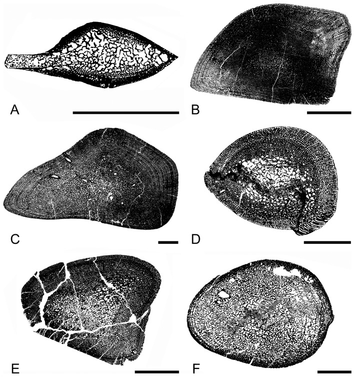
Mixosaurus sections differ from those of the other taxa analyzed. The sections essentially consist of a loose spongiosa surrounded by a layer of compact cortical bone (Microanatomical Type [MiT] 0; see [13]; Fig. 1A). This rather compact cortical bone, its thickness and the looseness of the spongiosa (i.e., few trabeculae surrounding rather large intertrabecular spaces) differ from what is observed in the other taxa (notably the thinner and more numerous trabeculae surrounding smaller and more numerous intertrabecular spaces).
Figure 1. Schematic drawings illustrating the microanatomical types observed in ichthyosaur humeri.
A, Mixosaurus PIMUZ T 2046; B, Ichthyosaurus SMNS Unnumbered; C, Ophthalmosaurus SMNS 10170; D, Ichthyosaurus IPB R222; E, Stenopterygius SMNS 81194; F, Stenopterygius SMNS A; A: Microanatomical type (MiT) 0; B–C: MiT1; D–E: MiT2; F: MiT3. Scale bars equal 10 mm.
Concerning the other taxa, variation also occurs: Some sections almost exclusively consist of a relatively loose spongiosa with randomly shaped (especially in the medullary region) intertrabecular spaces, surrounded by a relatively thin compact peripheral layer exhibiting rather small cavities (Fig. 1F). Conversely, other sections correspond to a relatively compact spongiosa with small cavities (even in the core of the section) displaying a circumferential organization in the outer and inner cortex and being randomly shaped and oriented in the core (Fig. 1B–C). Various sections are intermediate between these two ‘extremes’ with a variable percentage of the medullary region consisting of a relatively loose poorly organized spongiosa, whereas the surrounding spongiosa exposes a rather laminar organization (Fig. 1D–E).
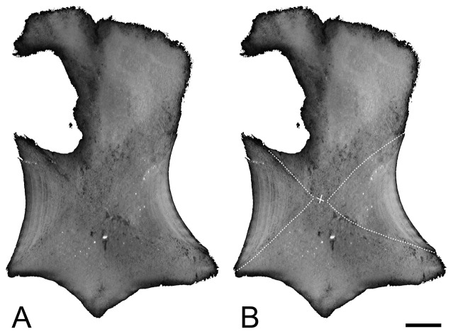
These various patterns are usually observed within a single genus and are thus not correlated with taxonomy. Moreover, they are correlated neither with species size, nor with ontogeny (size being estimated from section maximal diameter; see Table 1). Observation of two sections taken at a very short distance at bone mid-diaphysis highlighted already significant differences in the respective proportion of the unorganized versus laminar spongiosae and thus suggested important variability along the diaphysis. Indeed, if all sections are mid-diaphyseal, they probably do not all exactly correspond to the same homologous plane. The reference plane, or ‘perfect’ mid-diaphyseal section, is the one intercepting the point where growth originated and where all the bone originally consisted of periosteal bone. Virtual longitudinal and transverse sections from the specimens scanned highlighted the important difference in the thickness of the compact bone layer of periosteal origin along the diaphysis and the important resulting differences in microanatomical organization (Fig. 2). The parts where the spongiosa is looser are naturally less resistant during diagenesis and, as a result, are often crushed.
Figure 2. Virtual longitudinal sections of the humerus of Ophthalmosaurus ULg 2013-11-19I.
The dotted lines indicate the transition between the osseous tissues of periosteal (left-right) and endochondral (top-bottom) origin. Note the visible LAGs on the primary periosteal bone. The cross indicates the point of origin of growth. Scale bars equal 10 mm.
Compactness indices for the humerus vary from 55.3% in the Stenopterygius section SMNS A to 87.5% in the Ichthyosaurus section SMNS Unnumbered A.
Femora
The organization of the few femora available appears similar to that observed in the humeri and the same variations seem to occur (Fig. 3A–B). Compactness indices range from 51.3 to 68.3%.
Figure 3. Sections of ichthyosaur long bones.
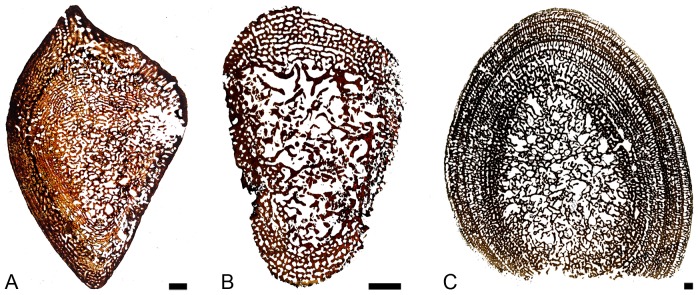
A–B, Ichthyosaurus femora; A, LO 11904t; B, SMNS Unnumbered. C, Pessopteryx epipodial PMU uncatalogued. Scale bars equal 10 mm.
Epipodials
Pessopteryx epipodials show an organization similar to that observed in the humeri analyzed (except Mixosaurus; Fig. 3C). Compactness indices were estimated between 54.0% and 60.5%.
(b) Histological features
Various histological features are observed depending on the sections. As differences between the different types of bones appear rather inconsequential, all bones are hereafter described together.
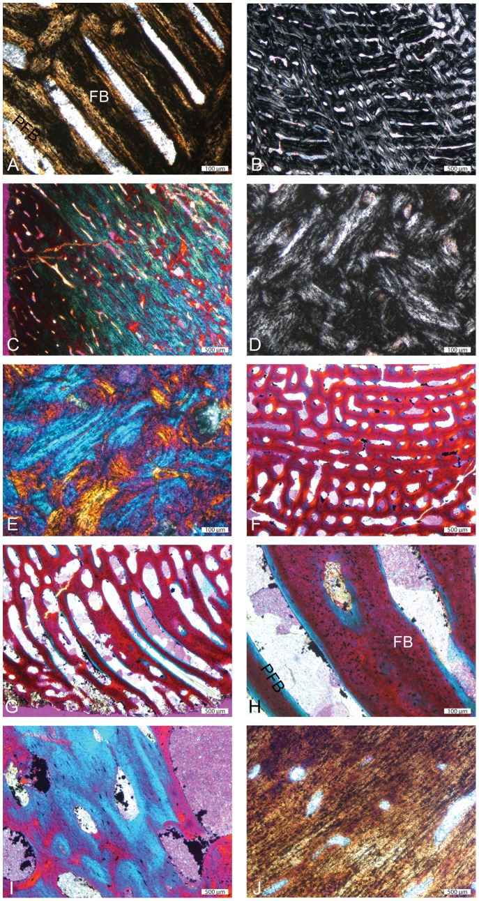
We first focus on the most compact sections, with no or almost no central area of rather loose spongiosa, which are therefore considered to expose only spongiosa of periosteal origin (MiT 1; e.g., Ichthyosaurus SMNS Unnumbered, Ophthalmosaurus SMNS 10170; see Table 1). In these sections, cortical bone consists of fibro-lamellar bone, i.e., a matrix of woven-fibered bone – as shown by the isotropy of the tissue and by the large irregularly shaped and randomly oriented osteocyte lacunae – with numerous primary osteons (Fig. 4A–B). The primary osteons are longitudinally oriented and organized in circumferential layers. Numerous anastomoses occur; they are, depending on the position on the section, essentially circular, or circular and radial, thus characterizing a laminar or plexiform tissue (see [39]; Fig. 4B). Locally, primary osteons can also be essentially radially oriented, thus characterizing radiating fibro-lamellar bone. Primary bone can also locally consist of ‘unusual parallel-fibered bone’ sensu [40] that is parallel-fibered bone with large, randomly shaped and oriented osteocyte lacunae (Fig. 4C). Resorption is limited in the outermost cortex, so that remains of primary bone are abundant (Fig. 4B), but increases toward the core of the section. Remodelling is generally important; secondary bone essentially consists of parallel-fibered bone. Numerous secondary osteons occur. Important centripetal bone deposits of lamellar or parallel-fibered bone fill the vascular and intertrabecular spaces, so that the spongiosa is secondarily compacted (Fig. 4D–E). As a result, most of the section almost exclusively consists of a dense network of primary and secondary bone in the outer cortex and of secondary bone with interstitial remains of woven-fibered bone in its core (Fig. 4D–E).
Figure 4. Histological features of ichthyosaur humeral sections.
A–C and D–E, Ichthyosaurus SMNS Unnumbered outer and inner parts of the section respectively. A, primary fibrolamellar bone (FLB) in natural light (NL); note the isotropic nature of the primary fibrous bone (FB); B, FLB in polarized light (PL) illustrating the variable orientations of the primary osteons; C, ‘unusual parallel-fibered bone’ (UPFB) in PL with gypsum filter; D–E, extremely compact core of the section made of almost exclusively secondary bone in PL and PL with gypsum filter. F–I, Ichthyosaurus IPB R222 outer cortex. J, Ichthyosaurus SMNS Unnumbered outer cortex. F–H, primary FLB in PL with gypsum filter; F, laminar organization; G, radiating FLB; H, important amount of primary FB in the osseous trabeculae. I–J, UPFB in I, PL with gypsum filter and J, NL respectively; note the occurrence of simple vascular canals. FB: fibrous bone; PFB: parallel-fibered bone.
In sections with a significant area of loose spongiosa, i.e. of supposed endochondral spongiosa (MiT 2; e.g., Ichthyosaurus R222; Pessopteryx epipodial; Table 1; Fig. 1D–E), primary bone also essentially consists of fibro-lamellar bone (Fig. 4F–H). However, the laminar or plexiform organization, as well as radiating fibro-lamellar bone, only occur in the outer cortex (Fig. 4F–G), i.e. in the spongiosa of periosteal origin. Important remains of primary woven bone are observed in the core of the trabeculae (Fig. 4H). As in MiT 1, parallel-fibered bone (or unusual parallel-fibered bone) also locally occurs (Fig. 4I–J). In the periphery, some vascular spaces are not yet filled with lamellar bone deposits and thus do not yet consist of primary osteons (Fig. 4J). In the core of the section, i.e., in the spongiosa of endochondral origin, remodelling is intense and characterized by an imbalance between bone resorption and reconstruction with a resorption prevalence. As a result, the deep spongiosa, where trabeculae are almost exclusively made of secondary lamellar bone, is loose. Secondary osteons occur in both areas.
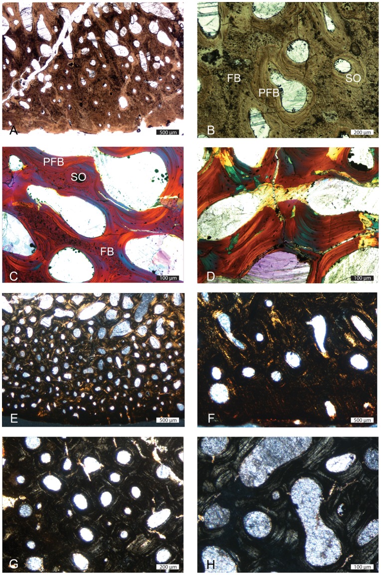
In some sections (MiT 3; Stenopterigius SMNS A; Table 1; Fig. 1F), the circumferential organization is absent or only occurs in the outermost cortex (Fig. 5A,E). The cortex is very thin and consists of primary woven-fibered bone with primary and secondary osteons rather randomly distributed and with random size and shapes (Fig. 5B). Remains of primary bone quickly diminish away from the bone periphery and are absent in the core of the section (Fig. 5C–D). Remodelling is very intense, even in the outer (but not outermost) cortex. In the Ichthyosaurus section PIMUZ A/III 843, the outer cortex essentially displays primary and numerous secondary osteons and restricted remains of primary bone (Fig. 5F). The latter diminish centripetally and are almost absent in the core of the section, where remodelling is characterized by a resorption prevalence, and which is thus much looser than the cortex (Fig. 5G–H). The core of the section corresponds to Haversian tissue. Such sections are considered as essentially exposing spongiosa of endochondral origin, surrounded by a very thin layer of periosteal bone. The outermost cortex is mostly compact, with areas deprived of any vascularization.
Figure 5. Histological features of ichthyosaur humeral sections.
A–D, Stenopterygius SMNS A; E–H, Ichthyosaurus Unnumbered. A,B,E,F outer cortex in natural light with numerous primary and secondary osteons. C, trabeculae slightly away from the bone periphery; note the remains of primary fibrous bone and the secondary lamellar and parallel-fibered bone; D, core of the section; the trabeculae are entirely secondary in origin. G–H, Haversian tissue in the core of the section. FB: fibrous bone; PFB: parallel-fibered bone; SO: secondary osteon.
Several sections (see Fig. 6) display evidence of cyclical bone deposition. Indeed, some layers with large intertrabecular spaces alternate with layers characterized by spaces of much lower size, thus probably illustrating a slowing in growth (Fig. 6A). These features are rarely observable on the whole section. They are generally localized, probably as a result of bone remodelling, which prevents their use for skeletochronological analyses. Some sections display in their outer cortex a vascularized layer deposited after an avascular one, which clearly suggests that growth resumed after a slow-down (Fig. 6B).
Figure 6. Cyclicity features in some ichthysaur humeri.
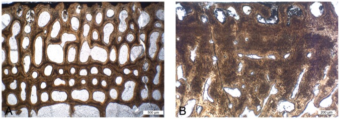
A, Temnodontosaurus PIMUZ SMNS 50329; note the alternation of layers with large and small intertrabecular spaces respectively; B, Stenopterygius SMNS 50093; note the vascularized layer at the bone periphery (top) following an almost avascular layer.
Discussion
(a) Histological features
The cortical spongiosa of Ichthyosaurus and Stenopterygius was described as resulting from the inner resorption of primary compact tissues, and thus as being secondary in origin, as opposed to that of Pessopteryx, which was assumed to be of primary origin [19]. Our study shows that primary bone in all the ichthyosaurian taxa sampled (to the possible exception of Mixosaurus, whose microanatomical organization appears peculiar within ichthyosaurs) is spongy in origin.
The presence of highly vascularized fibrolamellar bone confirms the previous observations to suggest high growth rates in ichthyosaurs (see [41] for details).
(b) Microanatomical variation along the diaphysis
Our analysis reveals an important diversity in microanatomical organization among ichthyosaur long bones, which is not correlated with size. The analysis of virtual longitudinal sections of the long bones scanned (see Material and Methods section) revealed an important change in microanatomy along the diaphysis, which probably explains the variations observed.
The transition from the rather compact to the looser spongiosa illustrates the transition between the spongiosa of periosteal and endochondral origin respectively. Such a variation in proportion, along the diaphysis, between the two types of spongiosa, exhibiting important differences in compactness, was already described in Pessopteryx [18]. However, even the periosteal spongiosa was not previously described as particularly compact.
The denser sections, exhibiting only a spongiosa of periosteal origin (MiT 1), are considered to correspond to the ‘perfect’ mid-diaphyseal sectional plane, i.e. the one intersecting the point of origin of growth. In these sections, remodelling is active, especially in the medullary area, and characterized by excessive secondary bone deposits filling the intertrabecular spaces, coupled with a slight inhibition in primary bone resorption, notably in the outer cortex, conferring to the whole section a high compactness. In the sections that are considered the further away from the ‘perfect’ mid-diaphyseal sectional plane (MiT 3), and which are assumed to essentially consist of a spongiosa of endochondral origin, remodelling is active and characterized, notably in the medullary area, by a reconstruction deficit, so that the spongiosa is more loosely organized. Bone remodelling varies thus strongly locally along the diaphysis, as these two transverse sectional planes are close in the ichthyosaur bones, which characteristically exhibit a short diaphysis.
A deficit in secondary bone deposits during remodelling generally characterizes what has been called an osteoporotic-like pattern, responsible for a decrease in bone mass [32]. Conversely, additional deposits filling the intertrabecular spaces correspond to one pattern of osteosclerosis, engendering bone mass increase (cf. [42]). Various bones from a single skeleton can display these two types of osseous specializations (e.g., bone mass increase in the rostrum of Mesoplodon; probably bone lightening in its long bones; [43]). However, the two types of remodelling patterns have never been described in a single bone yet. Our study thus raises questions about the definition of these specializations, notably based on the processes involved.
Based on MiT 3 sections, it was previously suggested that ichthyosaurs, like modern cetaceans, displayed osteoporotic-like bones [19], [32]. The lowest compactness indices obtained in our sample are slightly above 50% (51.3 and 52.4% in Ichthyosaurus and Mixosaurus femora, 54% in a Pessopteryx epipodial and 55.3% in a Stenopterygius humerus; see Table 1). These values, although among the lowest values within amniotes, are not particularly low, as several amniote taxa display similar compactness indices in their humeri and femora (cf. [44]). These bones thus do not seem to illustrate a true osteoporotic-like pattern. They are indeed not really characterized by a loss in bone mass, but rather by a spongious organization, with the absence of a medullary cavity. The highest compactness indices in the sections studied, range around 80–85% (83.3 and 87.5% in Ichthyosaurus, 78.1% in Mixosaurus, 78.1 and 76.5% in Ophthalmosaurus). These values are rather high within amniotes (cf. [44]) but, again, bones that are clearly osteosclerotic usually display much higher values (cf. [44]).
As a consequence, if based on one or another type of diaphyseal section it would be tempting to attribute an osteoporotic-like or osteosclerotic state to these bones, this would probably be a mistake. It would appear logical to determine the possible occurrence of a microanatomical specialization based on the whole bone general organization. Mid-diaphyseal sections are used as reference planes for long bones as they typically reflect the three-dimensional organization. However, this does not seem to be the case in ichthyosaurs, which complicates the understanding of their microanatomical specialization.
In ichthyosaurs, except in some specimens of Pessopteryx [18], [19], the long bones have clearly lost the medullary cavity. The general organization appears thus spongious, with no layer of highly compact bone, with the exception of a very thin one in the bone periphery of some specimens. If the spongiosa is much compacted in the ‘perfect’ mid-diaphyseal plane, it is much looser farther away from this plane.
Remodelling in the periosteal and endochondral areas appears thus characterized by an increase and decrease in bone compactness respectively. These antagonistic processes impede the attribution of a general type of specialization to the whole bone. It seems thus more cautious not to try to name this atypical microanatomy based on the specializations already described in other taxa.
As opposed to the condition described above, the microanatomical organization is overall homologous along the diaphysis in most amniotes, even in other efficient swimmers like cetaceans ([45], [46], [47]; A.H. pers. obs.). However, it must be pointed out that such a change also seems to occur in a few taxa, like the sea otter Enhydra lutris [47] or some plesiosaurs [48]. A compacted mid-shaft usually results from either an inhibition of primary periosteal bone resorption or from increased secondary bone deposition during remodelling. However, it is usually associated with an increase in compactness of the spongiosa of endochondral origin, which is not the case in ichthyosaurs. Our study reveals the interest of analyzing the possible occurrence of variations in microanatomical organization along the diaphysis in active swimmers characterized by short shafts, and notably the processes involved, in order to see if this phenomenon is specific to ichthyosaurs or not.
Bone microanatomy is generally considered to reflect the physical constraints of locomotion (see e.g., [8], [10], [11], [49]. Bone mass increase is considered to be an adaptation for hydrostatic buoyancy and body trim control in poorly active swimmers living in shallow water environments [42], whereas a spongious light organization generally characterizes active swimmers relying on a hydrodynamic control of buoyancy and body trim and requiring good manoeuvrability and acceleration abilities [32], [42]. A spongious organization with a compacted central area has never been described in any extant or extinct taxon so far. As a consequence, it appears too early to try to infer any specifically associated functional requirement.
(c) Specificity of Pessopteryx
All long bones of Pessopteryx (humerus, femur, tibia) were described as displaying a small medullary cavity [18], [19], which was interpreted as a specificity of this taxon among Ichthyosauria. However, we did not observe a medullary cavity in the epipodial bone of Pessopteryx analyzed.
Remodelling was described as relatively limited in Pessopteryx, as compared to the more derived Ichthyosaurus and Stenopterygius [19]. However, our analysis shows a high degree of remodelling in Pessopteryx epipodial bones, as in the other ichthyosaurs.
In addition, the bones of Pessopteryx were described as showing histological evidence of cyclic growth, which were considered absent in Ichthyosaurus and Stenopterygius [19]. The evidence of cyclic growth is suggested in sections of several ichthyosaurs, although the cycles are generally not continuous and thus cannot be used in skeletochronology (like the LAGs in Mixosaurus sections; see below, [13]). These observations nevertheless reveal that cyclical growth is a common pattern among ichthyosaurs but, as it is only observable in the primary spongiosa of periosteal origin, it is not seen in all sections, which probably resulted in this misinterpretation.
The absence of a marked difference between the histology of Pessopteryx and that of the other taxa in this study would be consistent with either a very basal position of this taxon among ichthyosaurs or with this taxon being a sister-group of Ichthyosauria (see above).
(d) Specificity of Mixosaurus
Our study also highlights the clear difference in microanatomical organization between Mixosaurus on the one hand, and the other ichthyosaurs from our sample on the other hand. Morphologically, Mixosaurus humeri characteristically show an anterior flange, as in many other Triassic ichthyosaurs [50]. But they also differ in their microanatomy. Mixosaurus long bones show a peripheral layer of compact cortex clearly distinct from the remainder of the section, which consists of a loose spongiosa [13]. Although it is not clear because of intense distortion, Utatsusaurus long bones seem to suggest a microanatomical organization closer to that of the non-Mixosaurus ichthyosaurs [23]. Further investigations are required to check the absence of a compacted mid-shaft area in Mixosaurus long bones. Another specificity of Mixosaurus is that it is the only taxon for which remains of calcified cartilage are observed in the core of sections of presumably new born and juvenile specimens. However, no specimen of similar ontogenetic stage has been analyzed for another taxa yet, so that this peculiarity should be interpreted with caution. Moreover, it is the only taxon showing LAGs [13], which remains unexplained.
It must be pointed out that Kolb et al. [13] described the inner compactness in Mixosaurus long bones as relatively high (essentially as a result of the compact outer cortex) and interpreted it as a possible characteristic of a near-shore form or shelf dweller. However, our study shows that Mixosaurus bones do not display a higher compactness than the other ichthyosaurs, which challenges this earlier interpretation. The peculiarity of Mixosaurus microanatomical features could nevertheless reflect some differences in locomotion mode, which needs further investigations to be specified.
Conclusions
Important variations are observed between the various ichthyosaur sections. The various patterns do not correlate with taxonomy (except maybe for Mixosaurus), species size, or ontogeny but seem to essentially illustrate a strong variability along the diaphysis.
Two types of remodelling patterns occur along the diaphysis, characterized by bone mass decrease and increase respectively, which has never been described in a single bone before. This result raises questions about the definition of the osseous specializations bone mass increase and osteoporosis, notably based on the processes involved. It suggests that none of these specializations truly occurs in ichthyosaur long bones and reveals the importance of analyzing the possible occurrence of variations in microanatomical organization along the diaphysis in other active swimmers, in order to see if this peculiarity is specific to ichthyosaurs or not.
Our study shows that primary bone in all the ichthyosaur taxa sampled (to the possible exception of Mixosaurus) is spongy in origin and that cyclical growth is a common pattern among these taxa.
Highly vascularized fibrolamellar bone is in accordance with previous assumptions of high growth rates in ichthyosaurs.
Acknowledgments
We thank N. Bardet (Museum National d'Histoire Naturelle, Paris, France) and M. Talevi (Universidad Nacional de Río Negro, Argentina) for fruitful comments that improved the manuscript, the Steinmann Institut (University of Bonn, Germany) and the European Synchrotron and P. Tafforeau (ESRF, Grenoble, France) for providing beamtime and support, and the Steinmann Institut and UMR 7207 CR2P MNHN CNRS UPMC-Paris6 for 3D imaging facilities.
Funding Statement
AH acknowledges financial support from the Alexander von Humboldt Foundation, TMS from the Swiss National Science Foundation (grant no. 31003A 146440), and CK from the DAAD (D/09/46969). The funders had no role in study design, data collection and analysis, decision to publish, or preparation of the manuscript.
References
- 1. Sander MP (2000) Ichthyosauria: their diversity, distribution, and phylogeny. Paläont Z 74: 1–35. [Google Scholar]
- 2.McGowan C, Motani R (2003) Handbook of Paleoherpetology. Part 8. Ichthyopterygia. Dr. Freidrich Pfeil Verlag, Munich. [Google Scholar]
- 3. Motani R (2009) The evolution of marine reptiles. Evo Edu Outreach 2: 224–235. [Google Scholar]
- 4. Motani R, You H, McGowan C (1996) Eel-like swimming in the earliest ichthyosaurs. Nature 382: 347–348. [Google Scholar]
- 5. Lingham-Soliar T (1998) Taphonomic evidence for fast tuna-like swimming in Jurassic and Cretaceous ichthyosaurs. N Jb Geol Paläont Abh 207: 171–183. [Google Scholar]
- 6.Motani R (2008) Combining uniformitarian and historical data to intrepret how earth environment influenced the evolution of ichthyopterygia. In: Kelley PH, Bambach RK, editors. From Evolution to Geobiology: Research Questions Driving Paleontology at the Start of a New Century: Paleontological Society Papers. pp. 147–164. [Google Scholar]
- 7. Buchholtz EA (2001) Swimming styles in Jurassic ichthyosaurs. J Vertebr Paleontol 21: 61–73. [Google Scholar]
- 8. Turner CH (1998) Three rules for bone adaptation to mechanical stimuli. Bone 23: 399–407. [DOI] [PubMed] [Google Scholar]
- 9. Huiskes R (2000) If bone is the answer, then what is the question? J Anat 197: 145–156. [DOI] [PMC free article] [PubMed] [Google Scholar]
- 10. Ruimerman R, Hilbers P, Rietbergen Bv, Huiskes R (2005a) A theoretical framework for strain-related trabecular bone maintenance and adaptation. J Biomech 38: 931–941. [DOI] [PubMed] [Google Scholar]
- 11. Ruimerman R, Rietbergen Bv, Hilbers P, Huiskes R (2005b) The effects of trabecular-bone loading variables on the surface signaling potential for bone remodeling and adaptation. Ann Biomed Engin 33: 71–78. [DOI] [PubMed] [Google Scholar]
- 12. Chappard D, Baslé MF, Legrand E, Audran M (2008) Trabecular bone microarchitecture: A review. Morphologie 92: 162–170. [DOI] [PubMed] [Google Scholar]
- 13. Kolb C, Sánchez-Villagra MR, Scheyer TM (2011) The palaeohistology of the basal ichthyosaur Mixosaurus Baur, 1887 (Ichthyopterygia, Mixosauridae) from the Middle Triassic: palaeobiological implications. C R Palevol 10: 403–411. [Google Scholar]
- 14.Kiprijanoff AV (1881–83) Studien Fiber die Fossilen Reptilien Russlands. Mémoires de l'Académie Impériale des Sciences, St Petersbourg 7, 1–144. [Google Scholar]
- 15.Fraas E (1891) Die Ichthyosaurier der süddeutschen Trias- und Jura-Ablagerungen. Laupp, Tübingen 1–81. [Google Scholar]
- 16. Seitz ALL (1907) Vergleichende Studien über den mikroskopischen Knochenbau fossiler und rezenter Reptilien, und dessen Bedeutung für das Wachstum und Umbildung des Knochengewebes im allgemeinen. Nova Acta, Abh. der Kaiserl. Leop.-Carol. Deutschen Akademie der Naturforscher 87: 230–370. [Google Scholar]
- 17. Gross W (1934) Die Typen des mikroskopischen Knochenbaues bei fossilen Stegocephalen und Reptilien. Z Anat Entwicklungsgesch 203: 731–764. [Google Scholar]
- 18. Buffrénil Vd, Mazin J-M, Ricqlès Ad (1987) Caractères structuraux et mode de croissance du fémur d'Omphalosaurus nisseri, ichthyosaurien du trias moyen du Spitzberg. Ann Paléontol 73: 195–216. [Google Scholar]
- 19. Buffrénil Vd, Mazin J-M (1990) Bone histology of the ichthyosaurs: comparative data and functional interpretation. Paleobiology 16: 435–447. [Google Scholar]
- 20. Talevi M, Fernandez M (2012) Unexpected skeletal histology of an ichthyosaur from the Middle Hurassic of Patagonia: implications for evolution of bone microstructure among secondary aquatic tetrapods. Naturwissenschaften 99: 241–244. [DOI] [PubMed] [Google Scholar]
- 21. Talevi M, Fernandez M, Salgado L (2012) Variacion ontogenetica en la histologia osea de Caypullisaurus bonapartei Fernandez, 1997 (Ichthyosauria: Ophthalmosauridae). Ameghiniana 49: 38–46. [Google Scholar]
- 22. Maxwell EE, Scheyer TM, Fowler DA (2014) An evolutionary and developmental perspective on the loss of regionalization in the limbs of derived ichthyosaurs. Geol Mag 151: 29–50 (In press) [Google Scholar]
- 23. Fernandez M, Talevi M (2014) Ophthalmosaurian (Ichthyosauria) records from the Aalenian–Bajocian of Patagonia (Argentina): an overview. Geol Mag 151: 49–59. [Google Scholar]
- 24. Nakajima J, Houssaye A, Endo H (2014) Osteohistology of Utatsusaurus hataii (Reptilia: Ichthyopterygia): Implications for early ichthyosaur biology. Acta Palaeontol Pol doi: 10.4202/app.2012.0045. (In press) [Google Scholar]
- 25. Wiman C (1910) Ichthyosaurier aus der Trias Spitzbergens. Bull Geol Instit Univ Uppsala 10: 124–148. [Google Scholar]
- 26. Sander PM, Faber C (2003) The Triassic marine reptile Omphalosaurus: osteology, jaw anatomy, and evidence for ichthyosaurian affinities. J Vert Paleontol 23: 799–816. [Google Scholar]
- 27. Maisch MW (2010) Phylogeny, systematics, and origin of the Ichthyosauria – the state of the art. Palaeodiv 3: 151–214. [Google Scholar]
- 28. Maxwell EE, Kear BP (2013) Triassic ichthyopterygian assemblages of the Svalbard archipelago: a reassessment of taxonomy and distribution. GFF 135: 85–94. [Google Scholar]
- 29. Sander MP, Faber C (1998) New finds of Omphalosaurus and a review of triassic ichthyosaur paleobiogeography. Paläont Z 72: 149–162. [Google Scholar]
- 30. Motani R (2000) Is Omphalosaurus ichthyopterygian? - A phylogenetic perspective. J Vertebr Paleontol 20: 295–301. [Google Scholar]
- 31. Buffrénil Vd, Schoevaert D (1988) On how the periosteal bone of the delphinid humerus becomes cancellous: ontogeny of a histological specialization. J Morphol 198: 149–164. [DOI] [PubMed] [Google Scholar]
- 32.Ricqlès Ad, de Buffrénil V (2001) Bone histology, heterochronies and the return of Tetrapods to life in water: where are we? In: Mazin JM, de Buffrénil V, editors. Secondary Adaptation of Tetrapods to life in Water. München. pp. 289–310. [Google Scholar]
- 33. Dumont M, Laurin M, Jacques F, Pellé E, Dabin W, et al. (2013) Inner architecture of vertebral centra in terrestrial and aquatic mammals: a two-dimensional comparative study. J Morphol 274: 570–584. [DOI] [PubMed] [Google Scholar]
- 34. Fischer V, Maisch MW, Naish D, Kosma R, Liston J, et al. (2012) New ophthalmosaurid ichthyosaurs from the European Lower Cretaceous demonstrate extensive ichthyosaur survival across the jurassic-Cretaceous boundary. Plos One 7 (1) e29234. [DOI] [PMC free article] [PubMed] [Google Scholar]
- 35. Canoville A, Laurin M (2010) Evolution of humeral microanatomy and lifestyle in amniotes, and some comments on palaeobiological inferences. Biol J Linn Soc 100: 384–406. [Google Scholar]
- 36. Quemeneur S, Buffrénil Vd, Laurin M (2013) Microanatomy of the amniote femur and inference of lifestyle in limbed vertebrates. Biol J Linn Soc 109: 644–655. [Google Scholar]
- 37. Abramoff MD, Magelhaes PJ, Ram SJ (2004) Image Processing with ImageJ. Biophoton Int 11: 36–42. [Google Scholar]
- 38. Sanchez S, Ahlberg PE, Trinajstic K, Mirone A, Tafforeau P (2012) Three dimensional synchrotron virtual paleohistology: a new insight into the world of fossil bone microstructures. Microsc Microanal 18: 1095–1105. [DOI] [PubMed] [Google Scholar]
- 39.Francillon-Vieillot H, de Buffrénil V, Castanet J, Geraudie J, Meunier FJ, et al.. (1990) Microstructure and Mineralization of Vertebrate Skeletal Tissues. In: Carter JG, editor. Skeletal Biomineralization: Patterns, Processes and Evolutionary Trends. New York. pp. 471–529. [Google Scholar]
- 40. Houssaye A, Lindgren J, Pellegrini R, Lee AH, Germain D, et al. (2013) Microanatomical and histological features in the long bones of mosasaurine mosasaurs (Reptilia, Squamata) – Implications for aquatic adaptation and growth rates. Plos One 8 (10) e76741. [DOI] [PMC free article] [PubMed] [Google Scholar]
- 41. Houssaye A (2013) Bone histology of aquatic reptiles: what does it tell us about secondary adaptation to an aquatic life. Biol J Linn Soc 108: 3–21. [Google Scholar]
- 42. Houssaye A (2009) “Pachyostosis” in aquatic amniotes: a review. Integrative Zool 4: 325–340. [DOI] [PubMed] [Google Scholar]
- 43. Buffrenil Vd, Casinos A (1995) Observations histologiques sur le rostre de Mesoplodon densirostris (Mammalia, Cetacea, Ziphiidae): le tissu osseux le plus dense connu. Ann Sci Nat, Zool 16: 21–32. [Google Scholar]
- 44. Hayashi S, Houssaye A, Nakajima Y, Chiba K, Inuzuka N, et al. (2013) Bone histology suggests increasing aquatic adaptations in Desmostylia (Mammalia, Afrotheria). Plos One 8 (4) e59146. [DOI] [PMC free article] [PubMed] [Google Scholar]
- 45. Wall WP (1983) The correlation between high limb-bone density and aquatic habits in recent mammals. J Paleontol 57: 197–207. [Google Scholar]
- 46. Stein BR (1989) Bone density and adaptation in semiaquatic mammals. J Mammal 70: 467–476. [Google Scholar]
- 47.Madar SI (1998) Structural Adaptations of Early Archaeocete Long Bones. In: Thewissen JGM, editor. The Emergence of Whales. New York: Plenum Press. pp. 353–378. [Google Scholar]
- 48. Liebe L, Hurum JH (2012) Gross internal structure and microstructure of plesiosaur limb bones from the Late Jurassic, central Spitsbergen. Norwegian J Geol 92: 285–309. [Google Scholar]
- 49. Liu XS, Bevill G, Keaveny TM, Sajda P, Guo XE (2009) Micromechanical analyses of vertebral trabecular bone based on individual trabeculae segmentation of plates and rods. J Biomech 42: 249–256. [DOI] [PMC free article] [PubMed] [Google Scholar]
- 50. Motani R (1999) Phylogeny of the Ichthypterygia. J Vertebr Paleontol 19: 473–496. [Google Scholar]


