Figure 5.
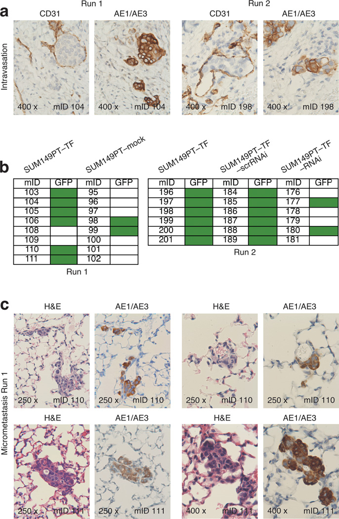
NKG2D enhances intravasation and micrometastasis in the orthotopic SUM149PT xenotransplant model. (a) Two micrograph pairs of SUM149PT–TF serial tumor tissue sections stained for mouse endothelial CD31, or human pan-cytokeratin AE1/AE3 as human tumor cell marker. Micrograph pairs were derived from run 1 and run 2 experiments and document examples of human tumor cells located within cross sections of CD31-positive peritumoral vessels. (b) Schematic representation of presence of absence of GFP signals in lungs explanted from the run 1 and run 2 experimental and control hosts. Green boxes represent positive GFP signals revealed by Typhoon Trio scanning. (c) Four micrograph pairs of serial lung tissue sections from run 1 SUM149PT–TF tumor hosts stained for H&E or human pan-cytokeratin AE1/AE3. Each two micrograph pairs were derived from two different mice and show examples of micrometastasic clusters of AE1/AE3-positive cells. Numbers in bottom left micrograph corners indicate microscopic magnification; numbers in bottom right micrograph corners specify mouse identifications (mIDs).

