Abstract
Splenic artery pseudoaneurysms can be caused by pancreatitis, trauma, or operation. Traditionally, the condition has been managed through surgery; however, nowadays, transcatheter arterial embolization is performed safely and effectively. Nevertheless, several complications of pseudoaneurysm embolization have been reported, including coil migration. Herein, we report a case of migration of the coil into the jejunal lumen after transcatheter arterial embolization of a splenic artery pseudoaneurysm. The migrated coil was successfully removed by performing endoscopic intervention.
Keywords: Splenic artery, False aneurysm, Therapeutic embolization, Migration, Endoscopy
INTRODUCTION
Splanchnic artery aneurysms and pseudoaneurysms can develop secondary to congenital, traumatic, and inflammatory pathologies.1 The splenic artery is the most common artery affected by pseudoaneurysms.2 When a pseudoaneurysm ruptures, hemodynamic instability due to bleeding can occur, which is life-threatening. Transcatheter arterial embolization (TAE) is a safe and effective treatment for pseudoaneurysms.3 However, some complications have been reported, such as bleeding, pseudoaneurysm recurrence, and postembolization syndrome.4 Coil migration from a visceral artery pseudoaneurysm is a rare complication of TAE.1
We describe a case of coil migration into the jejunal lumen after the embolization of a splenic pseudoaneurysm, which was successfully removed by means of an endoscopic intervention.
CASE REPORT
A 63-year-old man was found to have advanced gastric cancer on a routine cancer screening examination. Preoperative abdominal computed tomography (CT) was performed, and there was no specific finding besides advanced gastric cancer. He underwent total gastrectomy. The tumor was located at the posterior wall of the high body. The gross type of the tumor was Bormann type II. The size of the tumor was 4.5×3.8×0.8 cm, and the depth of invasion was at the subserosal level (pT2b). Sixty-six lymph nodes were dissected, and no metastases were observed (pN0). Lymphatic, venous, and perineural invasions were present.
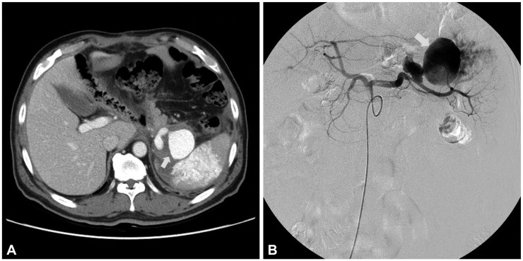
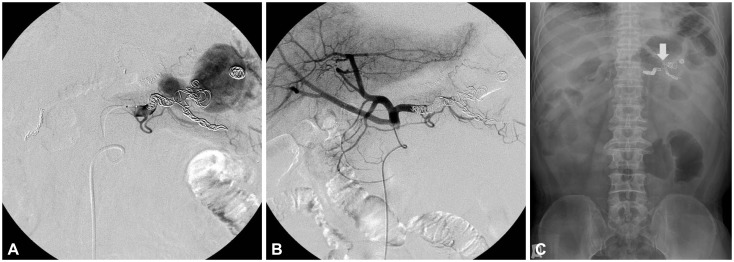
One month after the surgery, he came to the emergency department with complaints of a febrile sense and abdominal pain. An abdominal CT scan was carried out, and a pseudoaneurysm of the splenic artery and a small amount of hemoperitoneum were detected (Fig. 1A). The total gastrectomy performed 1 month ago was thought to be the cause of the pseudoaneurysm. The size of the pseudoaneurysm was 2.7×1.9×3.6 cm. On the day after admission, a celiac angiography was performed. A large pseudoaneurysm of the splenic artery was observed on angiography (Fig. 1B). Extravasation of the contrast agent was not observed, and there was no evidence of ongoing bleeding or rupture of the pseudoaneurysm. However, the risk of rupture seemed high, and we decided to perform embolization. The splenic artery was selected by using a microcatheter, and coils (Tornado embolization microcoil; Cook, Bloomington, IN, USA) were inserted to obliterate the pseudoaneurysm. The large size of the pseudoaneurysm and the fast blood flow required the use of 22 coils. After the coils were placed, splenic angiography was done and successful embolization of the pseudoaneurysm was confirmed (Fig. 2). He was discharged without any complication. As a routine surveillance, an abdominal CT scan was taken 5 months after the embolization, and it showed no evidence of cancer recurrence or any complication associated with embolization.
Fig. 1.
(A) Computed tomography showed a pseudoaneurysm of the splenic artery (arrow) and a splenic infarct. (B) Celiac angiography revealed a pseudoaneurysm of the splenic artery (arrow).
Fig. 2.
(A) Embolization of the pseudoaneurysm was done by using coils. (B) Angiography after coil embolization revealed total occlusion of the pseudoaneurysm. (C) Simple abdominal radiograph taken just after embolization showed the multiple coils (arrow) placed within the pseudoaneurysm and splenic artery.
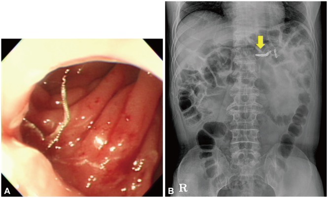
Nine months after the embolization of the pseudoaneurysm, the patient presented to the emergency department with epigastric pain. On upper gastrointestinal endoscopy, several strands of wire were noted at the inferolateral side of the esophagojejunal anastomosis (Fig. 3). A simple abdominal radiograph showed that the wires protruding through the jejunal wall were part of a coil used for pseudoaneurysm embolization (Fig. 4). We tried to cut the wires by using endoscissors (FS-5L-1; Olympus, Tokyo, Japan); however, it was difficult because the wires were thick and stiff. Thus, we tried to use hot biopsy forceps (Radial Jaw 3; Boston Scientific, Natick, MA, USA) for cutting the wires, which proved to be easy and effective. The hot biopsy forceps were used with an electrosurgical generator (VIO-300D; ERBE Elektromedizin, Tübingen, Germany) in the Endocut Q mode (effect 2, cut duration 2, and cut interval 2) (Fig. 5). It took about 1 to 2 seconds to cut each wire, and five wires were cut in total. Residual thin strands of wires were removed with endoscissors. There was no immediate complication during and after the procedure.
Fig. 3.
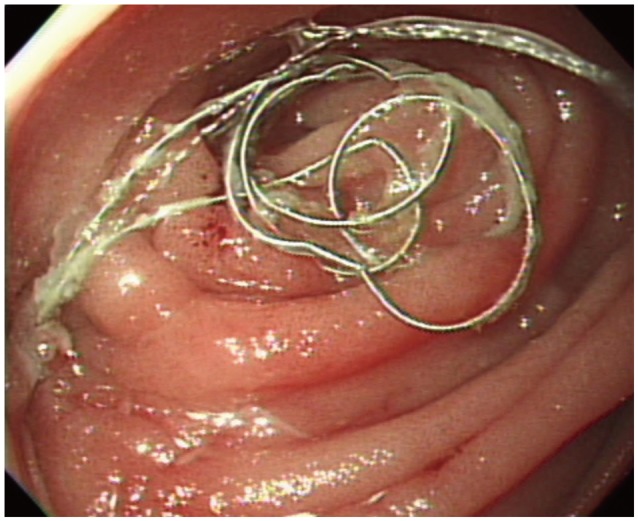
Endoscopy showed several strands of wire protruding through the jejunal lumen below the esophagojejunal anastomosis.
Fig. 4.
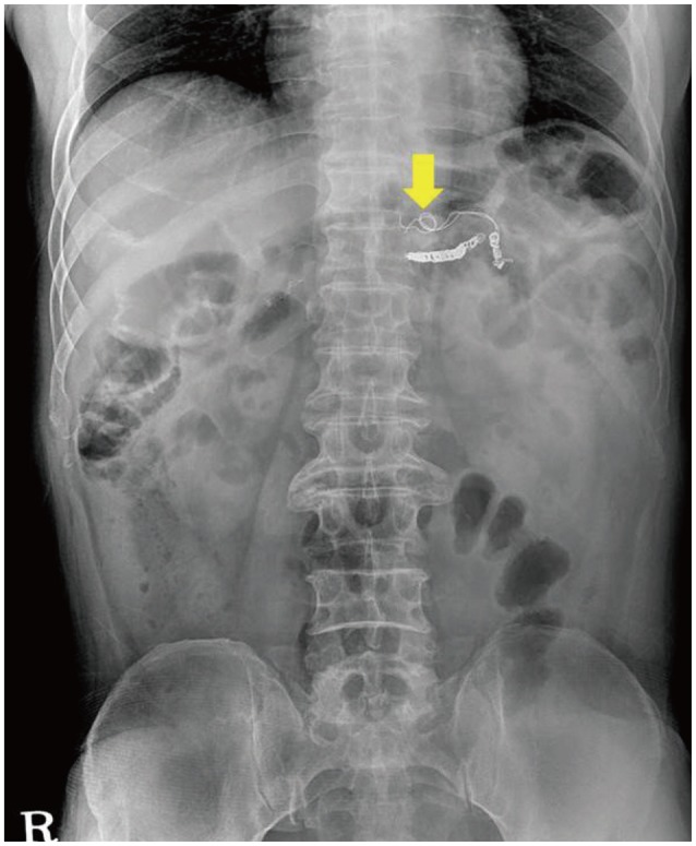
A migrated coil (arrow) was seen on simple abdominal radiograph.
Fig. 5.
(A) The wires were nearly removed by using hot biopsy forceps with an electrosurgical generator. (B) Simple abdominal radiograph showed the removed wires (arrow) protruding through the jejunal wall.
The patient was discharged from the hospital without epigastric pain and remained asymptomatic. He was followed up with upper gastrointestinal endoscopy 3 months after the endoscopic coil removal, and annually thereafter. Abdominal CT scans were performed every 6 months. There had been no symptom or complication related to the coil during the 2-year follow-up period.
DISCUSSION
Splanchnic artery aneurysms and pseudoaneurysms are uncommon; however, incidental detection of such cases has been increasing in recent decades.5 It is important to recognize splanchnic artery aneurysms and pseudoaneurysms because their risk of rupture is up to 25%, and if ruptured, the mortality rate is between 25% and 75%.6 The artery most commonly affected by pseudoaneurysms is the splenic artery.2 Causes of splenic artery pseudoaneurysms are chronic pancreatitis (46%), trauma (29%), acute pancreatitis (6%), postoperative complication (3%), and peptic ulcer disease (2%).2 In our case, a total gastrectomy performed 1 month before presentation might have been the cause of the pseudoaneurysm.
Pseudoaneurysms can be treated with surgery or endovascular intervention.7 Traditionally, pseudoaneurysms were managed surgically by means of ligation of the artery or aneurysm, or excision of the lesion. In recent decades, endovascular techniques such as TAE and covered stent placement were introduced.8 Angiography is a useful tool because it could achieve diagnosis and concurrent treatment. Splenic artery embolization is a minimally invasive procedure that involves the use of metallic coils, hydrogel particles, gel foam, acrylic glue, or a combination of these materials.9 The success rate of embolization in the treatment of splenic artery aneurysms and pseudoaneurysms is approximately 90%.4,7,8 Several complications have been reported, including bleeding, pseudoaneurysm recurrence, postembolization syndrome, and infarction.4 Coil migration is also reported after embolization of visceral arteries.1
Coil migration is one of the rare complications of endovascular management of a pseudoaneurysm. According to a cumulative review of the literature, there are only 10 reports documenting the migration of endovascular coils from visceral arteries (Table 1).1,10,11,12,13,14,15,16,17,18 In two cases, the coils migrated from the splenic artery, as in our case, one into the stomach10 and the other into the rectum.11 In the former case, the splenic pseudoaneurysm resulting from chronic pancreatitis bled into pseudocyst, and steel-wire coils were placed inside the aneurysm cavity. Several weeks later, some of the coils dislodged through a gastropseudocystic fistula, and open surgery was performed to remove the coils, pseudocyst, and fistula. In the latter case, the patient underwent embolization of a splenic artery pseudoaneurysm caused by chronic pancreatitis. After 3 weeks of intervention, two coils were passed through the rectum. No fistula was found on imaging studies, and it was postulated that the coils passed through a preexisting enteric fistula. As the migrating coils had already passed, no further management was performed.
Table 1.
Clinical Findings of Reported Coil Migration from Visceral Arteries

M, male; RUQ, right upper quadrant; F, female; CBD, common bile duct; UV, ureterovesical.
There are few reports about the risk factors associated with coil migration. Techniques associated with embolization and the presence of collaterals are assumed to influence the migration of coils.1 To reduce the chance of coil migration, occluding the normal portion of the splenic artery, both distal and proximal to the pseudoaneurysm, is preferred over filling the inside of a pseudoaneurysm.10 By occluding both sides of the artery, backflow from collateral circulation could be prevented.1 In our case, the radiologists tried to occlude both the afferent and efferent arteries; however, the blood flow was so fast that some of the coils migrated into the pseudoaneurysm. This may be the reason why the coil migration occurred.
No treatment strategy has been established for migrated coils. According to previous case reports, some coils were removed surgically or were spontaneously passed. Others, in the case of coils migrating into the common bile duct, were eliminated by using percutaneous or endoscopic cholangiography. What is special in our case is that the migrated coil was successfully removed by endoscopic intervention. To our knowledge, endoscopic removal of a migrated coil from the bowel lumen has not been described before. First, we tried to cut the wires with endoscissors, but it was unsuccessful. Before this case, we already had experience in the endoscopic removal of complicated suture materials at the anastomosis site by using hot biopsy forceps. Therefore, we used hot biopsy forceps for cutting the wires. The wires were cut easily without complication. Endoscopic removal seems to have many advantages over surgical treatment: it is less invasive and more cost-effective because not only is the procedure itself possibly less expensive but also the length of hospital stay might be shorter than with operation. The procedure might also be less painful for the patient than surgery. However, careful evaluation should be performed for the presence of conditions requiring surgery such as a fistula, perforation, and pseudocyst before choosing the treatment modality. Our patient had no relevant condition requiring surgery and was successfully treated with endoscopic intervention. He was followed up for 15 months after coil removal without complication.
In summary, we report a case of coil migration into the jejunal lumen after TAE of a splenic artery pseudoaneurysm. This case shows the feasibility of successful coil removal by means of endoscopic intervention.
Footnotes
The authors have no financial conflicts of interest.
References
- 1.Skipworth JR, Morkane C, Raptis DA, et al. Coil migration: a rare complication of endovascular exclusion of visceral artery pseudoaneurysms and aneurysms. Ann R Coll Surg Engl. 2011;93:e19–e23. doi: 10.1308/003588411X13008844298652. [DOI] [PMC free article] [PubMed] [Google Scholar]
- 2.Tessier DJ, Stone WM, Fowl RJ, et al. Clinical features and management of splenic artery pseudoaneurysm: case series and cumulative review of literature. J Vasc Surg. 2003;38:969–974. doi: 10.1016/s0741-5214(03)00710-9. [DOI] [PubMed] [Google Scholar]
- 3.Golzarian J, Nicaise N, Devière J, et al. Transcatheter embolization of pseudoaneurysms complicating pancreatitis. Cardiovasc Intervent Radiol. 1997;20:435–440. doi: 10.1007/s002709900189. [DOI] [PubMed] [Google Scholar]
- 4.Gaba RC, Katz JR, Parvinian A, et al. Splenic artery embolization: a single center experience on the safety, efficacy, and clinical outcomes. Diagn Interv Radiol. 2013;19:49–55. doi: 10.4261/1305-3825.DIR.5895-12.1. [DOI] [PubMed] [Google Scholar]
- 5.Røkke O, Søndenaa K, Amundsen S, Bjerke-Larssen T, Jensen D. The diagnosis and management of splanchnic artery aneurysms. Scand J Gastroenterol. 1996;31:737–743. doi: 10.3109/00365529609010344. [DOI] [PubMed] [Google Scholar]
- 6.Pasha SF, Gloviczki P, Stanson AW, Kamath PS. Splanchnic artery aneurysms. Mayo Clin Proc. 2007;82:472–479. doi: 10.4065/82.4.472. [DOI] [PubMed] [Google Scholar]
- 7.McDermott VG, Shlansky-Goldberg R, Cope C. Endovascular management of splenic artery aneurysms and pseudoaneurysms. Cardiovasc Intervent Radiol. 1994;17:179–184. doi: 10.1007/BF00571531. [DOI] [PubMed] [Google Scholar]
- 8.Loffroy R, Guiu B, Cercueil JP, et al. Transcatheter arterial embolization of splenic artery aneurysms and pseudoaneurysms: short- and long-term results. Ann Vasc Surg. 2008;22:618–626. doi: 10.1016/j.avsg.2008.02.018. [DOI] [PubMed] [Google Scholar]
- 9.Venkatesh SK, Kumar S, Baijal SS, Phadke RV, Kathuria MK, Gujral RB. Endovascular management of pseudoaneurysms of the splenic artery: experience with six patients. Australas Radiol. 2005;49:283–288. doi: 10.1111/j.1440-1673.2005.01466.x. [DOI] [PubMed] [Google Scholar]
- 10.Takahashi T, Shimada K, Kobayashi N, Kakita A. Migration of steel-wire coils into the stomach after transcatheter arterial embolization for a bleeding splenic artery pseudoaneurysm: report of a case. Surg Today. 2001;31:458–462. doi: 10.1007/s005950170141. [DOI] [PubMed] [Google Scholar]
- 11.Shah NA, Akingboye A, Haldipur N, Mackinlay JY, Jacob G. Embolization coils migrating and being passed per rectum after embolization of a splenic artery pseudoaneurysm, "the migrating coil": a case report. Cardiovasc Intervent Radiol. 2007;30:1259–1262. doi: 10.1007/s00270-007-9166-7. [DOI] [PubMed] [Google Scholar]
- 12.Turaga KK, Amirlak B, Davis RE, Yousef K, Richards A, Fitzgibbons RJ., Jr Cholangitis after coil embolization of an iatrogenic hepatic artery pseudoaneurysm: an unusual case report. Surg Laparosc Endosc Percutan Tech. 2006;16:36–38. doi: 10.1097/01.sle.0000202189.65160.ef. [DOI] [PubMed] [Google Scholar]
- 13.Dinter DJ, Rexin M, Kaehler G, Neff W. Fatal coil migration into the stomach 10 years after endovascular celiac aneurysm repair. J Vasc Interv Radiol. 2007;18:117–120. doi: 10.1016/j.jvir.2006.10.003. [DOI] [PubMed] [Google Scholar]
- 14.Ozkan OS, Walser EM, Akinci D, Nealon W, Goodacre B. Guglielmi detachable coil erosion into the common bile duct after embolization of iatrogenic hepatic artery pseudoaneurysm. J Vasc Interv Radiol. 2002;13:935–938. doi: 10.1016/s1051-0443(07)61778-3. [DOI] [PubMed] [Google Scholar]
- 15.Reed A, Suri R, Marcovich R. Passage of embolization coil through urinary collecting system one year after embolization. Urology. 2007;70:1222.e17–1222.e18. doi: 10.1016/j.urology.2007.09.007. [DOI] [PubMed] [Google Scholar]
- 16.Van Steenbergen W, Lecluyse K, Maleux G, Pirenne J. Successful percutaneous cholangioscopic extraction of vascular coils that had eroded into the bile duct after liver transplantation. Endoscopy. 2007;39(Suppl 1):E210–E211. doi: 10.1055/s-2007-966314. [DOI] [PubMed] [Google Scholar]
- 17.Kao WY, Chiou YY, Chen TS. Coil migration into the common bile duct after embolization of a hepatic artery pseudoaneurysm. Endoscopy. 2011;43(Suppl 2 UCTN):E364–E365. doi: 10.1055/s-0030-1256687. [DOI] [PubMed] [Google Scholar]
- 18.AlGhamdi HS, Saeed MA, Altamimi AR, O'Hali WA, Khankan AA, Altraif IH. Endoscopic extraction of vascular embolization coils that have migrated into the biliary tract in a liver transplant recipient. Dig Endosc. 2012;24:462–465. doi: 10.1111/j.1443-1661.2012.01307.x. [DOI] [PubMed] [Google Scholar]


