Abstract
Purpose
The aim of this study is to describe our initial experience and assess the feasibility and safety of robotic and laparoscopic lateral pelvic node dissection (LPND) in advanced rectal cancer.
Methods
Between November 2007 and November 2012, extended minimally invasive surgery for LPND was performed in 21 selected patients with advanced rectal cancer, including 11 patients who underwent robotic LPND and 10 who underwent laparoscopic LPND. Extended lymphadenectomy was performed when LPN metastasis was suspected on preoperative magnetic resonance imaging even after chemoradiation.
Results
All 21 procedures were technically successful without the need for conversion to open surgery. The median operation time was 396 minutes (range, 170-581 minutes) and estimated blood loss was 200 mL (range, 50-700 mL). The median length of stay was 10 days (range, 5-24 days) and time to removal of the urinary catheter was 3 days (range, 1-21 days). The median total number of lymph nodes harvested was 24 (range, 8-43), and total number of lateral pelvic lymph nodes was 7 (range, 2-23). Six patients (28.6%) developed postoperative complications; three with an anastomotic leakages, two with ileus and one patient with chyle leakage. Two patients (9.5%) developed urinary incontinence. There was no mortality within 30 days. During a median follow-up of 14 months, two patients developed lung metastasis and there was no local recurrence.
Conclusion
Robotic and laparoscopic LPND is technically feasible and safe. Minimally invasive techniques for LPND in selected patients can be an acceptable alternative to an open LPND.
Keywords: Rectal neoplasms, Lymph node excision, Laparoscopy, Robotics
INTRODUCTION
The role of lateral pelvic node dissection in rectal cancer surgery remains uncertain. Lateral pelvic lymph node (LPLN) involvement occurs in 10%-25% of patients with rectal cancers and is associated with increased local recurrence and poor survival rates. A recent meta-analysis comparing extended lymphadenectomy with conventional therapy for rectal cancer showed no difference in 5-year overall or disease free survival/local recurrence [1]. However, this was based on retrospective studies performed over a long period with significant heterogeneity between groups. In Japan, where preoperative radiotherapy is not popular, LPND is being performed for stage 2/3 rectal cancers below the peritoneal reflection (Rb) as the LPLN is considered a regional lymph node. Involvement of internal iliac/external iliac lymph nodes has a prognosis similar to N2a/N2b mesorectal node involvement and better than that of stage IV cancers [2]. Even with nerve preservation, LPND is associated with increased blood loss and urinary and sexual dysfunction. Therefore, a trial comparing total mesorectal excision (TME) alone and LPND with TME is being carried out in Japan for stage II/III rectal cancer with extramesorectal nodes less than 1 cm in size [3]. However, most LPND is still performed using an open approach.
In the West, the presence of LPLN is accepted as a prognostic factor in patients undergoing primary therapy, but seems to have no impact on survival of patients receiving preoperative chemoradiotherapy (CRT) [4,5]. In other words, preoperative CRT can substitute for LPND. However LPLNs do present as local recurrence and require surgery [6-8]. While there is no concrete evidence supporting this practice, LPND is performed by selected surgeons for persistent LPLN after administration of neoadjuvant CRT [9-11]. While the aim here is to avoid local recurrence, this practice probably needs to be evaluated in a clinical trial.
In South East Asia, although TME is being performed with minimally invasive surgery (laparoscopic/robotic), extended lymphadenectomy is usually performed using open approach. With the aim of extending the scope of minimal invasive surgery for LPND and TME, we evaluated our experience of laparoscopic and robotic extended lymphadenectomy in terms of short-term outcomes.
METHODS
Between November 2007 and November 2012, extended minimally invasive surgery for LPND was performed in 21 selected patients with advanced rectal cancer, including 11 patients who underwent robotic LPND and 10 who underwent laparoscopic LPND.
LPND for advanced rectal cancer was indicated when pelvic node metastasis was suspected on preoperative magnetic resonance imaging (MRI) even after preoperative chemoradiation. The dissection of these lymph nodes was performed according to the classification of the lateral pelvic area: aortic bifurcation, common iliac, external iliac, internal iliac, obturator, and median sacral regions. Information regarding patient demographics, tumor characteristics, and clinical outcomes was obtained. This also included data regarding American Society of Anesthesiologists grade, body mass index, tumor size, tumor distance from the anal verge, and preoperative serum carcinoembryonic antigen levels. Perioperative details included operative time, estimated blood loss, days to first flatus, and days to first soft diet. Perioperative mortality, morbidity, length of hospital stay, and histopathological findings were recorded.
Surgical technique of robotic-assisted pelvic node dissection for advanced rectal cancer
For bowel preparation, colonic lavage was performed the day before the operation using 4L of Colyte. All patients were given antibiotic prophylaxis. The operation was carried out under general anesthesia with the patient in the lithotomy position. Patients were put in the Trendelenberg position at 30° and tilted right-side-down at an angle of 10°-15°. A total of five robotic ports (one for a 12-mm camera and four additional 8-mm robotic ports) and one assistant port were placed. The operation was divided into two stages: colonic phase (stage 1) and pelvic phase (stage 2). Our technique for robotic TME (i.e. single docking dual phase) has been described previously [12]. Using the Da Vinci S/SI system (Intuitive Surgical Inc., Sunnyvale, CA, USA), our technique involves two robotic instruments on the right side (arms 1 and 3) (Figs. 1, 2). LPND was performed after TME was completed and the rectum transected. In nodal dissection, the double fenestrated forceps (arm 3) was used to maintain steady traction either on vessel or fat while dissection was performed using hot shears (arm 1) and bipolar forceps (arm 2).
Fig. 1.
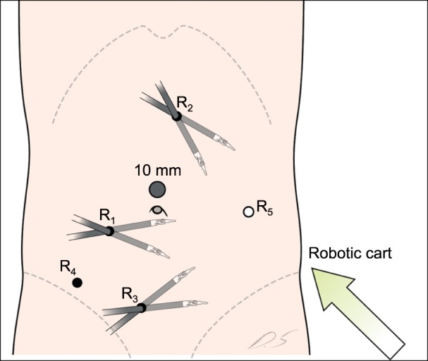
Setup for the colonic phase in a robotic lateral pelvic node dissection for advanced rectal cancer.
Fig. 2.
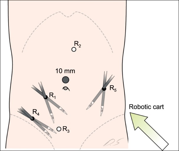
Setup for the pelvic phase in a robotic lateral pelvic node dissection for advanced rectal cancer.
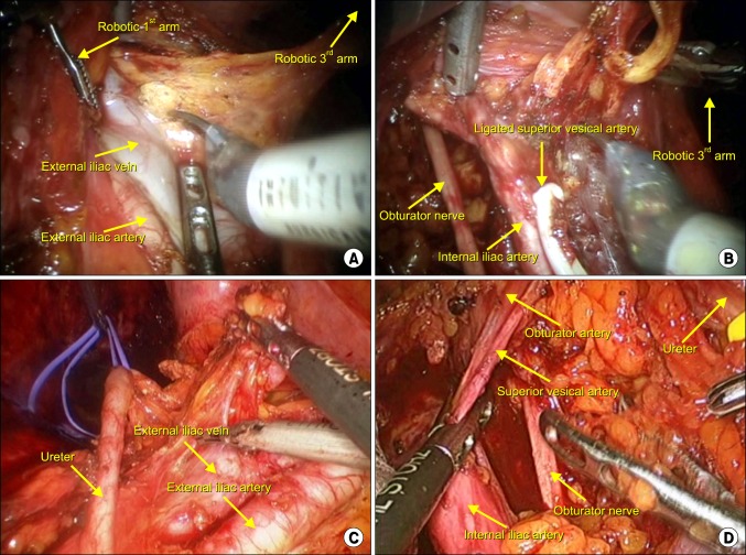
The first step in LPND was dissection and isolation of the ureters and inferior hypogastric nerves with a silastic loop. Lymph nodes and fatty tissue were dissected from the bifurcation of the aorta extending to the common iliac area. The external iliac artery and vein were exposed and the lymphatic tissues including lymph nodes were resected from external iliac vessels (Fig. 3A). The internal iliac vessels were then cleared from lymphatic tissue at a safe distance from the lateral side of the pelvic plexus. During the dissection, the obturator nerve and vessels were identified medial to the external iliac vein and lateral to the superior vesical artery. The obturator lymph nodes were resected leaving the obturator nerve and vessel in the obturator fossa (Fig. 3B). The internal iliac vessels were then cleared from fatty tissue including lymph nodes and the superior vesical artery was resected only when necessary. The dissection was extended to the area of the middle rectal lymph node. The entire dissection was performed in the surgical plane between the pelvic nerves (medial plane) and ureter (lateral plane). After, completion of LPND, only the external vessels, internal iliac vessels and their branches, the obturator nerve, and pelvic plexus remained. The specimen was extracted through the left lower trocar incision, and end-to-end intracorporeal anastomosis was performed with a double stapling technique.
Fig. 3.
A Intraoperative views during a laparoscopic and robotic-assisted lateral pelvic node dissection. (A) Dissection of a lymph node around the external iliac vessels. (B) Dissection of a lymph node around the internal iliac artery. (C) Isolation of ureter with a silastic loop and dissection along the external iliac vessels. (D) Isolation of the obturator nerve and dissection of a lymph node in the obturator fossa.
Surgical technique of laparoscopic-assisted pelvic node dissection for advanced rectal cancer
We perform extended lymph node dissection using the same port placement as for TME, a 12-mm umbilical port for the 30° camera, and four ports in all quadrants. Colorectal dissection using tumor-specific mesorectal excision principles and the operative procedure of the LPND was the same in both groups (Fig. 3C, D).
RESULTS
Patient characteristics
From the database we identified 1,686 patients undergoing TME for lower rectal cancer with curative intent from November 2007 to April 2012. Among these patients, 92 (5.4%) underwent TME and LPND, and of these, 11 (12%) underwent robotic LPND and 10 (11%) underwent laparoscopic LPND for advanced rectal cancer. The baseline demographics of patients who underwent robotic and laparoscopic LPND are tabulated in Table 1. The median age of the patients was 56 years (range, 37-75 years) with median tumor distance from the anal verge of 6 cm (range, 1-12 cm). Of the 21 patients who underwent LPND, 18 (86%) underwent preoperative chemoradiation.
Table 1.
Patient characteristics
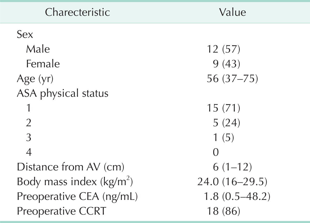
Values are presented as number (%) or median (range).
ASA, American Society of Anesthesiologists; AV, anal verge; CEA, carcinoembryonic antigen CCRT, concurrent chemoradiation therapy.
Perioperative clinicopathological outcomes
All 21 procedures were technically successful without the need for conversion to open surgery (Table 2). The procedures included 3 abdominoperineal resection, 14 low anterior resections, and 4 ultralow anterior resections and protective loop ileostomy was performed in 14 of the 18 patients (77.8%). LPND was bilateral in 7 patients (33%) and unilateral in 14 patients (67%). The median operation time was 396 minutes (range, 170-581 minutes) and median estimated blood loss was 200 mL (range, 50-700 mL). The median number of days to first gas passing was 3 (range, 1-9) and the median number of days to a soft diet was 4 (range, 2-13). The median length of stay was 10 days (range, 5-24 days) and time to removal of the urinary catheter was 3 days (range, 1-21 days). Six patients (28.6%) developed postoperative complications including three with anastomosis leakage, two with ileus, and one with chyle leakage. Two patients (9.5%) developed urinary incontinence. There was no mortality within 30 days.
Table 2.
Perioperative outcomes
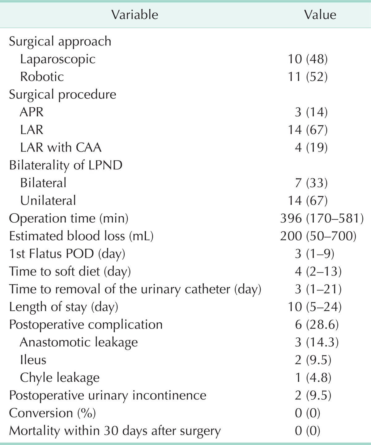
Values are presented as number (%) or median (range).
APR, abdominoperineal resection; LAR, low anterior resection; CAA, coloanal anastomosis; LPND, lateral pelvic node dissection; POD, postoperative day.
Pathologic characteristics are displayed in Table 3. The median total number of lymph nodes harvested was 24 (range, 8-43), and the total number of LPLNs was 7 (range, 2-23).During a median follow-up of 14 months, there was no death. Three patients developed lung metastasis and there was no local recurrence.
Table 3.
Postoperative pathologic outcomes
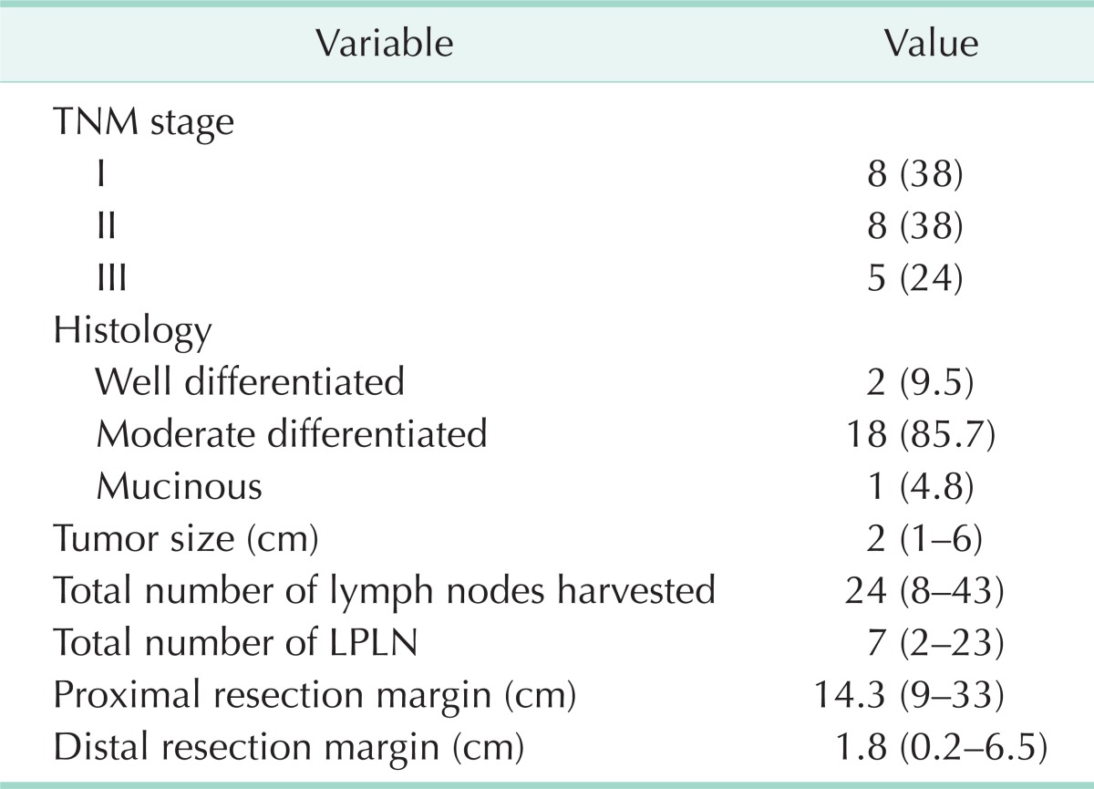
Values are presented as number (%) or median (range).
LPLN, lateral pelvic lymph nodes.
DISCUSSION
The role of LPND in advanced low rectal cancer remains undefined. LPND is not widely used in Western countries because it is associated with a longer operation time and greater blood loss than the conventional operation and may severely impair both urinary and sexual function and thus compromise the social life of the patient [1]. Additionally, in those countries LPLN metastasis is generally considered a systemic metastatic disease. In contrast, in Japan surgeons generally consider LPLN metastasis to be a regional disease, and TME with LPND is the standard operation for locally advanced low rectal cancer with the aim of improving survival and minimizing local recurrence [2,13,14]. Although we fully recognize that there is controversy regarding the indication and oncologic outcome of LPND for advanced low rectal cancer, the current paper does not address this important issue.
Laparoscopic and robotic TME is being increasingly used for the treatment of rectal cancer [15-18]. In comparison with conventional open surgery, laparoscopic colorectal resection has favorable short-term outcomes in terms of less pain, earlier return of bowel motility, and a shorter hospital stay [19]. Robotic systems are an emerging technology that may help overcome the limitations of conventional laparoscopic surgery while working in a narrow space such as the pelvis. These systems have technical advantages over traditional laparoscopy including increased maneuverability of instruments (endowrist), a 3-dimensional high definition screen, and increased precision and accuracy of anatomical dissection. Control of the camera in an ergonomical position with the third arm providing steady traction avoids fatigue during long operations and tremor elimination while scaling movements is very useful when dissecting close to major vessels.
Laparoscopic LPND has been reported in selected centers performing high-volume surgery [9,11,20,21]. However, colorectal surgeons do not perform these techniques as frequently as urologists or gynecologists. In fact, Liang [11] opined that this is a technically demanding operation and should be performed by highly experienced laparoscopic surgeons on carefully selected patients only. Robotic surgery, with its advantage of enhanced vision, precision, and control, may facilitate the practice of LPND by colorectal surgeons. Indeed, Park et al. [22] presented their initial experience with robotic LPND found it to be safe, feasible, and convenient.
In our institution, if LPLNs are detected on preoperative staging the patients usually undergo neoadjuvant CRT, and if LPLNs are persistent on MRI performed 6 weeks post-CRT they are considered candidates for LPND. Despite CRT there were no significant intraoperative events or conversions in any patients, similar to other reported studies [9,11,22]. Obara et al. [21] compared laparoscopic LPND with open LPND in a small series and found equivalent lymph node yield with less blood loss in the laparoscopic approach.
Most patients in our series had CRT and the median lateral lymph node yield in our series was seven, comparable to the series studied by Park et al. (4.1 nodes robotic, 9.1 nodes laparoscopic) [9,12] and Liang (6 nodes) [11]. However, Japanese surgeons who do not administer neoadjuvant CRT harvest a high lymph node count during LPND [23], which is probably related to the lack of radiotherapy, a better technique, or both [20]. Although we did not find any metastasis in LPLNs in our small series, there does seem to be wide variation in LPLN involvement after CRT in the literature, with rates ranging from 38%-71% [9,11,12].
Although this study was not comparative study, we evaluated short-term outcomes of robotic and laparoscopic LPND comparing with open surgery. The median number of days to first gas passing, soft diet, removal of the urinary catheter and hospital stay were significantly shorter in the minimally invasive group than in the open group (robotic and laparoscopic vs. open: 3, 4, 3, 10 days vs. 4, 6, 4, 12 days). These results show the advantages of the minimally invasive surgery and that the addition of LPND to TME does not compromise the minimally invasive approach.
With regards to morbidity, there were no significant complications related to LPND itself such as ureter, obturator nerve injuty or massive bleeding from major vessels and no mortality within 30 days. In colon cancer laparoscopic or open resection II trial [19], the leak rates were 13% in laparoscopic surgery and 10% in open surgery and in the laparoscopy group, 59% and 32% of the patients had preoperative radiotherapy and chemotherapy, respectively. Park et al. [23] reported that male sex, low anastomosis, preoperative chemoradiation, advanced tumor stage, perioperative bleeding, and multiple firings of the linear stapler increased the risk of anastomosis leakage after laparoscopic surgery for rectal cancer. While Liang [11] defunctioned all patients after laparoscopic extended lymphadenectomy following CRT with low leak rates, 14 of the 18 patients had covering stomas in our study. In this study, the rate of anastomotic leak was 14.3% and of the 21 patients who underwent LPND, 18 patients (86%) underwent chemoradiation, which may be reasonable leak rate based on our experience.
Our study does have the limitations of being a small series with no long-term oncological and limited functional outcome. However, it confirms the feasibility of laparoscopic/robotic LPND performed in selected patients in a high-volume center. Robotic LPND similar to the series by Park et al. [22] was found to very effective and may be a better tool for such complex surgery.
There is a need for a clinical trial to compare the role of TME with LPND versus standard TME dissection for persistent lymphadenopathy after neoadjuvant CRT. Our study confirms that such a trial can be carried out safely using minimally invasive techniques (both laparoscopic and robotic).
In conclusion, minimally invasive techniques for LPND in selected patients with advanced rectal cancer can be performed safely by high-volume surgeons with acceptable results.
ACKNOWLEDGEMENTS
The authors thank D. S. Jang for his excellent support with medical illustration.
Footnotes
No potential conflict of interest relevant to this article was reported.
References
- 1.Georgiou P, Tan E, Gouvas N, Antoniou A, Brown G, Nicholls RJ, et al. Extended lymphadenectomy versus conventional surgery for rectal cancer: a meta-analysis. Lancet Oncol. 2009;10:1053–1062. doi: 10.1016/S1470-2045(09)70224-4. [DOI] [PubMed] [Google Scholar]
- 2.Akiyoshi T, Watanabe T, Miyata S, Kotake K, Muto T, Sugihara K, et al. Results of a Japanese nationwide multi-institutional study on lateral pelvic lymph node metastasis in low rectal cancer: is it regional or distant disease? Ann Surg. 2012;255:1129–1134. doi: 10.1097/SLA.0b013e3182565d9d. [DOI] [PubMed] [Google Scholar]
- 3.Fujita S, Akasu T, Mizusawa J, Saito N, Kinugasa Y, Kanemitsu Y, et al. Postoperative morbidity and mortality after mesorectal excision with and wi-thout lateral lymph node dissection for clinical stage II or stage III lower rec-tal cancer (JCOG0212): results from a mul-ticentre, randomised controlled, non-infe-riority trial. Lancet Oncol. 2012;13:616–621. doi: 10.1016/S1470-2045(12)70158-4. [DOI] [PubMed] [Google Scholar]
- 4.MERCURY Study Group. Shihab OC, Taylor F, Bees N, Blake H, Jeyadevan N, et al. Relevance of magnetic resonance ima-ging-detected pelvic sidewall lymph node involvement in rectal cancer. Br J Surg. 2011;98:1798–1804. doi: 10.1002/bjs.7662. [DOI] [PubMed] [Google Scholar]
- 5.Dharmarajan S, Shuai D, Fajardo AD, Birnbaum EH, Hunt SR, Mutch MG, et al. Clinically enlarged lateral pelvic lymph nodes do not influence prognosis after neoadjuvant therapy and TME in stage III rectal cancer. J Gastrointest Surg. 2011;15:1368–1374. doi: 10.1007/s11605-011-1533-7. [DOI] [PubMed] [Google Scholar]
- 6.Hara J, Yamamoto S, Fujita S, Akasu T, Moriya Y. A case of lateral pelvic lymph node recurrence after TME for submu-cosal rectal carcinoma successfully treat-ed by lymph node dissection with en bloc resection of the internal iliac vessels. Jpn J Clin Oncol. 2008;38:305–307. doi: 10.1093/jjco/hyn011. [DOI] [PubMed] [Google Scholar]
- 7.Sueda T, Noura S, Ohue M, Shingai T, Imada S, Fujiwara Y, et al. A case of iso-lated lateral lymph node recurrence occurring after TME for T1 lower rectal cancer treated with lateral lymph node dissection: report of a case. Surg Today. 2013;43:809–813. doi: 10.1007/s00595-012-0266-x. [DOI] [PubMed] [Google Scholar]
- 8.Kim TH, Jeong SY, Choi DH, Kim DY, Jung KH, Moon SH, et al. Lateral lymph node metastasis is a major cause of locoregional recurrence in rectal cancer treated with preoperative chemoradiotherapy and curative resection. Ann Surg Oncol. 2008;15:729–737. doi: 10.1245/s10434-007-9696-x. [DOI] [PubMed] [Google Scholar]
- 9.Park JS, Choi GS, Lim KH, Jang YS, Kim HJ, Park SY, et al. Laparoscopic extended lateral pelvic node dissection following total mesorectal excision for advanced rectal cancer: initial clinical experience. Surg Endosc. 2011;25:3322–3329. doi: 10.1007/s00464-011-1719-9. [DOI] [PubMed] [Google Scholar]
- 10.Min BS, Kim JS, Kim NK, Lim JS, Lee KY, Cho CH, et al. Extended lymph node dissection for rectal cancer with ra-diologically diagnosed extramesenteric lymph node metastasis. Ann Surg Oncol. 2009;16:3271–3278. doi: 10.1245/s10434-009-0692-1. [DOI] [PubMed] [Google Scholar]
- 11.Liang JT. Technical feasibility of laparo-scopic lateral pelvic lymph node dissec-tion for patients with low rectal cancer after concurrent chemoradiation therapy. Ann Surg Oncol. 2011;18:153–159. doi: 10.1245/s10434-010-1238-2. [DOI] [PubMed] [Google Scholar]
- 12.Park YA, Kim JM, Kim SA, Min BS, Kim NK, Sohn SK, et al. Totally robotic surgery for rectal cancer: from splenic flexure to pelvic floor in one setup. Surg Endosc. 2010;24:715–720. doi: 10.1007/s00464-009-0656-3. [DOI] [PubMed] [Google Scholar]
- 13.Moriya Y, Sugihara K, Akasu T, Fuji-ta S. Importance of extended lympha-denectomy with lateral node dissection for advanced lower rectal cancer. World J Surg. 1997;21:728–732. doi: 10.1007/s002689900298. [DOI] [PubMed] [Google Scholar]
- 14.Mori T, Takahashi K, Yasuno M. Radical resection with autonomic nerve preser-vation and lymph node dissection tech-niques in lower rectal cancer surgery and its results: the impact of lateral lymph node dissection. Langenbecks Arch Surg. 1998;383:409–415. doi: 10.1007/s004230050153. [DOI] [PubMed] [Google Scholar]
- 15.Baik SH, Gincherman M, Mutch MG, Birnbaum EH, Fleshman JW. Laparoscopic vs open resection for patients with rectal cancer: comparison of perioperative out-comes and long-term survival. Dis Colon Rectum. 2011;54:6–14. doi: 10.1007/DCR.0b013e3181fd19d0. [DOI] [PubMed] [Google Scholar]
- 16.Leroy J, Jamali F, Forbes L, Smith M, Rubino F, Mutter D, et al. Laparoscopic total mesorectal excision (TME) for rectal cancer surgery: long-term outcomes. Surg Endosc. 2004;18:281–289. doi: 10.1007/s00464-002-8877-8. [DOI] [PubMed] [Google Scholar]
- 17.Kang J, Yoon KJ, Min BS, Hur H, Baik SH, Kim NK, et al. The impact of robotic sur-gery for mid and low rectal cancer: a case-matched analysis of a 3-arm comparison--open, laparoscopic, and robotic surgery. Ann Surg. 2013;257:95–101. doi: 10.1097/SLA.0b013e3182686bbd. [DOI] [PubMed] [Google Scholar]
- 18.Kim NK, Kang J. Optimal total mesorectal excision for rectal cancer: the role of ro-botic surgery from an expert's view. J Korean Soc Coloproctol. 2010;26:377–387. doi: 10.3393/jksc.2010.26.6.377. [DOI] [PMC free article] [PubMed] [Google Scholar]
- 19.van der Pas MH, Haglind E, Cuesta MA, Furst A, Lacy AM, Hop WC, et al. Lapa-roscopic versus open surgery for rectal cancer (COLOR II): short-term outcomes of a randomised, phase 3 trial. Lancet Oncol. 2013;14:210–218. doi: 10.1016/S1470-2045(13)70016-0. [DOI] [PubMed] [Google Scholar]
- 20.Konishi T, Kuroyanagi H, Oya M, Ueno M, Fujimoto Y, Akiyoshi T, et al. Multimedia article. Lateral lymph node dissection with preoperative chemoradiation for locally advanced lower rectal cancer through a laparoscopic approach. Surg Endosc. 2011;25:2358–2359. doi: 10.1007/s00464-010-1531-y. [DOI] [PubMed] [Google Scholar]
- 21.Obara S, Koyama F, Nakagawa T, Naka-mura S, Ueda T, Nishigori N, et al. Lapa-roscopic lateral pelvic lymph node dis-section for lower rectal cancer: initial clinical experiences with prophylactic dissection. Gan To Kagaku Ryoho. 2012;39:2173–2175. [PubMed] [Google Scholar]
- 22.Park JA, Choi GS, Park JS, Park SY. In-itial clinical experience with robotic lateral pelvic lymph node dissection for advanced rectal cancer. J Korean Soc Coloproctol. 2012;28:265–270. doi: 10.3393/jksc.2012.28.5.265. [DOI] [PMC free article] [PubMed] [Google Scholar]
- 23.Park JS, Choi GS, Kim SH, Kim HR, Kim NK, Lee KY, et al. Multicenter analysis of risk factors for anastomotic leakage after laparoscopic rectal cancer excision: the Korean laparoscopic colorectal surgery study group. Ann Surg. 2013;257:665–671. doi: 10.1097/SLA.0b013e31827b8ed9. [DOI] [PubMed] [Google Scholar]


