Applications
Diagnosis of fungal infections when findings of clinical examination and Wood lamp examination are inconclusive.
Equipment necessary
Microscope slide and cover glass
20% potassium hydroxide (KOH)
Gauze
Microscope with 10 × and 40 × objectives
Set-up
Depending on the type of sample being tested, there are various ways to yield a specimen. One of the easiest ways in the outpatient setting is to obtain a skin scraping using a small scalpel blade. Set up the microscope and turn the power on.
Procedure
Place the specimen on a clean glass slide. Add 1 drop of 20% KOH.
Place the cover glass on top of the slide and gently press to get rid of any air bubbles. Blot excess solution from the finished slide preparation with the gauze.
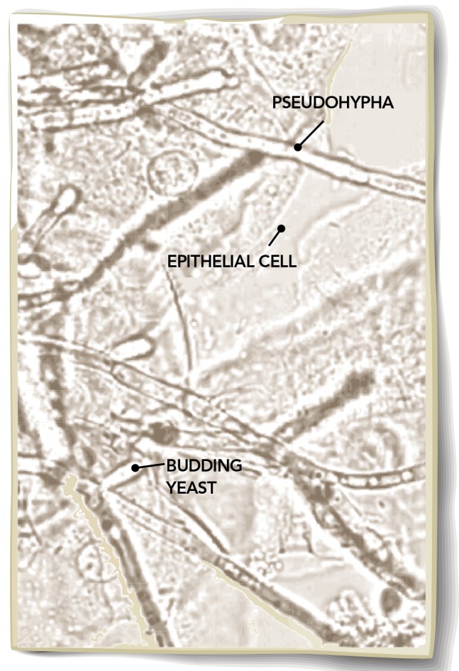
Place slide on the microscope stage and start with a low-power (10 ×) examination. To make epithelial cells visible, reduce illumination by lowering the condenser.
Examine for fungal structures such as hyphae or yeast. If any look suspicious, use the 40 × setting (high-dry objective) to investigate further, as hyphae or budding yeast suggest fungus.
Evidence
One study found a likelihood ratio of 2.86 with regard to finding fungal infection using KOH preparation.1 This represents a clinically useful value in the context of a common diagnosis. Moreover, the set-up required for this diagnostic tool is modest (KOH and a microscope), and the procedure can be performed quickly at the point of care. Culture is the criterion standard test used to determine the presence of a fungal infection. Given that culture has a likelihood ratio that is only moderately superior (3.28) to KOH preparation, is expensive, and takes 6 weeks to yield a result, teaching microscopic KOH preparation appears to be reasonable during residency. However, a negative KOH preparation result should still prompt a culture.
Diagnostic confirmation
Fungal culture or referral to a dermatologist for biopsy or periodic acid–Schiff stain might be necessary to confirm the diagnosis.

The physical examination is facing extinction in modern medicine. The Top Ten Forgotten Diagnostic Procedures series was developed as a teaching tool for residents in family medicine to reaffirm the most important examination-based diagnostic procedures, once commonly used in everyday practice. For a complete PDF of the Top Ten Forgotten diagnostic procedures, go to http://dl.dropbox.com/u/24988253/bookpreview%5B1%5D.pdf.
Reference
- 1.Weinberg JM, Koestenblatt EK, Tutrone WD, Tishler HR, Najarian L. Comparison of diagnostic methods in the evaluation of onychomycosis. J Am Acad Dermatol. 2003;49(2):193–7. doi: 10.1067/s0190-9622(03)01480-4. [DOI] [PubMed] [Google Scholar]


