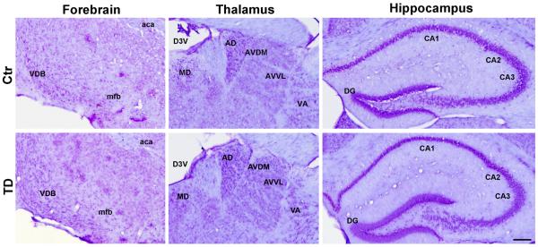
Figure 4.
Nissl staining of brain sections from control and TD mice which were sacrificed on the 26th day. General structure and neuronal morphology were examined by conventional Nissl staining as described under the Experimental Procedures. VDB, nucleus of the vertical limb of the diagonal band; mfb, medial forebrain bundle; aca, anterior commissure anterior; MD, mediodorsal thalamic nucleus; AD, anterodorsal thalamic nucleus; AVDM, anterovent thalamic nucleus of dorsomedial part; AVVL, anterovent thalamic nucleus of ventrolateral part; VA, ventral anterior thalamic nucleus; D3V, dorsal 3rd ventricle; CA1, field CA1 hippocampus; CA2, field CA2 hippocampus; CA3, field CA3 hippocampus; DG, dentate gyrus.

