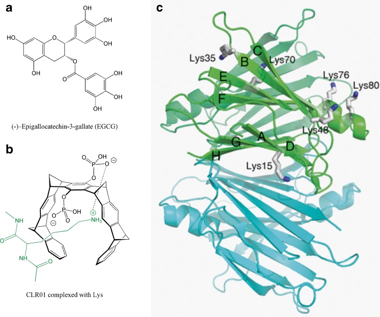
Fig. 6.
(A) Chemical structure of epigallocatechin gallate (EGCG) and (B) CLR01 complexed with lysine. (C) Crystal structure of transthyretin [PDB 2H4E] highlighting the lysine residues in one of the monomers. Each monomer has a β-sandwich structure composed of two β-sheets (DAGH and CBEF) and a short helix. Lys9 and Lys126 are located in the disordered N- and C-terminus (not shown)

