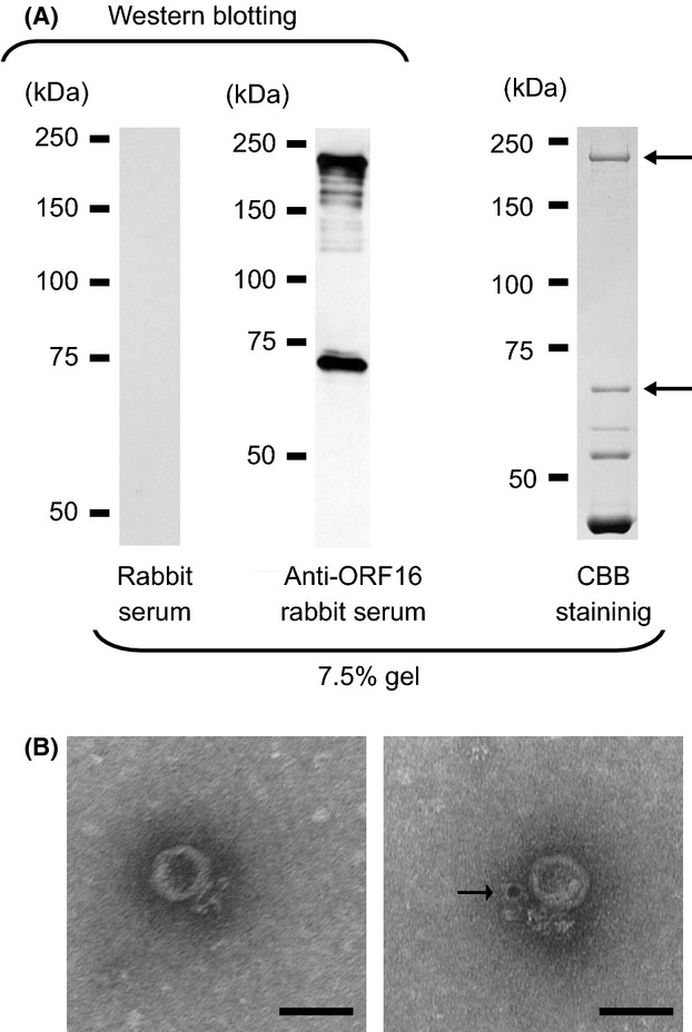
Figure 2.

(A) Analysis of the structural proteins of phage S24-1. Western blot analysis of ORF16 against the structural proteins of phage S24-1 (left and middle). The proteins separated by SDS-PAGE were visualized by CBB staining (right). The protein bands indicated by arrows were identified as ORF16 by mass spectrometry (see Fig. S2). (B) Immunoelectron microscopic analysis of ORF16 against phage S24-1. A control electron micrograph is shown on the left. An electron micrograph of phage S24-1 treated with the anti-ORF16 rabbit antibody is shown on the right, where a gold particle appears to be attached to the vicinity of the phage tail, which is indicated by an arrow. The bars represent 50 nm.
