
Figure 7. High LIF expression levels are associated with a poor relapse free survival of breast cancer patients.
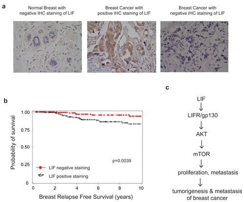
(a) The expression of LIF in a cohort of 374 human breast cancer specimens was determined by IHC staining of LIF. Representative images of LIF IHC staining in normal and breast cancer specimens are shown. (b) Kaplan-Meier curves for relapse free survival in breast cancer patients with or without positive LIF staining. Positive LIF staining (>10% cells stained with LIF) in breast cancer specimens is significantly correlated with a poorer relapse free survival and a higher breast cancer relapse risk (p=0.0039). (c) Schematic model depicting the activation of the AKT-mTOR signaling by LIF which in turn promotes proliferation and metastasis of breast cancer.