Abstract
The ability to move on solid surfaces provides ecological advantages for bacteria, yet many bacterial species lack this trait. We found that Xanthomonas spp. overcome this limitation by making use of proficient motile bacteria in their vicinity. Using X. perforans and Paenibacillus vortex as models, we show that X. perforans induces surface motility, attracts proficient motile bacteria and ‘rides' them for dispersal. In addition, X. perforans was able to restore surface motility of strains that lost this mode of motility under multiple growth cycles in the lab. The described interaction occurred both on agar plates and tomato leaves and was observed between several xanthomonads and motile bacterial species. Thus, suggesting that this motility induction and hitchhiking strategy might be widespread and ecologically important. This study provides an example as to how bacteria can rely on the abilities of their neighboring species for their own benefit, signifying the importance of a communal organization for fitness.
Keywords: Paenibacillus, surface motility, Xanthomonas
Bacteria colonize surfaces of various environments and are often dependent on surface motility for survival. Such a mode of motility allows bacteria to escape local stresses, translocate to a better nutritional environment and efficiently invade host tissue (Fraser and Hughes, 1999; Rashid and Kornberg, 2000; Harshey, 2003). However, despite the benefits, for many bacterial species the ability to migrate on solid surfaces does not exist or is dependent on high surface wetness and thus, restricted to specific environmental conditions (Wang et al., 2005; Kearns, 2010). The latter is true for the phytopathogenic bacteria from the genus Xanthomonas, which rely on high relative humidity for movement (Diab et al., 1982). We examined the interaction of X. perforans with several bacterial species that posses the ability to migrate across solid surfaces, focusing on Paenibacillus vortex. Paenibacillus spp. are found in many diverse environments and are often associated with plants (McSpadden Gardener, 2004, this study). Many of them including P. vortex are proficiently motile, able to migrate on solid surfaces containing ⩾1.5% agar, with no dependence on media type (Ingham and Ben Jacob, 2008).
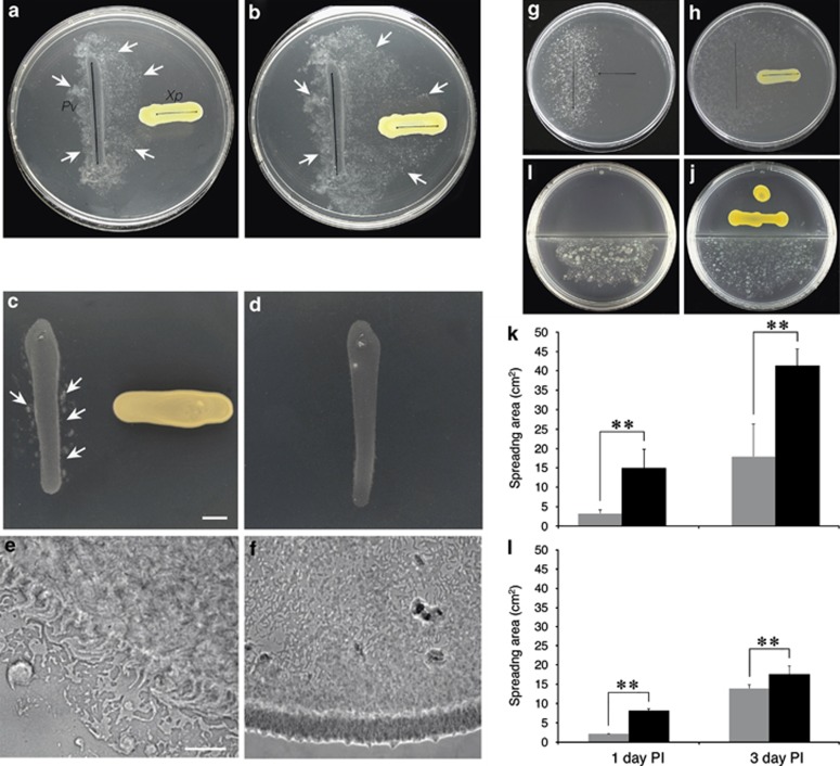
On co-inoculation P. vortex exhibited a strong migration toward X. perforans. Directional migration was observed on rich (Figures 1a and b), as well as poor media (Supplementary Figure S1). Examination of P. vortex movement over time indicated that the migration speed of both control and exposed colonies was not significantly different (Supplementary Table S1). However, in the presence of X. perforans, P. vortex colonies commenced movement earlier (Figures 1c–f) and spread to greater distances (2.56±0.4-fold) than control colonies (Figures 1g, h and k). Notably, the induction of movement in exposed colonies compared with control also occurred when the two bacterial colonies were separated by a plastic barrier of a bipartite plate, indicating that the effect of X. perforans on P. vortex surface motility was mediated by an airborne substance (Figures 1i, j and l; Supplementary Figure S2).
Figure 1.
The effect of X. perforans on P. vortex surface motility: directional migration of P. vortex toward X. perforans at 20 (a) and 48 h (b) post P. vortex inoculation. White arrows designate P. vortex colony edges. (c–f) Movment commencement of P. vortex in the presence (c) or absence of X. perforans (d). Light microscope image of P. vortex colony's edge in the presence (e) or absence of X. perforans (f). Migrational area of P. vortex in the presence (h) or (g) absence of X. perforans in regular (g, h and k) and (i, j and l) bipartite petri plates. In (k and l) black bar, presence of X. perforans; gray bar, control. (**) in (k) 1 day P=0.00003; 3 days P=0.007. (**) in (l) 1 day P=0.02, 3 days P=0.005. Mean±s.e.m. plotted; n=5; two-tailed t-test performed. Plate diameter 9 cm. (c and e) Scale bars are 0.5 cm and 100 μm, respectively.
Bacterial cell density could have a profound effect on the time point of movement commencement. In order to examine whether the induced movement of exposed P. vortex colonies was due to an increased growth rate and thus a higher cell density, P. vortex cells of exposed and control colonies were extracted from agar and analyzed by an image-stream cytometer, measuring both cell number and length of thousands of cells in each run. Interestingly, although P. vortex cells exposed to X. perforans headspace commenced movement earlier than control, cell numbers of exposed samples were actually lower (2.7±0.7-fold) than those of control samples (Supplementary Figure S3a). Conversely, mean cell length was higher in P. vortex colonies exposed to X. perforans headspace (Supplementary Figure S3b). Such attributes could result from incomplete cell division and elongation of a subpopulation, a phenomenon previously shown to be characteristic of migrating P. vortex colonies (Ingham and Ben Jacob, 2008). Indeed, image-stream cytometer analysis indicated that the fraction of cells longer than 15 μm was significantly higher (3.0±0.2-fold) in exposed P. vortex colonies compared with control (Supplementary Figure S3c).
The fact that X. perforans active substances induced motility of P. vortex in rich as well as poor media and that P. vortex cell numbers were not higher in exposed colonies compared with control, suggest that the affecting substance is not involved in P. vortex metabolism and likely serves as a cue. This hypothesis was further strengthened by the fact that X. perforans was able to induce surface motility in P. vortex colonies that stopped exhibiting this mode of motility under laboratory conditions (Supplementary Movie S1). Loss of surface motility after multiple growth cycles in the laboratory has been reported for several bacteria (Velicer et al., 1998; Henderson et al., 1999; Kearns et al., 2004). The mechanisms responsible for this loss are beyond the scope of this study. Nonetheless, the fact that a non-motile culture regained this ability on exposure to X. perforans points out that the switch between the two phenotypes can be modulated by external factors, and can work in both directions as proposed to occur in phase variations (Velicer et al., 1998; Henderson et al., 1999; Kearns and Losick, 2003; Kearns et al., 2004). The possibility that the external factors affecting surface motility can be signals produced by neighboring species raises many new questions regarding the mechanism that governs surface motility in nature.
In order to assess the prevalence of the described interaction, we examined the effect of additional bacteria from various genera, as well as of additional Xanthomonas species from differing pathovars on P. vortex. Response of additional bacterial species to X. perforans was also examined. All xanthomonads tested affected P. vortex surface motility similarly to X. perforans (Supplementary Table S1), but phytopathogenic bacteria from other genera had no effect (Supplementary Figure S4). Surface motility of the following bacterial species was not affected by X. perforans: Bacillus subtilis 3610, swrA/sfp complement strain of B. subtilis 168, Escherichia coli K-12 and Azospirillum brasilense, all of which typically swarm on surfaces containing less than 1% agar (data not shown). However, a positive effect was observed with Paenibacillus dendritiformis (data not shown) and Proteus mirabilis cells, which have the ability to disperse on relatively dry solid surfaces of ⩾1.5% agar (Supplementary Figure S5). Moreover, the active substance in X. perforans headspace was able to restore swarming motility in P. mirabilis colonies that lost this phenotype under laboratory conditions, in a similar manner to that described for P. vortex (Supplementary Figure S6). We also performed isolations of bacteria from tomato plants. Only bacteria that were able to move on 1.5% agar plates were examined. Two bacterial strains were isolated and according to their 16SrDNA sequences were identified as Flavobacterium sp. and Paenibacillus sp. (Supplementary Table S1). Out of these two isolates, only Paenibacillus sp. exhibited enhanced surface motility in response to X. perforans (data not shown).
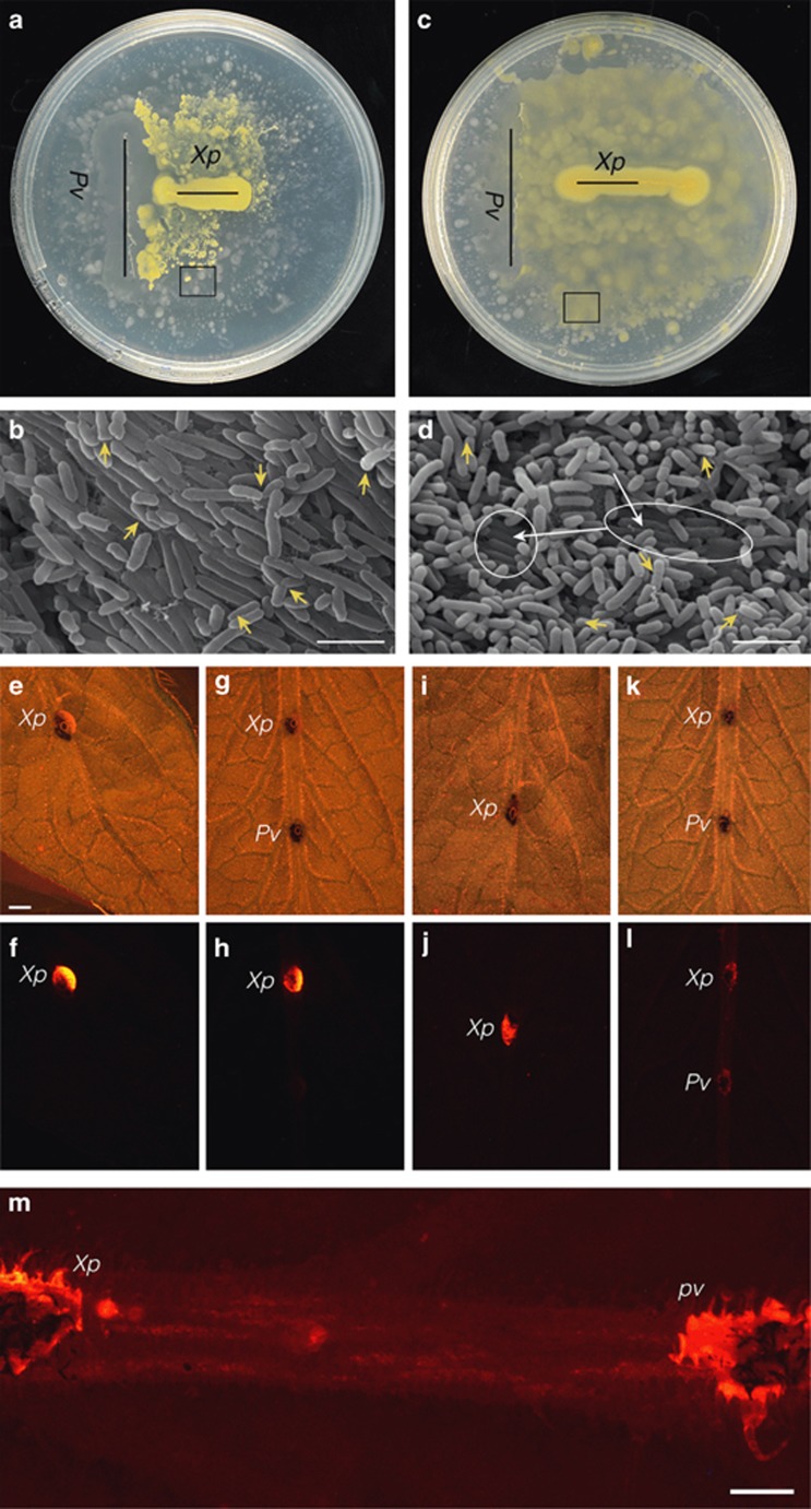
The induction of surface motility and attraction of such proficiently motile bacteria could hold a benefit, visible on a petri dish, for X. perforans. This bacterium lacks the ability to migrate across nutrient 1.5% agar plates (Shen et al., 2001, also see Figure 1). However, in co-inoculation experiments with P. vortex, X. perforans began to massively spread on the plates (Figures 2a and c). In scanning electron microscope images of the co-migration areas on the plate, X. perforans cells (slightly wider in diameter and shorter in length) were visible on top of the P. vortex rafts appearing as single cells on top of these rafts at the edges of the colonies but more as aggregates in the denser areas of both colonies (Figures 2b and d).
Figure 2.
Co-inoculation of X. perforans and P. vortex results in dispersal of X. perforans cells. Co-inoculation on the 1.5% nutrient agar plate (a and b) at 60 and 90 h (c and d) post P. vortex inoculation. (b and d) Scanning electron microscope images of areas in squares depicted in (a) and (c), respectively. Scale bar, 2.5 μm. White arrows indicate P. vortex rafts; yellow arrows indicate X. perforans ‘riders'. Co-inoculation on tomato leaves. Bright-field (e, g, i and k) and fluorescent (f, h, j, l and m) images of fluorescent stained X. perforans (Xp), inoculated on the surface of the major vein of tomato leaves, in the presence (g, h, k, l and m) or absence (e, f, i and j) of P. vortex (Pv). (e–h) Images taken at 2 days post inoculation. (i–m) Images taken at 5 days post bacterial inoculation. (e) Scale bar, 2 mm; (m) scale bar, 1 mm. (m) A higher magnification of (l) showing the fluorescent trail between the colonies. Brown dots visible in the bright-field images are colors marked on the leaf to indicate the point of inoculation.
In addition, scanning electron microscope analysis of X. perforans and P. vortex colonies on detached tomato leaf surface (major hosts for X. perforans infection) indicated that when both species were co-inoculated on leaves, a massive spread of cells occurred between the two colonies by 6 days post inoculation, (Supplementary Figure S7). Whereas when X. perforans cells were inoculated alone on the leaf, no bacterial cell spreading was observed at that time (data not shown). Notably, scanning electron microscope analysis could not provide definite proof for migration of both species, as when on leaves, both bacterial species were covered with a thick matrix, making species differentiation based on cell size and spatial organization not accurate (Supplementary Figures S7a and c). However, by using membrane-fluorescent stained X. perforans cells inoculated on tomato leaves, we were able to show X. perforans cell dispersion dependent on P. vortex presence (Figures 2e–m; Supplementary Figure S8). When inoculated alone or in the presence of E. coli bacteria, fluorescent X. perforans cells remained at the point of inoculation, even during prolong incubations of up to 7 days (Figures 3i and j; Supplementary Figures S8a and b). However, after 5 days of co-inoculation with non-fluorescent P. vortex on the major leaf vein, a spread of fluorescent X. perforans cells was visible between the two colonies (Figures 2l and m). When co-inoculation was performed between the minor leaf veins, a dispersal zone of fluorescent X. perforans cells was visible after 7 days (Supplementary Figure S8d).
The importance of surface motility for epiphytic survival and host infection was demonstrated in several studies (Haefele and Lindow, 1987; Harshey, 2003; Lindow and Brandl, 2003). Thus, the benefit that this interaction could provide to X. perforans is clear. As to the responding bacteria the consequences of this relationship is less clear. It was shown that resident bacteria on leaves could enhance the survival of immigrant bacteria by modifying the microenvironment of the leaf surface (Monier and Lindow, 2005; Poza-Carrion et al., 2013). It is possible that by ‘helping' X. perforans reach openings on the leaf surface, P. vortex might obtain more nutrients owing to the increased activity of plant-degrading exoenzymes employed by X. perforans on infection. This, yet to be proven, could determine whether this interaction is commensal or mutualistic.
Surface motility is a costly trait; this study suggests that in natural environments the interactions between bacteria that are able to migrate on solid surfaces and those unable, are not random but involve coordinated events, which enable a group of bacteria to spare the cost but obtain the end product of surface motility. Such interactions emphasize the importance of a community's phenotypic richness in addition to that of the individual's for the benefit of its members.
Acknowledgments
We thank Reut Shavit for her help in identifying isolated bacterial strains, the Photography Department at the Weizmann Institute for their professional help in imaging and figure preparation and the Otto Warburg Minerva Center for Agricultural Biotechnology for use of their facilities. Thanks to Professor Sigal Ben Yehuda for kindly providing Bacillus subtilis strain 3610 and Dr Avigor Eldar for kindly providing swrA/sfp complement strain of B. subtilis 168. This work was supported by the Israeli Science Foundation (ISF) and the Research Centre for Agriculture, Environment and Natural Resources.
The authors declare no conflict of interest.
Footnotes
Supplementary Information accompanies this paper on The ISME Journal website (http://www.nature.com/ismej)
Author contributions
YH led the project, designed experiments and wrote the paper together with SS. EH designed and performed experiments and wrote the paper. RD, TH-B EZ and ZP designed and performed experiments.
Supplementary Material
References
- Diab S, Bashan Y, Okon Y, Henis Y. Effects of relative humidity on bacterial scab caused by Xanthomonas campestris pv. vesicatoria on pepper. Phytopathology. 1982;72:1257–1260. [Google Scholar]
- Fraser GM, Hughes C. Swarming motility. Curr Opin Microbiol. 1999;2:630–635. doi: 10.1016/s1369-5274(99)00033-8. [DOI] [PubMed] [Google Scholar]
- Haefele DM, Lindow SE. Flagellar Motility Confers Epiphytic Fitness Advantages upon Pseudomonas syringae. Appl Environ Microbiol. 1987;53:2528–2533. doi: 10.1128/aem.53.10.2528-2533.1987. [DOI] [PMC free article] [PubMed] [Google Scholar]
- Harshey RM. Bacterial motility on a surface: many ways to a common goal. Annu Rev Microbiol. 2003;57:249–273. doi: 10.1146/annurev.micro.57.030502.091014. [DOI] [PubMed] [Google Scholar]
- Henderson IR, Owen P, Nataro JP. Molecular switches—the ON and OFF of bacterial phase variation. Mol Microbiol. 1999;33:919–932. doi: 10.1046/j.1365-2958.1999.01555.x. [DOI] [PubMed] [Google Scholar]
- Ingham CJ, Ben Jacob E. Swarming and complex pattern formation in Paenibacillus vortex studied by imaging and tracking cells. BMC Microbiol. 2008;8:36. doi: 10.1186/1471-2180-8-36. [DOI] [PMC free article] [PubMed] [Google Scholar]
- Kearns DB, Losick R. Swarming motility in undomesticated Bacillus subtilis. Mol Microbiol. 2003;49:581–590. doi: 10.1046/j.1365-2958.2003.03584.x. [DOI] [PubMed] [Google Scholar]
- Kearns DB, Chu F, Rudner R, Losick R. Genes governing swarming in Bacillus subtilis and evidence for a phase variation mechanism controlling surface motility. Mol Microbiol. 2004;52:357–369. doi: 10.1111/j.1365-2958.2004.03996.x. [DOI] [PubMed] [Google Scholar]
- Kearns DB. A field guide to bacterial swarming motility. Nature. 2010;8:634–644. doi: 10.1038/nrmicro2405. [DOI] [PMC free article] [PubMed] [Google Scholar]
- Lindow SE, Brandl MT. Microbiology of the phyllosphere. Appl Environ Microbiol. 2003;69:1875–1883. doi: 10.1128/AEM.69.4.1875-1883.2003. [DOI] [PMC free article] [PubMed] [Google Scholar]
- McSpadden Gardener BB. Ecology of Bacillus and Paenibacillus spp. in Agricultural Systems. Phytopathology. 2004;94:1252–1258. doi: 10.1094/PHYTO.2004.94.11.1252. [DOI] [PubMed] [Google Scholar]
- Monier JM, Lindow SE. Aggregates of resident bacteria facilitate survival of immigrant bacteria on leaf surfaces. Microb Ecol. 2005;49:343–352. doi: 10.1007/s00248-004-0007-9. [DOI] [PubMed] [Google Scholar]
- Poza-Carrion C, Suslow T, Lindow S. Resident bacteria on leaves enhance survival of immigrant cells of Salmonella enterica. Phytopathology. 2013;103:341–351. doi: 10.1094/PHYTO-09-12-0221-FI. [DOI] [PubMed] [Google Scholar]
- Rashid MH, Kornberg A. Inorganic polyphosphate is needed for swimming, swarming, and twitching motilities of Pseudomonas aeruginosa. Proc Natl Acad Sci USA. 2000;97:4885–4890. doi: 10.1073/pnas.060030097. [DOI] [PMC free article] [PubMed] [Google Scholar]
- Shen Y, Chern M, Silva FG, Ronald P. Isolation of a Xanthomonas oryzae pv. oryzae flagellar operon region and molecular characterization of flhF. Mol Plant Microbe Interact. 2001;14:204–213. doi: 10.1094/MPMI.2001.14.2.204. [DOI] [PubMed] [Google Scholar]
- Velicer GJ, Kroos L, Lenski RE. Loss of social behaviors by Myxococcus xanthus during evolution in an unstructured habitat. Proc Natl Acad Sci USA. 1998;95:12376–12380. doi: 10.1073/pnas.95.21.12376. [DOI] [PMC free article] [PubMed] [Google Scholar]
- Wang QF, Suzuki A, Mariconda S, Porwollik S, Harshey RM. Sensing wetness: a new role for the bacterial flagellum. Embo J. 2005;24:2034–2042. doi: 10.1038/sj.emboj.7600668. [DOI] [PMC free article] [PubMed] [Google Scholar]
Associated Data
This section collects any data citations, data availability statements, or supplementary materials included in this article.


