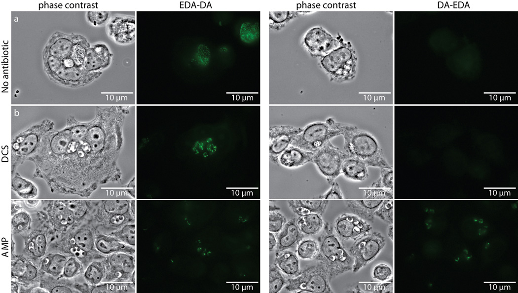
Extended Data Figure 7. D-cycloserine (DCS) and ampicillin (AMP) influence labeling of C. trachomatis PG by dipeptide probes EDA-DA and DA-EDA.
Phase contrast and epifluorescence microscopy of L2 cells infected with C. trachomatis 18 hours post infection. Cells were grown in the presence of either EDA-DA or DA-EDA (1 mM) and were either untreated (a), or treated with 294 µM DCS (b) or 2.8 µM AMP (c). Subsequent binding of the probe to a modified Alexa Fluor 488 (green) was achieved via click chemistry. The image used for EDA-DA labeling in the absence of antibiotics is the same image from Extended Data Figure 6b and experiments were all conducted in parallel on the same day. Images showing labeling by EDA-DA and DA-EDA in the presence or absence of DCS are representative of the vast majority of over 100 inclusions measured 18 hours post-infection. Labeling by EDA-DA in the presence of ampicillin is representative of 97% (73/75) total aberrant bodies while labeling by DA-EDA in the presence of ampicillin is representative of 95% (73/77) total aberrant bodies, as viewed by epifluorescence microscopy. Experiments were conducted in technical duplicates and represent at least three biological replicates.

