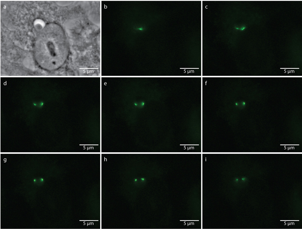
Extended Data Figure 8. Punctate labeling of aberrant bodies due to enlarged bacteria encompassing multiple focal planes.
Phase contrast (a) and epifluorescence microscopy (b–i) of an 18 hour, EDA-DA labeled, ampicillin-induced aberrant body. Images were taken through sequential focal planes in order to show how the ring-like, PG structure is maintained in aberrant bodies and can appear punctate when viewed via an epifluorescence microscope. Images are representative of between 3–5 fields viewed per technical replicate, comprising over 20 independent biological replicates, and each experiment was conducted in technical duplicates.

