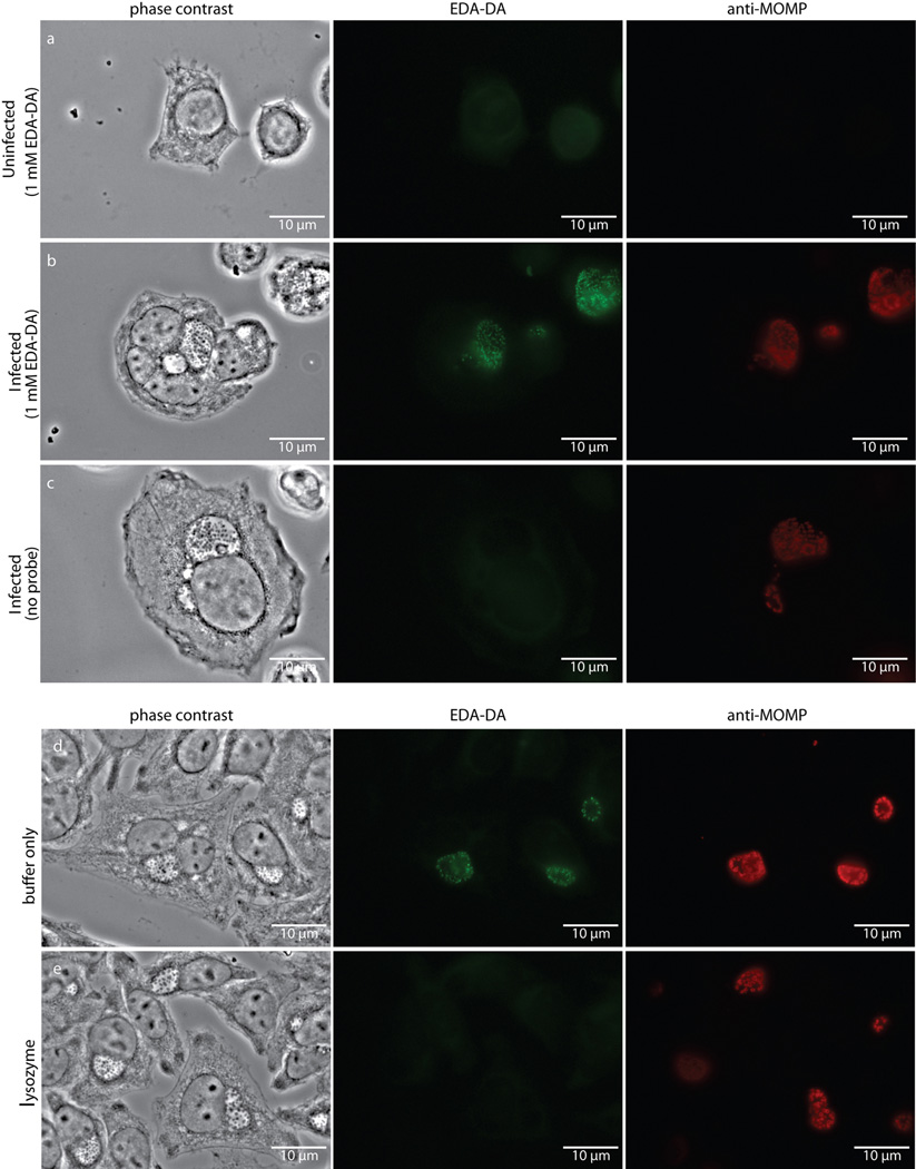
Extended Data Figure 6. Fluorescence is specific to chlamydial infected cells in the presence of the dipeptide probe EDA-DA, and lysozyme treatment is capable of removing the label from fixed bacteria.
Phase contrast and epifluorescence microscopy was conducted on (a) uninfected L2 cells grown in the presence of 1 mM EDA-DA, (b) 18 hour C. trachomatis-infected cells in the presence of 1 mM EDA-DA, and (c) 18 hour C. trachomatis-infected cells grown in the absence of probe. Subsequent binding of the probe to a modified Alexa Fluor 488 (green) was achieved via a click chemistry reaction. For lysozyme treatments, 18 hour C. trachomatis-infected cells (fixed and labeled as described above) were suspended in either (d) buffer (25 mM NaPO4 pH 6.0, 0.5 mM MgCl2) or (e) buffer and lysozyme (200 µg/mL) for two hours. Cells were subsequently washed, blocked, and counter labeled with anti-MOMP, as described previously. Images are representative of between 3–5 fields examined (with ~1–10 inclusions viewed per field) per technical replicate, each condition conducted in technical duplicates, and experiments represent a total of three biological replicates.

