

Abstract
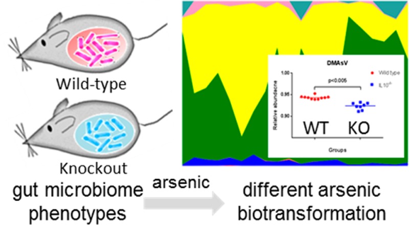
Large individual differences in susceptibility to arsenic-induced diseases are well-documented and frequently associated with different patterns of arsenic metabolism. In this context, the role of the gut microbiome in directly metabolizing arsenic and triggering systemic responses in diverse organs raises the possibility that gut microbiome phenotypes affect the spectrum of metabolized arsenic species. However, it remains unclear how host genetics and the gut microbiome interact to affect the biotransformation of arsenic. Using an integrated approach combining 16S rRNA gene sequencing and HPLC-ICP-MS arsenic speciation, we demonstrate that IL-10 gene knockout leads to a significant taxonomic change of the gut microbiome, which in turn substantially affects arsenic metabolism.
Exposure to arsenic affects large human populations worldwide, with contamination of drinking water by geological sources of inorganic arsenic being the primary route of exposure. Hundreds of millions of people drink water with arsenic levels that far exceed the 10 μg/L guideline established by the World Health Organization (WHO) and U.S. Environmental Protection Agency (EPA).1 For example, approximately 25 million Americans are estimated to drink water from private wells with an arsenic level above EPA guidelines.2 Arsenic exposure has been associated with a number of diseases, such as skin, bladder, lung, and liver cancers and diabetes as well as cardiovascular disorders and reproductive defects.1 It is well-documented that there are large differences in susceptibility to arsenic-induced diseases among individuals at similar exposure levels,3 with several underlying mechanisms, such as genetic polymorphisms, epigenetics, and nutrition homeostasis, being proposed. Individual susceptibility is frequently associated with different spectra of arsenic metabolism. The metabolic pathway of inorganic arsenic consists of alternating reduction from pentavalent arsenic to trivalent and oxidative methylation of the trivalent arsenic metabolites. The six arsenic species involved in this pathway in humans are inorganic arsenic (iAsV and iAsIII), monomethylarsonic acid (MMAsV), monomethylarsonous acid (MMAsIII), dimethylarsinic acid (DMAsV), and dimethylarsinous acid (DMAsIII). The abundance and patterns of arsenic species have long been closely associated with toxicity in humans exposed to arsenic, with the MMAsV/DMAsV ratio showing a positive association with arsenic toxicity in several studies.4
Accumulating evidence indicates that perturbations of the gut microbiome and its functions may play an important role in the development of human diseases.6 The essential role of the gut microbiome in the metabolism of xenobiotics raises the possibility that gut microbiome phenotypes affect the biotransformation of arsenic; a few experiments have documented this using gut microbiota from animal models or in vitro simulators of the human intestinal microbial ecosystem.7 In particular, our group has recently demonstrated that bacterial infection-induced gut microbiome perturbations impact the biotransformation of inorganic arsenic.9 The composition of the gut microbiome is highly diverse, and this diversity can be modulated by many factors, such as environment, diet, bacterial/viral infection, and antibiotics. However, it remains unclear whether host genetics define the gut microbiome and subsequently affect the metabolism of arsenic. Therefore, in this study, we employed a mouse model with different gut microbiome phenotypes driven by gene knockout to address this question. The selection of interleukin-10 (IL-10) was based on its function in immune response, which is intrinsically intertwined with the gut flora.10
The experimental workflow and detailed methods are described in the Supporting Information and Figure S1. Briefly, the gut microbiome difference between wild-type and IL-10–/– mice were first determined using 16S rRNA gene sequencing (Figure 1). Next, wild-type and knockout mice were treated with arsenic (10 ppm in drinking water) for 4 weeks followed by arsenic-speciation analysis using HPLC-ICP-MS to quantify major arsenic metabolites (Figure 2). Finally, correlations between the gut microbiome changes and altered arsenic metabolites in urine were examined to establish the functional impacts of gene-driven gut microbiome changes on arsenic biotransformation (Figure 3).
Figure 1.
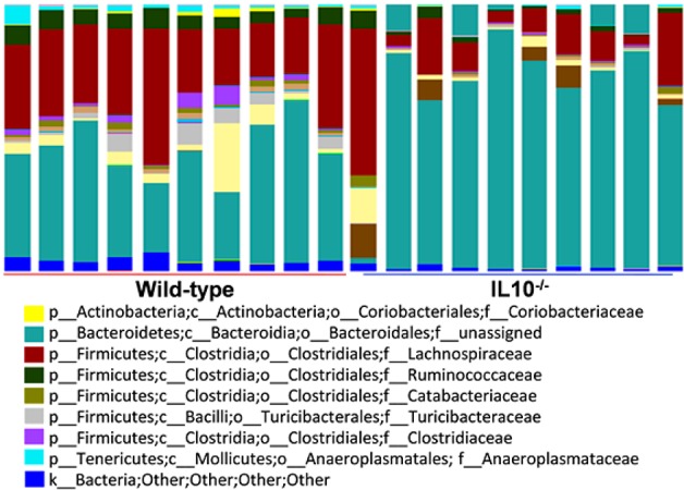
Gut microbiome composition profiles at the family level in wild-type and knockout mice (p, phylum; c, class; o, order; and f, family).
Figure 2.
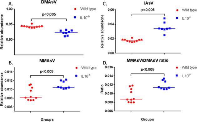
Gene-knockout-driven gut microbiome changes affect arsenic biotransformation in mice: (A) DMAsV, (B) MMAsV, (C) iAsV, and (D) ratio of MMAsV/DMAsV.
Figure 3.
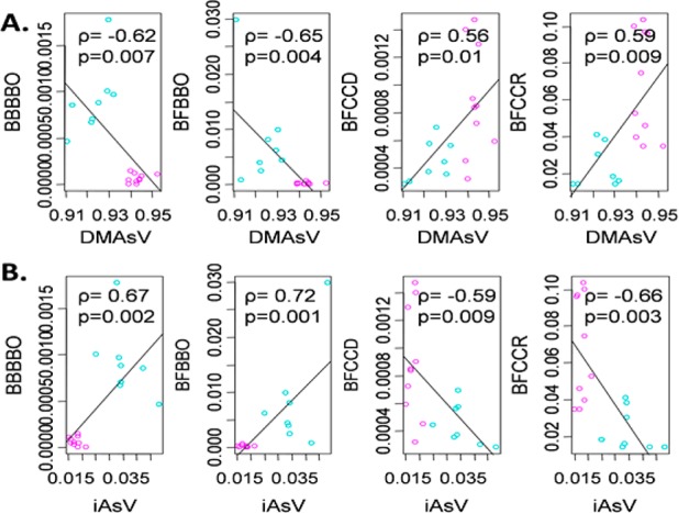
Correlation plots, calculated by Pearson’s correlation coefficient, demonstrating the functional correlation between altered gut bacteria families (relative abundance given on the y axis) and DMAsV (A) and iAsV (B) (relative abundance given on the x axis) (BBBBO: p_Bacteroidetes, c_Bacteroidia, o_Bacteroidales, and other; BFBBO: p_Firmicutes, c_Bacilli, o_Bacillales, and other; BFCCD: p_Firmicutes, c_Clostridia, o_Clostridiales, and f_Dehalobacteriaceae; and BFCCR: p_Firmicutes, c_Clostridia, o_Clostridiales, and f_Ruminococcaceae).
Figure 1 shows the gut microbiome difference between wild-type and knockout mice. Identified gut bacteria assigned at the family level from 16S rRNA sequencing reads are illustrated, with each color representing an individual bacterial family. Clearly, IL-10 knockout induces a significant taxonomic perturbation in the gut microbiome of mice, with Bacteroidetes (teal) and Firmicutes (brown) being remarkably increased and decreased, as shown in Figure 1. Detailed information about changed gut bacterial families is listed in Table S1. The difference in the gut microbiome patterns arising from gene knockout is also readily differentiated using multivariate statistical analysis, as shown by the PCoA plot in Figure S2. The jackknifed β diversity and hierarchical-clustering analysis via unweighted pair group method with arithmetic mean (UPGMA) demonstrate that all wild-type and knockout animals cluster in their own groups (Figure S2). In addition, histological analysis revealed that gene knockout and arsenic did not induce statistically significant differences between the wild-type and knockout mice with regard to inflammation, edema, epithelial defects, crypt atrophy, hyperplasia, and dysplasia in multiple regions of the colon and liver during the experimental period (Figure S3).
We next performed arsenic-speciation analysis in urine (Figure S4) and compared the relative abundance of measured arsenic species to examine the impact of gene-driven gut microbiome alterations on arsenic biotransformation. Among six arsenic metabolites quantified, DMAsV was significantly decreased in urine after 4 weeks of exposure to arsenic (Figure 2A), whereas MMAsV and iAsV were concurrently increased in the knockout group (Figure 2B,C), suggesting that gut microbiome changes arising from gene knockout interfere with the detoxification of inorganic arsenic. In addition, the ratio of MMAsV/DMAsV was increased in knockout mice (Figure 2D), resembling those observed in humans who are associated with higher arsenic toxicity.11
To explore the functional correlation between the gut microbiome phenotypes and arsenic metabolites, a correlation matrix was generated by calculating the Pearson’s correlation coefficient (Figure S5). Clear correlations were identified between the gut microbiome and altered arsenic species (ρ > 0.5 or < −0.5, p < 0.05) in exposed mice. Figure 3 lists several typical gut bacterial components that are highly negatively or positively correlated with DMAsV and iAsV. These results show that gut microbiome phenotypes driven by gene knockout substantially alter the metabolite profile of arsenic.
Previously, we have shown that bacterial infection-induced gut microbiome perturbations alter arsenic metabolism.9 This study further reveals that gene-knockout-driven gut microbiome phenotypes affect the biotransformation of inorganic arsenic, highlighting the role of the interaction between host genetics and the gut microbiome in xenobiotics metabolism. It has been reported that there are large individual variations in the gut microbiome and metagenomic genotypes in humans,12 with mechanisms that have not been identified yet. In addition to external factors, animal studies suggest that the host’s genetic background may play a role in shaping gut-flora phenotypes.14 Of particular importance, gut microbiome phenotypes are associated with different individuals’ capacity to metabolize xenobiotics, as the gut microbiome is not only involved in direct metabolism of compounds but also triggers systemic effects beyond the gut, such as altering diverse enzymes in the liver, including P450s and other enzymes responsible for the metabolic activation of xenobiotics.15 Therefore, it would provide novel mechanistic insights into individual susceptibility to decipher complex interactions among host genetics, gut microbiome composition/function, and xenobiotics metabolism and subsequent toxic effects.
In summary, we have combined an animal model with gene-driven gut microbiome phenotypes, 16S rRNA gene sequencing, and HPLC-ICP-MS arsenic speciation to delineate the impact of gut microbiome phenotypes on the biotransformation of arsenic. Several major arsenic species were significantly modulated by gut microbiome changes resulting from gene knockout. These data show that gut microbiome compositions play a role in affecting the abundance of specific toxic species, supporting the hypothesis that gut microbiome phenotypes affect the spectra of arsenic metabolites and individual response to exposure. These findings also emphasize the importance of host genetics in shaping the gut microbiome. Taken together, this study serves as the first demonstration of interaction among host genetics, gut microbiome, and xenobiotics metabolism and highlights the gene-driven gut microbiome phenotype as a potential novel risk factor associated with individual susceptibility to diverse environmental chemicals such as inorganic arsenic.
Glossary
Abbreviations
- ICP-MS
inductively coupled plasma mass spectrometry
- iAs
inorganic arsenic
- MMAsV
monomethylarsonic acid
- MMAsIII
monomethylarsonous acid
- DMAsV
dimethylarsinic acid
- DMAsIII
dimethylarsinous acid
- MMAsV
monomethylarsonic acid
- PCoA
principal coordinate analysis
Supporting Information Available
Experimental workflow and detailed methods; gut bacterial patterns revealed by 16S rRNA sequencing; histological analysis for the wild-type and knockout animals; arsenic metabolism and speciation by HPLC-ICP-MS; and correlation analysis between arsenic metabolites and gut bacterial families. This material is available free of charge via the Internet at http://pubs.acs.org.
Author Contributions
The manuscript was written through contributions of all authors. All authors have given approval to the final version of the manuscript.
We thank the MIT Center for Environmental Health Sciences for financial support through a pilot project under NIEHS grant P30 ES002109 and the UNC Center for Environmental Health and Susceptibility for partial financial support by NIEHS grant P30 ES010126. We also thank the UGA for CPH internal grant and an FRG grant.
The authors declare no competing financial interest.
Funding Statement
National Institutes of Health, United States
Supplementary Material
References
- Hughes M. F.; Beck B. D.; Chen Y.; Lewis A. S.; Thomas D. J. (2011) Arsenic exposure and toxicology: A historical perspective. Toxicol. Sci. 123, 305–332. [DOI] [PMC free article] [PubMed] [Google Scholar]
- Kozul C. D.; Ely K. H.; Enelow R. I.; Hamilton J. W. (2009) Low-dose arsenic compromises the immune response to influenza A infection in vivo. Environ. Health Perspect. 117, 1441–1447. [DOI] [PMC free article] [PubMed] [Google Scholar]
- Steinmaus C.; Yuan Y.; Kalman D.; Rey O. A.; Skibola C. F.; Dauphine D.; Basu A.; Porter K. E.; Hubbard A.; Bates M. N.; Smith M. T.; Smith A. H. (2010) Individual differences in arsenic metabolism and lung cancer in a case-control study in Cordoba, Argentina. Toxicol. Appl. Pharmacol. 247, 138–145. [DOI] [PMC free article] [PubMed] [Google Scholar]
- Ahsan H.; Chen Y.; Kibriya M. G.; Slavkovich V.; Parvez F.; Jasmine F.; Gamble M. V.; Graziano J. H. (2007) Arsenic metabolism, genetic susceptibility, and risk of premalignant skin lesions in Bangladesh. Cancer Epidemiol., Biomarkers Prev. 16, 1270–1278. [DOI] [PubMed] [Google Scholar]
- Kinross J. M.; Darzi A. W.; Nicholson J. K. (2011) Gut microbiome-host interactions in health and disease. Genome Med. 3, 14–25. [DOI] [PMC free article] [PubMed] [Google Scholar]
- Kubachka K. M.; Kohan M. C.; Herbin-Davis K.; Creed J. T.; Thomas D. J. (2009) Exploring the in vitro formation of trimethylarsine sulfide from dimethylthioarsinic acid in anaerobic microflora of mouse cecum using HPLC-ICP-MS and HPLC-ESI-MS. Toxicol. Appl. Pharmacol. 239, 137–143. [DOI] [PubMed] [Google Scholar]
- Lu K.; Cable P. H.; Abo R. P.; Ru H.; Graffam M. E.; Schlieper K. A.; Parry N. M.; Levine S.; Bodnar W. M.; Wishnok J. S.; Styblo M.; Swenberg J. A.; Fox J. G.; Tannenbaum S. R. (2013) Gut microbiome perturbations induced by bacterial infection affect arsenic biotransformation. Chem. Res. Toxicol. 26, 1893–1903. [DOI] [PMC free article] [PubMed] [Google Scholar]
- Maharshak N.; Packey C. D.; Ellermann M.; Manick S.; Siddle J. P.; Huh E. Y.; Plevy S.; Sartor R. B.; Carroll I. M. (2013) Altered enteric microbiota ecology in interleukin 10-deficient mice during development and progression of intestinal inflammation. Gut Microbes 4, 316–324. [DOI] [PMC free article] [PubMed] [Google Scholar]
- Yu R. C.; Hsu K. H.; Chen C. J.; Froines J. R. (2000) Arsenic methylation capacity and skin cancer. Cancer Epidemiol., Biomarkers Prev. 9, 1259–1262. [PubMed] [Google Scholar]
- Schloissnig S.; Arumugam M.; Sunagawa S.; Mitreva M.; Tap J.; Zhu A.; Waller A.; Mende D. R.; Kultima J. R.; Martin J.; Kota K.; Sunyaev S. R.; Weinstock G. M.; Bork P. (2013) Genomic variation landscape of the human gut microbiome. Nature 493, 45–50. [DOI] [PMC free article] [PubMed] [Google Scholar]
- Vijay-Kumar M.; Aitken J. D.; Carvalho F. A.; Cullender T. C.; Mwangi S.; Srinivasan S.; Sitaraman S. V.; Knight R.; Ley R. E.; Gewirtz A. T. (2010) Metabolic syndrome and altered gut microbiota in mice lacking Toll-like receptor 5. Science 328, 228–231. [DOI] [PMC free article] [PubMed] [Google Scholar]
- Claus S. P.; Ellero S. L.; Berger B.; Krause L.; Bruttin A.; Molina J.; Paris A.; Want E. J.; de Waziers I.; Cloarec O.; Richards S. E.; Wang Y.; Dumas M. E.; Ross A.; Rezzi S.; Kochhar S.; Van Bladeren P.; Lindon J. C.; Holmes E.; Nicholson J. K. (2011) Colonization-induced host-gut microbial metabolic interaction. mBio 2, e00271-1–e00271-10. [DOI] [PMC free article] [PubMed] [Google Scholar]
Associated Data
This section collects any data citations, data availability statements, or supplementary materials included in this article.