Abstract
Increased consumption of fresh vegetables that are high in polyphenols has been associated with a reduced risk of oxidative stress-induced disease. The present study aimed to evaluate the antioxidant effects of spinach in vitro and in vivo in hyperlipidemic rats. For measurement of in vitro antioxidant activity, spinach was subjected to hot water extraction (WE) or ethanol extraction (EE) and examined for total polyphenol content (TPC), oxygen radical absorbance capacity (ORAC), cellular antioxidant activity (CAA), and antigenotoxic activity. The in vivo antioxidant activity of spinach was assessed using blood and liver lipid profiles and antioxidant status in rats fed a high fat-cholesterol diet (HFCD) for 6 weeks. The TPC of WE and EE were shown as 1.5±0.0 and 0.5±0.0 mg GAE/g, respectively. Increasing the concentration of the extracts resulted in increased ORAC value, CAA, and antigenotoxic activity for all extracts tested. HFCD-fed rats displayed hyperlipidemia and increased oxidative stress, as indicated by a significant rise in blood and liver lipid profiles, an increase in plasma conjugated diene concentration, an increase in liver thiobarbituric acid reactive substances (TBARS) level, and a significant decrease in manganese superoxide dismutase (Mn-SOD) activity compared with rats fed normal diet. However, administration of 5% spinach showed a beneficial effect in HFCD rats, as indicated by decreased liver TBARS level and DNA damage in leukocyte and increased plasma conjugated dienes and Mn-SOD activity. Thus, the antioxidant activity of spinach may be an effective way to ameliorate high fat and cholesterol diet-induced oxidative stress.
Keywords: spinach, comet assay, liver TBARS, tail DNA, hyperlipidemic rat
INTRODUCTION
Hyperlipidemia, a condition characterized by high levels of circulating fats, is regarded as a modifiable risk factor for cardiovascular disease and cerebrovascular disease. The incidence of these vascular disorders among South Koreans is increasing at a rapid rate due in part to adoption of a more Western lifestyle and increased consumption of fatty foods (1). A high-fat diet has been shown to increase oxidative stress in a variety of tissues, a side effect that may contribute to the development of numerous degenerative diseases (2–5).
In order to prevent or moderate oxidation-related diseases, it is necessary to sequester and eliminate free radicals from the body (6). Antioxidants may offer some resistance to oxidative stress by scavenging free radicals, inhibiting cell membrane damage, and suppressing lipid peroxidation, thus preventing the onset of chronic disease (7). There is increasing interest in the antioxidant activity of the phytochemicals present in our diet and in health food supplements (8), and a number of studies have demonstrated that antioxidant supplementation prevents or delays hyperlipidemia-related disease (9,10).
Spinach, which is cultivated globally, is an important dietary vegetable and a common raw material in the food processing industry (11,12). Spinach is a proven source of essential nutrients such as carotene (a precursor of vitamin A), ascorbic acid, and several types of minerals. Although a number of studies have been conducted on the antioxidant activities of spinach (12–14), there is still a relative lack of information available regarding its hypolipidemic activity.
The purpose of this study was to determine the in vitro antioxidant effects of spinach and evaluate the potential benefits of spinach supplementation in hyperlipidemic rats.
MATERIALS AND METHODS
Sample preparation
Spinach, in dried powder form, was obtained from the Spinach Cluster Agency (Jeonnam, Korea) after being cultivated in Shinan Island, Jeonnam, Korea. For preparation of the hot water extracts (WE), 5 g of spinach powder was put in a glass bottle with 100 mL of deionized water, extracted with an autoclave, and freeze dried (Ilshin, Yangju, Korea). The ethanol extracts (EE) were prepared as follows: 5 g of spinach powder was extracted with 100 mL of ethyl alcohol and concentrated by evaporator (N-1110S-W, EYELA, Tokyo, Japan). WE and EE were redissolved in dimethyl sulfoxide to a concentration of 50 mg/mL and aliquots were kept at 20°C until used.
Measurement of total phenolic contents
Total polyphenol content (TPC) was measured by the method of Park et al. (15). Briefly, WE was mixed with 2 mL of 1 N Folin-Ciocalteu reagent and incubated at room temperature. Then 2 mL of 10% Na2CO3 were added and the mixture was incubated at room temperature. The absorbance was measured at 690 nm with an enzyme-linked immunosorbent assay (ELISA) reader (Tecan, Grödig, Salzburg, Austria). TPC was expressed as gallic acid equivalents (GAE).
Oxygen radical absorbance capacity assay
The oxygen radical absorbance capacity (ORAC) assay was carried out on a FLUOstar OPTIMA fluorescence plate reader (BMG LABTECH Gmbh, Ortenberg, Germany) with fluorescent filters (excitation wavelength 485 nm, emission wavelength 535 nm) according to the method of Park et al. (15). The results were calculated based on the difference in the area under the fluorescence decay curves between the blank and each sample. ORAC ROO· was expressed as μmol of Trolox equivalents (TE).
Cellular antioxidant activity assay
HepG2 cells were seeded at a density of 5×10⁴ cells/mL on 96-well microplates. Twenty-four hours after seeding, the growth medium was removed, and the wells were washed with phosphate buffered saline (PBS). Triplicate wells were treated for 1 h with 100 μL of extracts dissolved in treatment medium. Wells were washed with 100 μL of PBS, and 80 μM of AAPH in 100 μL of PBS plus 40 μM DCFH-DA was applied to the cells. Following the addition of the AAPH solution, the 96-well microplate was placed into a FLUOstar OPTIMA fluorescence plate reader at 37.8°C. Emission at 535 nm was measured with excitation at 485 nm. Triplicate control and blank wells were included on each plate: control wells contained cells treated with DCFH-DA and an oxidant, AAPH; blank wells contained cells treated with PBS without AAPH.
DNA damage determination by alkaline comet assay
The alkaline comet assays were conducted by the modified method (16) described by Singh et al. (17). For the measurement of antigenotoxic activity of spinach in vitro, human leukocytes were incubated for 30 min at 37°C with varying concentrations of WE or EE dissolved in DMSO (1, 10, 50, and 100 μg/mL). Following incubaton, leukocytes were resuspended in PBS with 200 μM H2O2, incubated on ice for 5 min, and washed with PBS. The slides were then immersed in a lysis solution and incubated for 1 h at 4°C. Next, the slides were equilibrated in electrophoresis buffer for 40 min. For electrophoresis of the DNA, an electric current of 25 V/300±3 mA was applied for 20 min at 4°C. The slides were washed three times with a neutralizing buffer and treated with ethanol for another 5 minutes before staining with 20 μL of ethidium bromide (20 μg/mL). A fluorescence microscope (Leica DMLB, Leica, Solms, Germany) was used to image the slides and Komet 4.0 software (Kinetic Imaging Ltd., Merseyside, UK) was used to analyze each image and determine the percent fluorescence in the tail (tail intensity [TI]; 50 cells from each of two replicate slides).
Animals and diets
All aspects of the in vivo experiment were conducted according to the guidelines provided by the Ethical Committee for Experimental Animal Care of Kyungnam University. Eight-week-old male Sprague-Dawley rats (SD, n=24) were purchased from KOATEC, Inc. (Pyeongtaek, Korea) and housed individually in a room with a 12-h light/12-h dark cycle, a temperature of 22~24°C, and a relative humidity of 50±5%. The rats were allowed free access to water and fed a commercially prepared diet for a 1 wk adjustment period. The rats were then randomly divided into three groups of eight animals each and fed an AIN-93 based normal diet (ND), a high fat-cholesterol diet (HFCD), or a HFCD supplemented with 5% freeze- dried spinach powder (HFCD+S). The HFCD contained 18% casein, 50% corn starch, 10% sucrose, 6.5% cellulose, 3.5% mineral mixture (AIN-93G), 1% vitamin mixture (AIN-93G), 0.2% choline bitartarate, 0.3% DL-methionine, 0.5% cholesterol, and 9.5% fat. Animals were monitored daily for general health. At the end of the experimental period, the rats were anesthetized with iso-flurane and blood was collected from the abdominal artery into a heparinized sterile tube. The plasma fraction was obtained from the blood samples by centrifugation at 450 g for 30 min and stored at −80°C until required for further analysis. The experiment was approved by Animal Care Committee, Kyungnam University, Gyeongnam, Korea.
Blood and hepatic lipid profiles
Plasma lipid profiles (i.e., total cholesterol, high-density lipoprotein [HDL]-cholesterol, triglycerides) were measured using assay kits from Bioclinical Systems (Anyang, Korea) and a photometric autoanalyzer (CH-100 plus; SEAC, Calenzano, Italy). Plasma low-density lipoprotein (LDL) cholesterol concentrations were calculated using the formula developed by Friedewald et al. (18). The concentration of total lipids in liver samples was determined using the method of Folch et al. (19). Total cholesterol (TC) and triglyceride concentrations in liver samples were analyzed with the same enzymatic kits used in the plasma analyses.
Baseline conjugated dienes in LDLs
The concentration of conjugated dienes in plasma was determined according to the method of Park et al. (16). Briefly, plasma was added to 700 μL of heparin citrate buffer and incubated for 10 min at room temperature. After centrifugation at 1,000 g for 10 min, the pellet was resuspended in 100 μL of 0.1 M Na-phosphate buffer containing 0.9% NaCl (pH 7.4). Lipids were extracted from 100 μL of the LDL suspension with chloroform/methanol (2:1), dried under nitrogen, and redissolved in cyclohexane. A spectrophotometer (UV-1205, Shimadzu, Tokyo, Japan) was used to determine the absorbance of the redissolved sample at 234 nm.
Hepatic thiobarbituric acid reactive substance (TBARS)
The thiobarbituric acid reactive substances (TBARS) level in plasma was estimated by the method of Buege and Aust (20). Briefly, 0.2 mL of plasma was mixed with 0.3 mL of distilled water and added to 1 mL of a mixture containing 0.38% thiobarbituric acid, 15% trichloroacetic acid, and 0.25 N HCl. The resulting mixture was incubated for 20 min in a boiling water bath prior to centrifugation at 2,000 g for 10 min. The absorbance of the supernatant was measured at 540 nm using 1,1,3,3- tetramethoxypropane (TMP) as standard. Lipid peroxidation was expressed as TBARS in nmol/mL plasma.
Liver lipid peroxidation levels were measured by the method of Ohkawa et al. (21) with some modification. Liver homogenates (0.4 mL) in 1.15% KCl, 0.1 mL of 8.1% SDS, 0.75 mL of 20% acetic acid, and 0.75 mL of 0.8% TBA were added to a test tube. The sample was vortexed and heated in a 95°C oil bath (OHB-2000, Tokyo Rikakikai Co., Tokyo, Japan) for 1 h. After cooling for 10 min, butanol-pyridine 15:1 (v/v) was added. The sample was mixed thoroughly and centrifuged at 3,515 g for 15 min. The fluorescence of the upper layer was measured at 552 nm. The amount of MDA present in the sample was converted to TBARS values using a TMP standard curve. A Pierce BCA protein assay kit (Thermo Scientific, Aalst, Belgium) was used to quantify the protein concentration of each liver sample.
Erythrocytic catalase
Erythrocytic hemolysates were prepared by the dilution of erythrocytes to 1:500 with distilled H2O. One hundred microliter of erythrocytic hemolysate was dissolved in 50 mM phosphate buffer 50 mL (pH 7), and 2 mL of the mixture was added to a cuvette. The reaction was initiated by the addition of 1 mL of H2O2 30 nM at 20°C. The H2O2 decomposition rate was measured at 240 nm for 30 sec using a spectrophotometer.
Hepatic antioxidant enzymes
To determine liver superoxide dismutase activity, liver samples were homogenized with 0.1 mL of 65 mM phosphate buffer (pH 7.8), and then centrifuged at 10,000 g for 20 min. Copper, zinc-superoxide dismutase (Cu/Zn-SOD) activity was measured in the resulting supernatant (i.e., the cytosolic fraction). The remaining pellet (i.e., the mitochondrial fraction) was dissolved in 0.1% triton and used for the determination of manganese-superoxide dismutase (Mn-SOD) activity. For the Mn-SOD activity assay, 4 mmol of KCN solution was added to the assay mixture to inhibit Cu/Zn-SOD. For Mn-SOD and Cu/Zn-SOD analyses, the samples were pre-incubated with 75 mM Na-xanthine and 10 mM hydroxylamine hydrochloride at 37°C for 10 min. Then 0.1 units of xanthine oxidase was added and samples were incubated at 37°C for an additional 20 min. The reaction was stopped by the addition of 1% sulphanilamide and 0.02% ethylenediamine dihydrochloride. After standing at room temperature for 20 min, the absorbance of the final mixture was measured at 540 nm. One nitrate unit (NU) of SOD activity was defined as the amount of protein required for 50% inhibition.
Catalase (CAT) activity was measured according to the method of Carrillo et al. (22). Liver tissue was homogenized in Na-K phosphate buffer (pH 7.0). The homogenates were centrifuged at 600 g for 10 min to obtain the supernatant. After centrifuging at 10,000 g for 20 min, the supernatant was discarded and the pellet was suspended in 1× RBC buffer. After incubating on ice for 10 min, the suspension was centrifuged at 10,000 g for 20 min. The pellet was washed in Na-K phosphate buffer (pH 7.0) and centrifugation was repeated. After washing, the pellet was resuspended in Na-K phosphate buffer. For the assay, Na-K phosphate buffer and sample were mixed in a quartz cuvette (QS, Hellma GmbH & Co., Müllheim, Germany) and the reaction was started by the addition of 300 μL of 30 mM H2O2 solution. The H2O2 decomposition rate was measured at 240 nm for 40 sec using a spectrophotometer.
Glutathione peroxidase (GSH-Px) activity was measured according to the method of Bogdanska et al. (23). Liver tissue was homogenized with 1 mL of 250 mM potassium phosphate buffer (pH 7.0), and then centrifuged at 10,000 g for 20 min. Twenty-five μL of supernatant, containing cytosolic fraction, was incubated with 10 mM EDTA, 10 mM NaN3, 10 mM GSH, 2 mM NADPH, and 1 unit glutathione reductase at room temperature for 5 min. The reaction was initiated by the addition of 25 μL of 2.5 mM H2O2. The H2O2 decomposition rate was measured at 340 nm for 70 sec using a spectrophotometer.
The protein concentration of each supernatant was determined by BCA protein assay. Samples containing equal amounts of protein (25 μg) were used for the enzyme activity assays.
DNA damage in rat leukocytes
Twenty μL of whole blood were suspended with 150 μL of 0.7% low melting agarose (LMA), and added to slides that had been pre-coated with agarose. The slides were then treated in PBS with 200 μM H2O2 for 5 min on ice and washed with PBS. The following steps were the same as done in the in vitro DNA damage determination by comet assay.
Statistical analysis
All measurements were analyzed using the SPSS package for Windows (Ver. 14.0; SPSS, Chicago, IL, USA). Mean values among concentrations/animal groups were compared using one-way analysis of variance (ANOVA) followed by Duncan’s multiple range tests (P<0.05).
RESULTS
Total polyphenol contents, antioxidant, and antigenotoxic effects of spinach
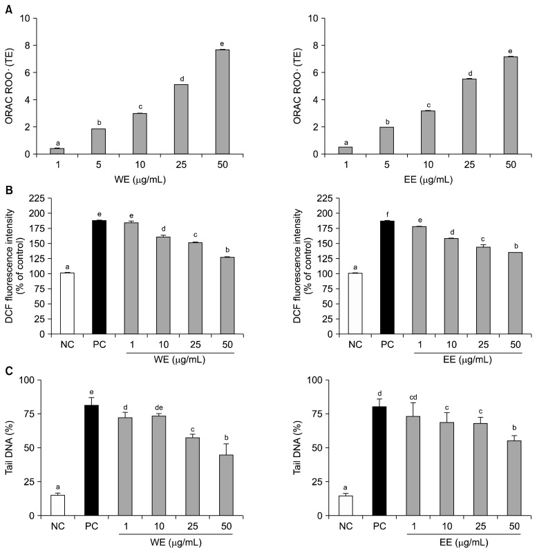
The TPC of WE and EE were 1.5±0.0 mg GAE/g and 0.5±0.0 mg GAE/g, respectively. The ORAC values of WE and EE increased in a concentration dependent manner (Fig. 1A). At the highest tested concentration (50 μg/mL), WE (7.6±0.1 TE) and EE (7.2±0.1 TE) showed similar ability to protect a fluorescent reporter from oxidative degeneration by AAPH. The viability of spinach-extract-treated HepG2 cells was 70% at the highest concentration tested (50 μg/mL, data not shown). These extracts reduced AAPH-induced oxidative stress in HepG2 cells (Fig. 1B), with higher concentrations producing greater increases in CAA. Pretreatment of the human leukocytes with spinach extracts for 30 min significantly reduced H2O2-induced oxidative stress (Fig. 1C), with higher concentrations showing increased ability to inhibit DNA damage.
Fig. 1.
ORAC value (A), CAA value (B), and antigenotoxic activity (C) of spinach. Values are mean±SD (n=3). Values not sharing the same letter are significantly different from one another (P<0.05) by Duncan’s multiple range test.
Effects of spinach on blood and hepatic lipid profiles
In the in vivo studies, no adverse reactions or clinical signs were observed in HFCD rats throughout the feeding period. As shown in Table 1, there was a marked increase in the levels of TC and LDL-C in the HFCD group compared with the ND group. Plasma HDL-C decreased significantly in the HFCD group compared to the ND group. Plasma TC, HDL-C, and LDL-C levels did not differ between HFCD and HFCD+S rats. Additionally, while hepatic total lipid, TC, and triglyceride levels were significantly higher in the HFCD rats than in the ND rats, these concentrations did not differ between HFCD and HFCD+S groups.
Table 1.
Effects of spinach on the plasma and hepatic lipid profiles of rats fed a high fat-cholesterol diet (HFCD)
| ND1) | HFCD | HFCD+S | |
|---|---|---|---|
| Plasma (mg/g dL) | |||
| Total cholesterol | 117.4±3.1a | 174.7±14.0b | 183.2±20.0b |
| Triglyceride | 57.3±2.6ns2) | 65.4±4.7 | 58.1±4.3 |
| HDL cholesterol | 67.14±3.7a | 37.0±3.2b | 28.8±1.5b |
| LDL cholesterol | 44.9±5.4a | 121.9±12.3b | 142.1±19.4b |
| Liver (mg/g wet sample) | |||
| Total lipid | 9.5±1.6a | 41.4±4.6b | 48.6±3.6b |
| Total cholesterol | 0.7±0.0a | 5.7±0.5b | 6.0±0.5b |
| Triglycerides | 0.6±0.0a | 2.8±0.1b | 3.0±0.2b |
Value are mean±SE (n=8).
Different letters indicate significant difference at P<0.05.
ND, normal diet; HFCD, high fat-cholesterol diet; HFCD+S, high fat-cholesterol diet supplemented with 5% spinach powder.
Not significant.
Effects of spinach on antioxidant status
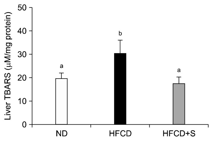
Liver TBARS levels were 79% higher in rats in the HFCD group versus the ND group, as shown in Fig. 2. Liver TBARS levels in rats that received the HFCD+S were not different from those of rats that received the ND. There was no difference in plasma TBARS level among treatment groups.
Fig. 2.
Effects of spinach on liver TBARS of rats fed a high fat-cholesterol diet. ND, normal diet; HFCD, high fat-cholesterol diet; HFCD+S, high fat-cholesterol diet supplemented with 5% spinach powder. Values are mean±SD (n=8). Values not sharing the same letter are significantly different from one another (P<0.05) by Duncan’s multiple range test.
Plasma conjugated diene concentrations were significantly higher in the HFCD group than in the ND group (Table 2). Plasma conjugated diene concentrations were decreased in the HFCD+S group compared to the HFCD group; however, this difference was not statistically significant.
Table 2.
Effects of spinach on blood and liver antioxidant metabolism of rats fed a high fat-cholesterol diet
| ND1) | HFCD | HFCD+S | |
|---|---|---|---|
| Plasma | |||
| Conjugated dienes (μM) | 4.0±0.2a | 4.9±0.3b | 4.3±0.2ab |
| TBARS (μM) | 100.0±4.3ns2) | 110.0±0.2 | 105.0±10.0 |
| Erythrocytes | |||
| Catalase (K/g Hb) | 1,159.0±85.7ns | 989.1±73.3 | 1,058.8±77.4 |
| Liver | |||
| Mn-SOD (U/mg protein) | 0.20±0.02a | 0.17±0.01b | 0.20±0.10ab |
| Cu/Zn-SOD (U/mg protein) | 277.8±70.0ns | 283.8±51.8 | 285.8±41.0 |
| Catalase (mM/mg protein) | 100.0±4.3ns | 78.3±11.2 | 72.5±6.7 |
| GSH-Px (μM/mg protein) | 100.0±25.0ns | 84.0±15.3 | 95.4±6.8 |
Value are mean±SD (n=8).
Different letters indicate significant difference at P<0.05.
ND, normal diet; HFCD, high fat-cholesterol diet; HFCD+S, high fat-cholesterol diet supplemented with 5% spinach powder.
Not significant.
The activity of Mn-SOD was significantly lower in the livers of rats in the HFCD group than in the livers of rats in the ND group, as shown in Table 2. Liver Mn-SOD activity was greater in the HFCD+S group than in the HFCD group; however, this difference was not statistically significant. There was no difference in plasma Cu/Zn-SOD activity among treatment groups.
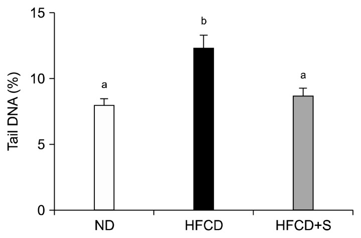
Tail DNA percentage was greater in the HFCD group than in the ND group, while tail DNA percentage in the HFCD+S group was not different from that of the ND group (Fig. 3).
Fig. 3.
Effects of spinach on H2O2 induced DNA damage in the leukocytes of rats fed a high fat-cholesterol diet. ND, normal diet; HFCD, high fat-cholesterol diet; HFCD+S, high fat-cholesterol diet supplemented with 5% spinach powder. Values are mean±SD (n=8). Values not sharing the same letter are significantly different from one another (P<0.05) by Duncan’s multiple range test.
DISCUSSION
In this study, we analyzed the TPC and antioxidant activity of spinach in vitro and the effect of spinach supplementation on antioxidant metabolism in a hyperlipidemic rat model. Previous reports have suggested that spinach, a vegetable with a high nutritional value, is a rich source of carotenoids, which are visually obscured by green chlorophyll (24). Several studies have indicated that spinach leaves contain several powerful and water-soluble natural antioxidants with potential biological activities (14,25–27). Polyphenols are now widely accepted as physiological antioxidants that have significant potential to protect against the numerous degenerative diseases linked to free radical-related tissue damage (28). The health benefits of polyphenols appear to arise from their antioxidant activities and capacity to protect critical macromolecules, such as chromosomal DNA, structural proteins and enzymes, LDL, and membrane lipids from damage resulting from exposure to ROS (29,30).
Because different solvent extractions yield different constituents, the spinach used in this study was extracted with hot water and ethanol. The TPC (147 mg/100 g) of WE was similar to that of carrot (156 mg/100 g) and onion (150 mg/100 g) (31), while the TPC of EE (51 mg/100 g) was similar to that of tomato (62 mg/100 g) and nectarine (57 mg/100 g). Our results demonstrate that WE and EE of spinach exhibit antioxidant activities that are attributable to their high TPC. The protective ability of spinach extracts against H2O2-induced DNA damage was assessed in normal human leukocytes by the comet assay. Pretreatment of the cells for 30 min with spinach extracts significantly reduced the genotoxicity of H2O2, as measured by DNA strand breaks.
Our in vitro experiments demonstrate that spinach extracts exert potent antioxidant activities; these effects were confirmed in hyperlipidemic rats. Many studies examining the in vitro antioxidant activity of extracts have also evaluated extract antioxidant activity in oxidative stress-induced animal models (32–34). We show here that hyperlipidemia induced by a fat and cholesterol-enriched diet increases blood and liver lipid levels in rats, thereby leading to oxidative stress that can be partly prevented by the antioxidant activities of spinach. Previous work by Lee et al. (35), showed that the blood lipid concentrations of cholesterol-fed rabbits are not improved by white ginseng extracts, although antioxidant enzyme activities are somewhat enhanced. Similarly, work by Kang et al. (36) revealed that the administration of plant extracts is not effective at lowering blood cholesterol in a hypercholesterolemic model. These findings can be attributed to the composition of the extract used in the experiments, as well as the dosages and experimental period. Hence, further investigation is necessary to determine the precise amount of supplementation necessary to achieve beneficial effects from spinach and prevent hyperlipidemia.
Remarkably, the addition of spinach to the diet was associated with reduced liver TBARS levels in the HFCD+S group compared to the HFCD group. As such, spinach treatment may reduce liver TBARS levels by inhibiting the oxidation of LDL; this would imply that spinach maintains antioxidant and antihyperlipidemic effects. The plasma concentration of conjugated dienes, a measure of lipid peroxidation, was dramatically increased the HFCD group compared to the ND group. However, administration of spinach reduced plasma lipid peroxidation, but the difference was not significant. Lee et al. (35) reported that the phenolic compounds present in ginseng extract reduce plasma and liver TBARS levels in hypercholesterolemic rats by increasing the activity of antioxidant enzymes. These results suggest that spinach may help improve antioxidant metabolism through the action of phenolic compounds.
Liver Mn-SOD activities of rats fed the HFCD were 28.1% lower than those of rats fed the ND, which is likely to aggravate existing oxidative stress. Mn-SOD activity was improved by administration of spinach, while Cu/Zn-SOD was not shown a significant difference among treatment groups. Previous work indicated that inhibition of antioxidant enzymes such as SOD occured in many degenerative diseases (37). Mn-SOD is active as an antioxidant enzyme in the mitochondria, while Cu/Zn-SOD is primarily responsible for the dismutation of superoxide anions in the cytosol (38). Differential regulation of Mn-SOD and Cu/Zn-SOD may occur under pathologic conditions (39,40). Oliveira et al. (41) reported that green juice reduced TBARS levels and catalase activity, suggesting that this type of supplementation may have a protective effect against reactive species.
Hydroxyl radicals, a kind of reactive species generated by oxidative stress, are also known to cause oxidation-induced breaks in DNA strands that promote open circular or relaxed DNA forms (42). WE and EE of spinach showed a robust and concentration-dependent reduction in the formation of nicked DNA and an increase in super-coiled DNA in in vitro studies, indicating a beneficial effect of spinach on the resistance of leukocyte DNA to oxidative damage. Our in vivo studies also confirmed that spinach protects leukocytes from oxidative stress; these effects are most likely related to the antioxidant properties of spinach. Kim et al. (43) reported that a leafy vegetable mix prevented lipid peroxidation and oxidative DNA damage in C57BL/6J mice fed a high fat and cholesterol diet.
To the best of our knowledge, this is the first study to show that dietary supplementation with spinach increases antioxidant activity and can improve antioxidant status in hyperlipidemic rats fed a HFCD. These results demonstrate that spinach alleviates oxidative stress through the modulation of antioxidant metabolism, resulting in a protective effect against HFCD-induced DNA damage.
CONCLUSIONS
Spinach extracts exhibit significant TPC, antioxidant activity (ORAC value and CAA), and antigenotoxic activity. In addition, spinach extract may improve the efficiency of the enzymatic antioxidant enzymatic system (i.e., Mn-SOD) following deactivation of the substrates for plasma conjugated dienes, in turn reducing liver TBARS levels and DNA damage in leukocyte of rats fed a high fat-cholesterol diet. Therefore, the results of the present study indicate that spinach extract is a potential source of natural antioxidants and its consumption improves antioxidant status.
Footnotes
AUTHOR DISCLOSURE STATEMENT
The authors declare no conflict of interest.
REFERENCES
- 1.Korean Statistical Information Service. Analysis on the actual conditions of deaths. [accessed September 2010]. http://kosis.kr/wnsearch/totalSearch.jsp.
- 2.Chen X, Zhong HY, Zeng JH, Ge J. The pharmacological effect of polysaccharides from Lentinus edodes on the oxidative status and expression of VCAM-1mRNA of thoracic aorta endothelial cell in high-fat-diet rats. Carbohyd Polym. 2008;74:445–450. [Google Scholar]
- 3.Lieber CS, Leo MA, Cao Q, Mak KM, Ren CL, Ponomarenko A. The combination of S-adenosylmethionine and dilinoleoylphosphatidylcholine attenuates nonalcoholic steatohepatitis produced by a high-fat diet in rats. Nutr Res. 2007;27:565–573. doi: 10.1016/j.nutres.2007.07.005. [DOI] [PMC free article] [PubMed] [Google Scholar]
- 4.Ma M, Liu GH, Yu ZH, Chen G, Zhang X. Effect of the Lycium barbarum polysaccharides administration on blood lipid metabolism and oxidative stress of mice fed high-fat diet in vivo. Food Chem. 2009;113:872–877. [Google Scholar]
- 5.Schreibelt G, van Horssen J, van Rossum S, Dijkstra CD, Drukarch B, de Vries HE. Therapeutic potential and biological role of endogenous antioxidant enzymes in multiple sclerosis pathology. Brain Res Rev. 2007;56:322–330. doi: 10.1016/j.brainresrev.2007.07.005. [DOI] [PubMed] [Google Scholar]
- 6.Hait-Darshan R, Grossman S, Bergman M, Deutsch M, Zurgil N. Synergistic activity between a spinach- derived natural antioxidant (NAO) and commercial anti-oxidants in a variety of oxidation system. Food Res Int. 2009;42:246–253. [Google Scholar]
- 7.Fu H, Xie B, Ma S, Zhu X, Fan G, Pan S. Evaluation of antioxidant activities of principal carotenoids available in water spinach (Ipomoea aquatica) J Food Compos Anal. 2011;24:288–297. [Google Scholar]
- 8.Bergman M, Parelman A, Dubinsky Z, Grossman S. Scavenging of reactive oxygen species by a novel glucurinated flavonoid antioxidant isolated and purified from spinach. Phytochemistry. 2003;62:753–762. doi: 10.1016/s0031-9422(02)00537-x. [DOI] [PubMed] [Google Scholar]
- 9.Xu J, Zhou X, Deng Q, Huang Q, Yang J, Huang F. Rapeseed oil fortified with micronutrients reduces atherosclerosis risk factors in rats fed a high-fat diet. Lipids Health Disease. 2011;10:96–103. doi: 10.1186/1476-511X-10-96. [DOI] [PMC free article] [PubMed] [Google Scholar]
- 10.Zhu L, Luo X, Jin Z. Effect of resveratrol on serum and liver lipid profile and antioxidant activity in hyperlipidemia rats. Asian-Aust J Anim Sci. 2008;21:890–895. [Google Scholar]
- 11.Aritomi M, Kawasaki T. Three highly oxygenated flavone glucuronides in leaves of Spinacia oleracea. Phytochemistry. 1984;23:2043–2047. [Google Scholar]
- 12.Gil MI, Ferreres F, Tomas-Barberan FA. Effect of postharvest storage and processing on the antioxidant constituents (flavonoids and vitamin C) of fresh-cut spinach. J Agric Food Chem. 1999;47:2213–2217. doi: 10.1021/jf981200l. [DOI] [PubMed] [Google Scholar]
- 13.Edenharder R, Keller G, Platt KL, Unger KK. Isolation and characterization of structurally novel antimutagenic flavonoids from spinach (Spinacia oleracea) J Agric Food Chem. 2001;49:2767–2773. doi: 10.1021/jf0013712. [DOI] [PubMed] [Google Scholar]
- 14.Lomnitski L, Carbonatto M, Ben-Shaul V, Peano S, Conz A, Corradin L, Maronpot RR, Grossman S, Nyska A. The prophylactic effects of natural water-soluble antioxidant from spinach and apocynin in a rat model of lipopoly-saccharide-induced endotoxemia. Toxicol Pathol. 2000;28:588–600. doi: 10.1177/019262330002800413. [DOI] [PubMed] [Google Scholar]
- 15.Park JH, Kim RY, Park E. Antioxidant and α-glucosidase inhibitory activities of different solvent extracts of skullcap (Scuellaria baicalensis) Food Sci Biotechnol. 2011;20:1107–1112. [Google Scholar]
- 16.Park JH, Seo BY, Lee KH, Park E. Onion supplementation inhibits lipid peroxidation and leukocyte DNA damage due to oxidative stress in high fat-cholesterol fed male rats. Food Sci Biotechnol. 2009;18:179–184. [Google Scholar]
- 17.Singh PN, McCoy MT, Tice RR, Schneider EL. A simple technique for quantitation of low levels of DNA damage in individual cells. Exp Cell Res. 1988;175:184–191. doi: 10.1016/0014-4827(88)90265-0. [DOI] [PubMed] [Google Scholar]
- 18.Friedewald WT, Levey RI, Fredrickson DS. Estimation of the concentration of low-density lipoprotein cholesterol plasma, without use of the preparative ultracentrifuge. Clin Chem. 1972;18:499–502. [PubMed] [Google Scholar]
- 19.Folch J, Lees M, Sloan-Stanley GH. A simple method for isolation and purification of total lipids from animal tissues. J Biol Chem. 1956;226:497–509. [PubMed] [Google Scholar]
- 20.Buege JA, Aust SD. Microsomal lipid peroxidation. Methods Enzymol. 1978;52:302–310. doi: 10.1016/s0076-6879(78)52032-6. [DOI] [PubMed] [Google Scholar]
- 21.Ohkawa H, Ohishi N, Yagi K. Assay for lipid peroxides in animal tissues by thiobarbituric acid reaction. Anal Biochem. 1979;95:351–358. doi: 10.1016/0003-2697(79)90738-3. [DOI] [PubMed] [Google Scholar]
- 22.Carrillo MC, Kanai S, Nokubo M, Kitani K. (−) deprenyl induces activities of both superoxide dismutase and catalase but not of glutathione peroxides in the striatum of young male rats. Life Sci. 1991;48:517–521. doi: 10.1016/0024-3205(91)90466-o. [DOI] [PubMed] [Google Scholar]
- 23.Bogdanska JJ, Korneti P, Todorova B. Erythrocyte superoxide dismutase, glutathione peroxidase and catalase activities in healthy male subjects in Republic of Macedonia. Bratisl Lek Listy. 2003;104:108–114. [PubMed] [Google Scholar]
- 24.Bunea A, Andjelkovic M, Socaciu C, Bobis O, Neacsu M, Verhé R, Camp JV, Bunea A, Andjelkovic M, Socaciu C, Bobis O. Total and individual carotenoids and phenolic acids content in fresh, refrigerated and processed spinach (Spinacia oleracea L.) Food Chem. 2008;108:649–656. doi: 10.1016/j.foodchem.2007.11.056. [DOI] [PubMed] [Google Scholar]
- 25.Lomnitski L, Foley J, Grossman S, Ben-Shaul V, Maronpot R, Moomaw C, Carbonatto M, Nyska A. Effects of apocynin and natural antioxidants from spinach on iNOS and COX-2 induction in LPS-induced hepatic injury in rat. Pharmacol Toxicol. 2000;87:18–25. doi: 10.1111/j.0901-9928.2000.870104.x. [DOI] [PubMed] [Google Scholar]
- 26.Lomnitski L, Nyska A, Ben-Shaul V, Maronpot RR, Haseman JK, Harrus TL, Bergman M, Grossman S. Effects of antioxidants apocynin and the natural water-soluble anti-oxidant from spinach on cellular damage induced by lipopolysaccaride in the rat. Toxicol Pathol. 2000;28:580–587. doi: 10.1177/019262330002800412. [DOI] [PubMed] [Google Scholar]
- 27.Nyska A, Lomnitski L, Spalding J, Dunson DB, Goldsworthy TL, Grossman S, Bergman M, Boorman G. Topical and oral administration of the natural water-soluble antioxidant from spinach reduces the multiplicity of papillomas in the Tg.AC mouse model. Toxicol Lett. 2001;122:33–44. doi: 10.1016/s0378-4274(01)00345-9. [DOI] [PubMed] [Google Scholar]
- 28.Zhu L, Luo X, Jin Z. Effect of resveratrol on serum and liver lipid profile and antioxidant activity in hyperlipidemia rats. Asian-Aust J Anim Sci. 2008;21:890–895. [Google Scholar]
- 29.Dreosti IE. Antioxidant polyphenols in tea, cocoa, and wine. Nutr. 2000;16:692–701. doi: 10.1016/s0899-9007(00)00304-x. [DOI] [PubMed] [Google Scholar]
- 30.Rice-Evans CA, Miller NJ, Paganga G. Structure-anti-oxidant activity relationships of flavonoids and phenolic acids. Free Radic Biol Med. 1996;20:933–956. doi: 10.1016/0891-5849(95)02227-9. [DOI] [PubMed] [Google Scholar]
- 31.Cieślik E, Gręda A, Adamus W. Contents of poly-phenols in fruit and vegetables. Food Chem. 2006;94:135–142. [Google Scholar]
- 32.Trajković LMH, Mijatović SA, Maksimović-Ivanić DD, Stojanović ID, Momčilović MB, Tufegdžić SJ, Maksimović VM, Marjanovi ZS, Stošić-Grujičić SD. Anticancer properties of Ganoderma lucidum methanol extracts in vitro and in vivo. Nutr Cancer. 2009;61:696–707. doi: 10.1080/01635580902898743. [DOI] [PubMed] [Google Scholar]
- 33.Lee HS, Won NH, Kim KH, Lee H, Jun W, Lee KW. Antioxidant effects of aqueous extract of Terminalia chebula in vivo and in vitro. Biol Pharm Bull. 2005;28:1639–1644. doi: 10.1248/bpb.28.1639. [DOI] [PubMed] [Google Scholar]
- 34.Celep E, Aydın A, Kırmızıbekmez H, Yesilada E. Appraisal of in vitro and in vivo antioxidant activity potential of cornelian cherry leaves. Food Chem Toxicol. 2013;11:448–455. doi: 10.1016/j.fct.2013.09.001. [DOI] [PubMed] [Google Scholar]
- 35.Lee LS, Cho CW, Hong HD, Lee YC, Choi UK, Kim YC. Hypolipidemic and antioxidant properties of phenolic compound-rich extracts from white ginseng (Panax ginseng) in cholesterol-fed rabbits. Molecules. 2013;18:12548–12560. doi: 10.3390/molecules181012548. [DOI] [PMC free article] [PubMed] [Google Scholar]
- 36.Kang SY, Kim SH, Schini VB, Kim ND. Dietary ginsenosides improve endothelium-dependent relaxation in the thoracic arota of hypercholesterolemic rabbit. Gen Pharmac. 1995;26:483–487. doi: 10.1016/0306-3623(95)94002-x. [DOI] [PubMed] [Google Scholar]
- 37.Moron MS, Dipierre JW, Mannervik B. Levels of glutathione reductase and glutathione-S-transferase activities in rat lung and liver. Biochim Biophys Acta. 1979;582:67–68. doi: 10.1016/0304-4165(79)90289-7. [DOI] [PubMed] [Google Scholar]
- 38.Qi X, Guy J, Nick H, Valentine J, Rao N. Increase of manganese superoxide dismutase, but not of Cu/Zn-SOD, in experimental optic neuritis proinvestigative. Invest Ophthalmol Visual Sci. 1997;38:1203–1212. [PubMed] [Google Scholar]
- 39.Bergeron C, Petrunka C, Weyer L. Copper/zinc super-oxide dismutase expression in the human central nervous system. Correlation with selective neuronal vulnerability. Am J Pathol. 1996;148:273–279. [PMC free article] [PubMed] [Google Scholar]
- 40.Yoneda T, Inagaki S, Hayashi Y, Nomura T, Takagi H. Differential regulation of manganese and copper/zinc superoxide dismutases by the facial nerve transection. Brain Res. 1992;582:342–345. doi: 10.1016/0006-8993(92)90153-z. [DOI] [PubMed] [Google Scholar]
- 41.Oliveira PS, Saccon TD, da Silva TM, Costa MZ, Dutra FS, de Vasconcelos A, Lencina CL, Stefanello FM, Barschak AG. Green juice as a protector against reactive species in rats. Nutr Hosp. 2013;28:1407–1412. doi: 10.3305/nh.2013.28.5.6505. [DOI] [PubMed] [Google Scholar]
- 42.Verma AR, Vijayakumar M, Rao CV, Mathela CS. In vitro and in vivo antioxidant properties and DNA damage protective activity of green fruit of Ficus glomerata. Food Chem Toxicol. 2010;48:704–709. doi: 10.1016/j.fct.2009.11.052. [DOI] [PubMed] [Google Scholar]
- 43.Kim MY, Cheong SH, Kim MH, Son C, Yook HS, Sok DE, Kim JH, Cho Y, Chun H, Kim MR. Leafy vegetable mix supplementation improves lipid profiles and antioxidant status in C57BL/6J mice fed a high fat and high cholesterol diet. J Med Food. 2009;12:877–884. doi: 10.1089/jmf.2008.1125. [DOI] [PubMed] [Google Scholar]


