
Figure 5. Organization of HUVECs on fibrin gel when cultured in macrophage-conditioned media for 4 days.
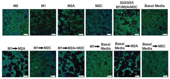
The behavior of endothelial cells in media conditioned by a single macrophage phenotype was compared to that of endothelial cells cultured in media that was switched after 24hrs from one macrophage phenotype to another. Cell nuclei were stained with DAPI (blue) and actin filaments were stained with fluorescent phalloidin (green). Experiments were repeated three times. Scale bars are 500μm.