Abstract
Background
The prevalence of lipoatrophy in children on antiretroviral therapy in Southern Africa is high, affecting around a third of children. Early diagnosis of lipoatrophy is essential for effective intervention to arrest progression.
Methods
Pre-pubertal children on antiretroviral therapy were recruited from a hospital-based family HIV clinic in Cape Town and followed up prospectively. Lipoatrophy was identified and graded by consensus between two HIV pediatricians. A dietician performed anthropometric measurements of trunk and limb fat. Anthropometric measurements in children with and without lipoatrophy were compared using multivariable linear regression adjusting for age and gender. The most discerning anthropometric indicators of lipoatrophy underwent Receiver Operating Characteristic curve analysis. The precision of anthropometric measurements performed by an inexperienced healthcare worker was compared to a research dietician.
Results
36/100 recruits had lipoatrophy at baseline and a further 9 developed lipoatrophy by 15 month follow-up. Annual incidence of lipoatrophy was 12% (CI: 5–20%) per person-year of follow-up. A biceps skin-fold thickness <5mm at baseline had a sensitivity of 89% (CI: 67–100%) and a specificity of 60% (CI: 46–75%) for predicting which children would go on to develop lipoatrophy by 15 month follow-up. Negative and positive predictive values were 97% (CI: 91–100%) and 32% (CI: 14–50%).
Conclusion
Biceps skin-fold thickness <5mm in pre-pubertal children exposed to thymidine analogue-based antiretroviral therapy may be a useful screening tool to identify children who are likely to go on to develop lipoatrophy. The variation in precision of measurements performed by an inexperienced healthcare worker only marginally impacted performance.
Suggested keywords: Lipoatrophy, Lipodystrophy, screening, biceps, skin-fold thickness, pre-pubertal, children, antiretroviral therapy, South Africa, HIV
Introduction
Long-term use of antiretroviral therapy (ART), particularly the nucleoside reverse transcriptase inhibitors stavudine and zidovudine, may result in disfiguring loss of subcutaneous fat, termed lipoatrophy1. While programmatic changes are starting to phase out stavudine use in adults, stavudine remains the most commonly used antiretroviral for HIV-infected children in sub-Saharan Africa1–2. Even in South Africa, while children initiating ART after 2010 have been initiated on abacavir, the majority of children treated for HIV remain on stavudine, and current guidelines state that children taking stavudine should continue unless side effects develop, at which stage the child is switched to abacavir3. In most other sub-Saharan African countries the cost of abacavir remains prohibitive and the most common alternative is zidovudine, which also causes lipoatrophy albeit less severely4–5.
The prevalence of lipoatrophy in children on ART appears to be rising over time: Studies from 2005 and 2006 estimated the prevalence of lipoatrophy at 8% to 11%6–7 whereas the most recent studies have found a prevalence of around 28%8. Accumulation of cases is not surprising since lipoatrophy changes may persist despite switching of ART regimen, and survival rates are high in medication-adherent children who are at highest risk of lipoatrophy. Recently we have shown that the prevalence of lipoatrophy in pre-pubertal African children on any ART in South Africa is 36%9.
Lipoatrophy looks very similar to AIDS wasting syndrome, termed “Slims disease” throughout Africa, and may confer the same stigmatization. In contrast to the developed world, stigmatization due to HIV in the communal cultures of sub-Saharan Africa may lead to loss of housing, loss of employment or livelihood, denial of schooling, denial of healthcare, secondary stigmatization of family members and physical violence10–11. Patients who develop recognizable ART-related fat distribution abnormalities may become non-adherent to ART in order to avoid stigmatization12–13, which will result in declining CD4 cells, development of opportunistic infections and possibly death. This is particularly true of adolescents who are piquantly concerned about body image and social acceptance, and who may become non-adherent even in the absence of ART-related body changes14. Previous reports of partial recovery occurred in the least severely affected individuals15–16. Severe lipoatrophy may not be reversible16–18. This is understandable since lipoatrophy is due to progressive apoptosis of adipocytes, which do not recover19, as opposed to nutritional wasting where adipocyte fat stores shrink but the cell survives. However, stigmatization only occurs when lipoatrophy is easily recognizable by the broader community. Communities with the highest prevalence of HIV are the most likely to recognize HIV-related or ART-related body changes. Southern Africa has the highest HIV prevalence in the world20. Stigmatization can be prevented if lipoatrophy is diagnosed early and appropriate ART switches are made, which will arrest lipoatrophy progression18.
Diagnosis of early lipoatrophy is difficult. While an objective case definition for lipoatrophy has been established for adults21, this has not been validated in children and the international gold standard for diagnosing lipoatrophy in children is the skilled visual assessment of subcutaneous limb and face fat performed by experienced pediatric HIV clinicians who have been specifically trained to do this22. In developed countries, serial magnetic resonance imaging (MRI) and serial dual emission X-ray absorptiometry (DXA) scanning are available to monitor the amount and distribution of subcutaneous fat in HIV-infected children exposed to thymidine analogue ART. Radiographic methods are not feasible in resource-constrained settings and pediatric-trained HIV specialists are scarce. A simple screening tool is needed to detect lipoatrophy in pre-pubertal children before it causes stigmatizing disfigurement, allowing appropriate antiretroviral switches to be made early to arrest progression. The tool should require minimal equipment or training and should be easy for primary care nurses and ancillary healthcare workers to use. Preliminary evidence from two previous studies has indicated that anthropometric measures of subcutaneous fat might be able to differentiate HIV-infected children with and without noticeable lipoatrophy7, 23. However, those studies had a number of limitations: Hartman et al included only 4 children with lipoatrophy, all of whom were pubertal, and data was presented as median z-scores without an indication of range23. Dzwonek et al included only 5 children with lipoatrophy and did not record pubertal stage7. Both studies relied on a visual assessment to identify lipoatrophy.
The aim of the current study was to develop an anthropometric screening tool to detect lipoatrophy in pre- pubertal children before it causes stigmatizing disfigurement. We performed a second study to test the impact of imprecision in anthropometric measurements performed by an inexperienced healthcare worker on the diagnostic performance of the screening tool.
Methods
The Family Clinic for HIV at Tygerberg Children’s Hospital is a public sector clinic providing ART to infants and children from the Northern suburbs of Cape Town. In this prospective study, children who were 3–12 years old, on antiretroviral therapy and pre-pubertal were recruited. Pre-pubertal status was defined as Tanner stage 1 for genital and pubic hair development in boys, and Tanner stage 1 for pubic hair development with Tanner stage 1 or 2 for breast development in girls. Using review of our electronic health record database, we identified 190 patients that potentially met inclusion criteria. Of these, 124 attended clinic during the study period and could be approached for screening. A total of 121 provisionally agreed to participate, however, 21 did not attend the study visit nor did they respond to attempts at further contact. 100 subjects were finally recruited. There was no difference in demographic characteristics of the 100 enrolled subjects and the 90 who were not recruited (p>0.20 for age, gender, cumulative stavudine exposure and CD4, data not shown). Sampling bias is therefore unlikely.
Lipoatrophy was identified and graded by consensus between two experienced HIV pediatricians using the following lipoatrophy grading scale as defined by existing literature21, 23–24: 0 – No fat changes; 1 – Possible minor changes, noticeable only on close inspection; 2 - Moderate changes, readily noticeable to an experienced clinician or a close relative who knows the child well; 3 – Major changes, readily noticeable to a casual observer. Face, arms, legs and buttocks were assessed for loss of subcutaneous fat resulting in abnormally prominent limb veins, a lean, muscular appearance of limbs and face, and loss of gluteal fat pad with reduction in buttock size and loss of gluteal contour. Where the assessment of the two investigators did not concur, the change was graded as the lower score. Lipoatrophy was defined as a score of 2 or 3. Lipoatrophy assessment was repeated at 15 month follow-up. The duration and details of prior ART and demographics were recorded from our electronic health record database. HIV RNA values and CD4 values were extracted from our central electronic laboratory results server. Doses of antiretroviral drugs followed nationally prescribed protocols. For stavudine this meant a minimum of 1mg/kg twice daily rounded up to the nearest practical dose25. A professional dietician performed formal dietary assessment and anthropometric measurements of trunk and limb fat using a non-stretchable tape-measure (model number F10-02DM, Muratec KDS Corporation, Kyoto, Japan), a high-precision Harpenden® skin-fold caliper (Baty International, West Sussex, United Kingdom), a ShorrBoard® stadiometer (Shorr Productions, Maryland, USA), and a precision weighing scale (model number UC-321, A&D Company, Tokyo, Japan) which was calibrated daily. The stated accuracy of the Harpenden® skin-fold caliper is 99%, with a dial graduation of 0.2mm and repeatability of 0.2mm26. Measurements included mid upper arm circumference (MUAC), mid- thigh circumference, chest circumference, waist circumference, hip circumference, biceps skin fold thickness (SFT), triceps SFT, iliac crest SFT, sub-scapular SFT, mid-thigh SFT, height and weight. All anthropometric measurements were performed three times and averaged. The following ratios were derived: waist to hip circumference ratio; body mass index; torso to arm SFT ratio ([subscapular + iliac crest SFT]/[biceps + triceps SFT]); waist-to-MUAC ratio, and weight-to-MUAC ratio. Diet assessment variables included in the analysis were total daily carbohydrate consumption, total daily fat consumption and total daily calorie consumption. The dietician graded each as inadequate, appropriate or excessive. Data was stored in a secure electronic database using REDCap® software (https://redcap.vanderbilt.edu, Vanderbilt University, Nashville, Tennessee).
Analysis
Baseline data were compared between patients with and without lipoatrophy using T-test for continuous variables and Chi-square test for categorical variables. Univariate Spearman’s correlation analysis was performed between maximum lipoatrophy grading score and each of the anthropometric measures. To adjust for age and gender, multiple linear regression models were conducted to assess the associations between each of the anthropometric measures and 4-point lipoatrophy grading scores. SFT data was log-transformed for analysis. The model used the anthropometric measure as the dependent variable and lipoatrophy grading score, age and gender as the independent variables. Partial Spearman’s correlations were calculated between the 4-point lipoatrophy score and each of the anthropometric measures adjusted for age and gender, using the variance-covariance matrix.
Receiver Operating Characteristic (ROC) curve analysis was performed on anthropomorphic variables that were significant in the multiple regression analyses. ROC analyses (using pROC package in R) were conducted to compare the performance of these anthropometric measures in predicting lipoatrophy. Two sets of analyses were performed. The first used baseline anthropometric measures to predict baseline prevalent lipoatrophy diagnosis. The second used baseline anthropometric measures to predict the 1-year incident lipoatrophy outcome. The latter analysis only included patients without lipoatrophy at baseline. For each variable, a threshold was determined at which the values of sensitivity and specificity for detecting early lipoatrophy were optimized. Positive predictive value (PPV) and negative predictive value (NPV) were calculated. For each of these anthropometric measures, empirical ROC curves and partial AUC (pAUC between 80% and 100% sensitivity) with correction by McClish27 were calculated. 95% confidence intervals were determined by bootstrap for ROC AUC, sensitivity and specificity, and by Gaussian approximation for NPV and PPV. Statistical analyses were performed using R version 2.10.0 (Bell Laboratories, New Jersey).
Second study to determine the precision of anthropometric measurements performed by an inexperienced observer
In a second study, anthropometric measurements performed by an inexperienced observer (an early-stage medical student) were compared to measurements performed by a highly skilled and experienced research dietician. Before the study began, the inexperienced observer received basic instruction on the anthropometric method for each measurement. Measurements were then performed separately by the inexperienced observer and the research dietician on the same children on the same day using the same equipment used in the parent study, without observation or communication between the research dietician and the inexperienced observer. Coefficients of variation were calculated. For each anthropometric variable, the difference between the inexperienced observer’s measurement and the research dietician’s measurement were calculated. This difference was then incorporated into the threshold anthropometric values used by the screening tool, in order to assess how the difference in precision might impact the effectiveness of the screening tool.
This study was designed in accordance with the guidelines of the International Conference on Harmonization for Good Clinical Practice and with the Declaration of Helsinki (version 2000), and approved and monitored by the Ethics Committee for Human Research of the Stellenbosch University, approval reference number N08/11/349. Written informed consent was obtained from each caregiver prior to participation, and informed assent was obtained from capable children. All patient-related data was stored in a password-secured database under a patient identifying number and kept strictly confidential.
Results
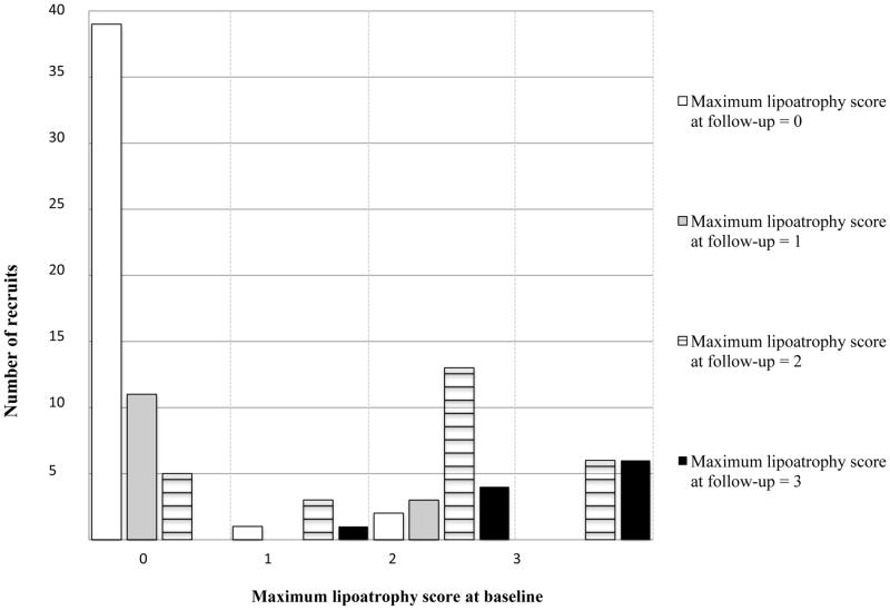
Thirty-six children had lipoatrophy at baseline and 64 did not. Baseline characteristics are presented in table 1. WHO clinical stage, CD4, HIV RNA values and formal dietary assessment variables were similar between those with and without lipoatrophy. One of the 15 girls with lipoatrophy and 2 of the 33 girls without lipoatrophy were Tanner stage 2 for breast development at recruitment. All others were Tanner stage 1. Twenty-nine of the 36 children with lipoatrophy at baseline had been diagnosed with lipoatrophy prior to enrolment, of which 23 had been switched to abacavir more than 6 months prior to assessment. Lipoatrophy grading scores at baseline and at follow-up are presented in figure 1 and table 2.
Table 1.
Comparison of HIV-infected children with and without visually obvious lipoatrophy
| Children with lipoatrophy N=36 |
Children without lipoatrophy N=64 |
Univariate p-value (two-tailed) | |
|---|---|---|---|
| Median age at antiretroviral therapy (ART) initiation, with inter-quartile range (IQR) | 24 (9 – 43) | 19 (9 – 37) | 0.74 |
| Median age at recruitment (months) (IQR) | 89 (71 – 112) | 71 (50 – 92) | 0.001 |
| Gender: Male/Female | 21 (58%)/15 (42%) | 31 (48%)/33 (52%) | 0.41 |
| Median nadir absolute CD4 before ART initiation (IQR) | 694 (439 – 798) | 802 (447 – 1131) | 0.29 |
| Median nadir CD4% before ART initiation (IQR) | 15% (7 – 21%) | 17% (14 – 25%) | 0.05 |
| Absolute CD4 at recruitment (IQR) | 1213 (919 – 1556) | 1129 (792 – 1524) | 0.57 |
| CD4% at recruitment (IQR) | 37% (30 – 40%) | 31% (26 – 37%) | 0.03 |
| Median log10 viral load at recruitment (IQR) | 1.85 (1.60 – 2.54) | 1.85 (1.60 – 2.54) | 0.10 |
| Number with HIV RNA viral load below 400copies/ml | 35 (97%) | 55 (86%) | 0.07 |
| Maximum WHO clinical stage ever reached: 1/2/3/4 | 25%/11%/39%/25% | 17%/9%/46%/28% | 0.78 |
| Median weight for age Z-score (IQR) | −1.0 (−1.8 – −0.5) | −1.0 (−1.6 – −0.3) | 0.39 |
| Median height for age Z-score (IQR) | −1.1 (−2.0 – −0.5) | −1.3 (−2.3 – −0.8) | 0.49 |
| Median body mass index Z-score (IQR) | −0.6 (−1.1 – 0.0) | −0.2 (−0.8 – 0.6) | 0.008 |
| Number on 2nd line therapy, defined as switch of ≥2 antiretroviral drugs (%) | 3 (8%) | 4 (6%) | 0.58 |
| Any antiretroviral exposure, median months (IQR) | 56 (44 – 75) | 43 (25 – 60) | 0.002 |
| Number ever exposed to stavudine (%) | 35 (97%) | 53 (83%) | 0.04 |
| Stavudine, median months (IQR) | 41 (27 – 48) | 30 (7 – 49) | 0.02 |
| Number ever exposed to zidovudine (%) | 17 (47%) | 16 (25%) | 0.02 |
| Zidovudine, median months (IQR) | 12 (5 – 21) | 0 (0 – 0) a | 0.56 |
| Lamivudine, median months (IQR) | 52 (41 – 72) | 41 (25 – 58) | 0.01 |
| Number ever exposed to lopinavir/r (%) | 22 (61%) | 50 (78%) | 0.07 |
| Lopinavir/r, median months (IQR) | 26 (0 – 56) | 36 (6 – 51) | 0.58 |
| Number ever exposed to efavirenz (%) | 17 (47%) | 19 (30%) | 0.09 |
| Efavirenz, median months (IQR) | 0 (0 – 44) a | 0 (0 – 4) a | 0.003 |
Since fewer than half of the subjects in these groups had been exposed to these drugs, the median exposure was 0 months. Mean efavirenz exposure was 23 months in children with lipoatrophy versus 7 months in children without lipoatrophy. Mean zidovudine exposure was 8 months in children with lipoatrophy versus 10 months in children without lipoatrophy.
Figure 1.
Lipoatrophy grading scores at baseline and at follow-up
Table 2.
Lipoatrophy grading scores at baseline and at follow-up
| Baseline | Follow-up | ||
|---|---|---|---|
| Maximum lipoatrophy score | n | Maximum lipoatrophy score | n |
| 0 | 58 | 0 | 39 |
| 1 | 11 | ||
| 2 | 5 | ||
| 3 | 0 | ||
| 1 | 6 | 0 | 1 |
| 1 | 0 | ||
| 2 | 3 | ||
| 3 | 1 | ||
| 2 | 24 | 0 | 2 |
| 1 | 3 | ||
| 2 | 13 | ||
| 3 | 4 | ||
| 3 | 12 | 0 | 0 |
| 1 | 0 | ||
| 2 | 6 | ||
| 3 | 6 | ||
Baseline anthropomorphic measures that were significantly different (p< 0.001) between those with versus without lipoatrophy were biceps SFT (3.8± 1.1 [mean, standard deviation] versus 5.3 ± 2.2, respectively), triceps SFT (6.6 ± 1.9 vs. 9.0 ± 3.0) and torso-to-arm SFT ratio (1.1 ± 0.3 vs. 0.9 ± 0.3) while body mass index Z-score and waist-to-hip circumference ratio were not different between groups. Separate multivariable models, using the three significant SFT measures as the dependent variables and the full 4- point lipoatrophy score, age and sex as independent variables, showed significant correlations between SFT and lipoatrophy score after adjusting for age and gender. When adjusted for age and gender, partial correlation coefficients between the three significant SFT measures and 4-point lipoatrophy score were 0.33 (biceps, p=0.0006), 0.37 (triceps, p=0.0001) and 0.39 (torso-to-arm ratio, p=0.0001).
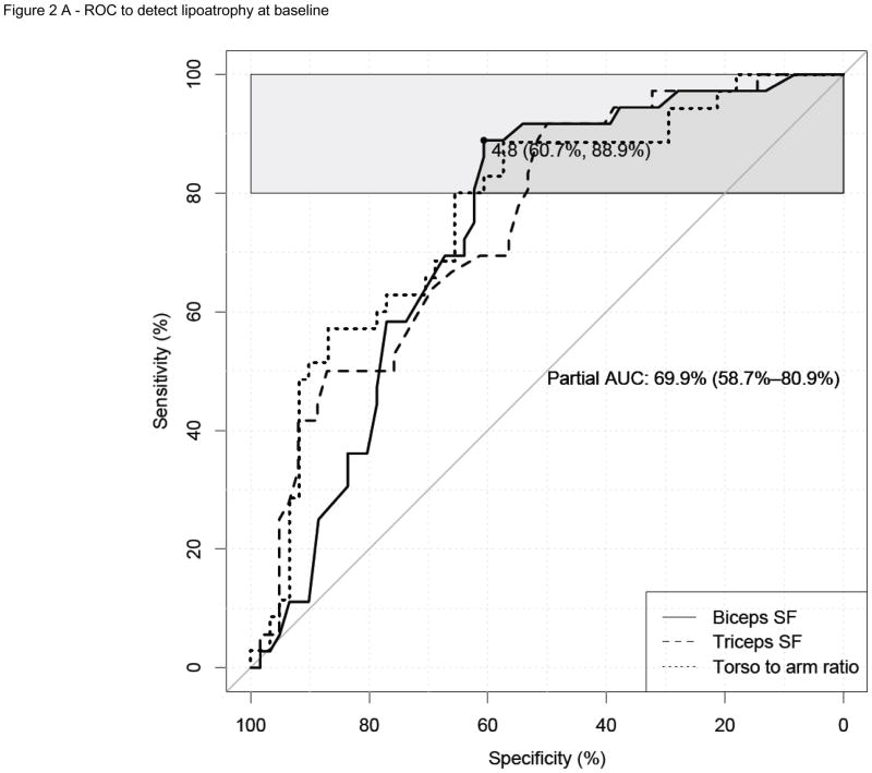
In order to determine the optimal differentiating anthropometric variable, partial ROC area-under-the-curve (pAUC), for 80%–100% sensitivity, were done; these were nominally higher for biceps (0.7; 95% CI: 0.59 – 0.80) than for triceps or torso-to-arm ratio SFT (0.68 and 0.66 respectively, figure 2A). As the biceps measurement is also clinically easier to perform, this was selected as the optimal measure. The total ROC area-under-curve value was 0.75 (CI: 0.64 – 0.84). An ROC area-under-curve measurement of 0.5 for a diagnostic test indicates zero diagnostic power, whereas a value of 1.0 indicates perfect diagnostic power. The biceps SFT ROC curve revealed that the threshold value for biceps SFT that gave optimal screening sensitivity and specificity for differentiating children with and without lipoatrophy at baseline was 4.85mm. A clinically measurable biceps SFT threshold of 5mm had a sensitivity of 89% (CI: 78 – 97%) and a specificity of 61% (CI: 48 – 72%) to detect lipoatrophy at baseline. NPV was 90% (CI: 80 – 99%) and PPV was 57% (CI: 42 – 68%).
Figure 2.
Figure 2A: ROC curves of biceps SFT (solid line), triceps SFT (dashed line), and torso-to-arm SFT ratio (dotted line) to detect prevalent lipoatrophy at baseline, n = 97. The horizontal light grey shape corresponds to the pAUC region. The pAUC of biceps SFT with 95% confidence interval is printed in the middle of the plot. The best threshold of 4.8mm with corresponding specificity (60.7%) and sensitivity (88.9%) for biceps SFT is also located in the plot.
ROC = Receiver Operating Characteristic curve; SFT = skin-fold thickness; pAUC = partial area-under-the-curve for sensitivity between 80% and 100%.
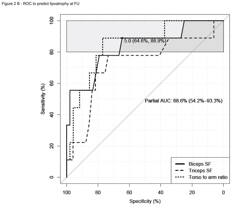
Figure 2B: ROC curves of biceps SFT (solid line), triceps SFT (dashed line), and torso-to-arm SFT ratio (dotted line) to predict which children will go on to develop new lipoatrophy by 15 month follow-up, n = 58. The horizontal light grey shape corresponds to the pAUC region. The pAUC of biceps SFT with 95% confidence interval is printed in the middle of the plot. The best threshold of 5mm with corresponding specificity (64.6%) and sensitivity (88.9%) for biceps SFT is also located in the plot.
ROC = Receiver Operating Characteristic curve; SFT = skin-fold thickness; pAUC = partial area-under-the-curve for sensitivity between 80% and 100%.
Follow-up was completed on 34 of the 36 children with lipoatrophy and 60 of the 64 children without lipoatrophy at baseline. Median follow-up time was 14.9 months (interquartile range: 14.5 – 15.6 months). At follow-up, 9/60 children had developed new lipoatrophy, giving an incidence rate of 12% (CI: 5 – 20%) per person-year of follow-up. Lipoatrophy had resolved in 5/34 children, all of whom had been switched to abacavir more than 18 months before and had no more than moderate (grade 2) lipoatrophy signs at baseline.
In children without lipoatrophy at baseline, ROC analysis of baseline biceps SFT revealed an area-under- curve value of 0.83 (CI: 0.67 – 1.00) and a partial ROC area-under curve value for sensitivity between 80% and 100% of 0.69 (CI: 0.54 – 0.93) for predicting which children would go on to develop lipoatrophy by 15 month follow-up (Figure 2B). In comparison, the ability of triceps SFT to predict lipoatrophy at follow-up was limited, with a partial ROC area-under-curve value for sensitivity between 80% and 100% of 0.56 (CI: 0.46 – 0.91). Figure 2B shows ROC curves of biceps SFT, triceps SFT and torso-to-arm SFT ratio to predict new lipoatrophy at follow-up. In children without lipoatrophy at baseline, a baseline biceps SFT <5mm yielded a sensitivity of 89% (CI: 67 – 100%) and a specificity of 60% (CI: 46–75%) for predicting which children would go on to develop lipoatrophy. NPV was 97% (CI: 91 – 100%) and PPV was 32% (14 – 50%). Post-test probabilities were as follows: In the presence of a negative test, the post-test probability of developing lipoatrophy by 15-month follow-up was 0.10 (CI: 0.02 – 0.18), whereas the probability of not developing lipoatrophy was 0.91 (CI: 0.84 – 0.99). In the presence of a positive test, the post-test probability of developing lipoatrophy by 15-month follow-up was 0.59 (CI: 0.46 – 0.71), whereas the probability of not developing lipoatrophy was 0.41 (CI: 0.29 – 0.54). Repeating the analyses without the three children with Tanner stage 2 breast development at baseline, marginally improved the performance of the screening tool both at baseline and at follow-up.
Second study to determine the precision of anthropometric measurements performed by an inexperienced observer
An additional 77 children from the same clinic were recruited to compare the precision of anthropometric measurements performed by an inexperienced observer to that of an experienced research dietician. These subjects had a median age of 3.0 years (interquartile range 2.0 – 3.8 years) and 37/77 were female. Two subsequently refused to co-operate with any measurements and six refused SFT measurements. SFT measurements were successfully completed on 69 children. The measured differences between the inexperienced observer and the research dietician are presented in table 3. The mean absolute difference in biceps SFT measurement between the inexperienced observer and the research dietician was 0.8mm. Taking this variability into account, the sensitivity, specificity, PPV and NPV of biceps SFT thresholds of <4mm and <6mm were calculated in order to determine how the variability in precision might affect the performance of the screening tool for predicting which children would have lipoatrophy at 15 month follow-up (table 4). The variation in precision of measurements performed by an inexperienced healthcare worker only marginally impacted performance.
Table 3.
Precision of anthropometric measurements performed by an inexperienced observer compared to an experienced research dietician (inter-observer variability)
| N = 69 | Measurements performed by inexperienced observer | Measurements performed by experienced research dietician | Measured pairwise absolute difference between inexperienced observer and research dietician | |||||
|---|---|---|---|---|---|---|---|---|
| Mean | SD | CV (%) | Mean | SD | CV (%) | Mean | SD | |
| Waist circumference (cm) | 50.4 | 3.9 | 8% | 50.1 | 3.3 | 7% | 0.9 | 0.9 |
| Hip circumference (cm) | 49.7 | 3.8 | 8% | 49.1 | 7.6 | 15% | 1.7 | 1.5 |
| Mid-upper arm circumference (cm) | 15.7 | 1.8 | 11% | 15.9 | 1.3 | 8% | 0.5 | 0.4 |
| Triceps SFT (mm) | 9.1 | 3.4 | 38% | 9.2 | 2.2 | 24% | 0.9 | 0.9 |
| Biceps SFT (mm) | 5.9 | 3.8 | 64% | 6.0 | 1.5 | 26% | 0.8 | 0.6 |
| Subscapular SFT (mm) | 6.5 | 4.1 | 64% | 6.3 | 1.7 | 27% | 0.5 | 0.4 |
| Iliac crest SFT (mm) | 8.2 | 4.5 | 54% | 6.7 | 2.9 | 44% | 1.7 | 1.5 |
SFT=skin-fold thickness; CV=coefficient of variation; SD=standard deviation
Table 4.
Effectiveness of biceps skin-fold thickness (SFT) threshold of <5mm with variations in precision of +/−1mm, for predicting any lipoatrophy at 15 month follow-up (incorporating resolved cases, persistent cases and new cases), n = 94.
| Biceps SFT threshold | Sensitivity | Specificity | Positive predictive value | Negative predictive value |
|---|---|---|---|---|
| <4mm | 68% | 75% | 65% | 78% |
| <5mm | 89% | 63% | 62% | 90% |
| <6mm | 95% | 43% | 53% | 92% |
Each anthropometric measurement had been performed three times and averaged. Table 5 uses the mean absolute difference between the three values to calculate the intra-observer variability for the inexperienced observer compared to the experienced research dietician. The intra-observer variability of the inexperienced observer compared favourably to that of the experienced research dietician.
Table 5.
Intra-observer variability of the inexperienced observer compared to the experienced research dietician, using mean absolute difference in repeated measurements.
| N = 69 | Inexperienced observer measurements | Experienced research dietician measurements | ||||
|---|---|---|---|---|---|---|
| Mean | SD | CV (%) | Mean | SD | CV (%) | |
| Waist circumference (cm) | 0.49 | 0.48 | 97% | 0.44 | 0.38 | 87% |
| Hip circumference (cm) | 0.33 | 0.27 | 82% | 0.34 | 0.32 | 95% |
| Mid-upper arm circumference (cm) | 0.13 | 0.11 | 84% | 0.13 | 0.10 | 78% |
| Triceps SFT (mm) | 0.42 | 0.33 | 79% | 0.38 | 0.38 | 102% |
| Biceps SFT (mm) | 0.29 | 0.26 | 90% | 0.24 | 0.26 | 106% |
| Subscapular SFT (mm) | 0.25 | 0.22 | 86% | 0.17 | 0.16 | 92% |
| Iliac crest SFT (mm) | 0.41 | 0.30 | 74% | 0.30 | 0.33 | 107% |
SFT=skin-fold thickness; CV=coefficient of variation; SD=standard deviation
Discussion
Children with and without lipoatrophy had similar immunologic and clinical presentation at ART initiation and at recruitment (p >0.2 for absolute CD4 and WHO clinical staging). They did not start ART earlier and a similar proportion was on second-line ART. The median age of children with lipoatrophy was higher than that of children without lipoatrophy (7.4 versus 5.9 years, p=0.001). This difference in age was expected since the risk of lipoatrophy increases with cumulative ART use9, which increases with age.
Some partial improvement of lipoatrophy may be experienced after drug switching, with the more severe cases experiencing the least improvement15. The biceps SFT at baseline was less informative for detecting existing cases than for predicting new cases at follow-up. Individuals known to have lipoatrophy prior to recruitment had been switched to abacavir months or years before recruitment and may have experienced some improvement in subcutaneous SFT before recruitment as a result of the switch. This may explain the limited performance of the screening tool in detecting lipoatrophy at baseline. The ROC analysis for predicting new cases at follow-up may be more helpful since new cases at follow-up had not been influenced by interventional drug switches.
The ROC analyses at baseline did not confirm that any of the three SFT contenders (biceps, triceps and arm- to-trunk ratio) was statistically superior to the others. However, the biceps SFT technique is the most simple to perform, and biceps SFT measurements had the least inter- and intra-observer variability. These strengths led us to choose biceps SFT over triceps SFT and torso-to-arm SFT ratio. Using a threshold of 5mm for biceps SFT yields a high sensitivity and NPV, making it effective for screening. The relatively low specificity is clinically acceptable; as the next step after screening is referral, not change of therapy. The screening tool can be employed by nurses and ancillary healthcare workers who can then refer children with suspected early lipoatrophy to pediatric HIV doctors for confirmation and antiretroviral switching as necessary. Experienced HIV pediatricians who have been specifically trained to identify early lipoatrophy will typically use the formal visual grading scale described above to confirm the diagnosis.
Lipoatrophy does not occur in all children exposed to ART8, 22, 24. Some show no signs of fat changes despite many years of stavudine or zidovudine exposure, whereas others develop lipoatrophy within 18 months of ART initiation16. This variation may be due to genetic differences that increase or decrease an individual’s susceptibility to lipoatrophy28–30, which would explain why viral load did not clearly differentiate between children with and without lipoatrophy in our study. Ethnic differences may also play a role8. In the face of this unpredictability, our screening tool provides an objective, easy-to-use method to identify children who may be developing lipoatrophy.
The current studies were performed using a Harpenden® calliper, which although highly precise, is expensive ($340). A Slimguide® skin-fold calliper (Vancouver: Rosscraft) is marginally less precise (repeatability 0.5mm31) but is cheap ($20) and durable, and may be a more suitable device to roll out in resource-limited primary healthcare settings in sub-Saharan Africa. The reason for using the more precise Harpenden® calliper rather than the Slimguide® in our second study was to isolate and quantify the imprecision of measurements performed by an inexperienced healthcare worker, and to be consistent with the parent study. Before implementation of the Slimguide® can be recommended, a further study is needed in which repeated measurements are performed by an experienced anthropometric dietician using both the Harpenden® and Slimguide® callipers to quantify the additional imprecision contributed by using the less precise device.
For broad implementation of this screening tool, a brief written description of how to perform a biceps SFT measurement may be all the training that is necessary. For children on ART, the effort required to perform a single annual biceps SFT measurement is likely to be logistically feasible even in busy community healthcare clinics. Serial annual biceps SFT measurements also give the added benefit of identifying changes from baseline: A biceps SFT that has dropped from a higher baseline to below 5mm, in the absence of an obvious nutritional or other cause, is probably a more convincing indication of impending lipoatrophy than a static measurement below 5mm.
Inclusion of girls with Tanner stage 2 breast development was allowed in this study since early minimal telarche may be a physiological in some girls32–33. Only three recruits had Tanner stage 2 breast development at baseline, and repeating the analyses without the three children did not significantly alter the performance of the screening tool.
Our data on the use of biceps SFT with a cut-off of 5mm as a screening tool has a number of limitations:
The current study had a limited sample size, resulting in broad confidence intervals and possibly misleading results. A follow-up study is needed to test the performance of this screening tool on a different cohort of pre-pubertal HIV-infected African children on ART.
The standard deviation for SFT measurements performed by the experienced research dietician was up to four times that of previously published findings34. This raises concern about the precision of those measurements, which were used as a reference for measurements performed by the inexperienced healthcare worker. The imprecision may have produced misleading results, which could have altered the assessment of the reliability of the screening tool in inexperienced hands.
The multivariable analyses demonstrated a significant effect of age and sex on the various anthropomorphic measures; the study’s small sample size did not allow for stratification of ROC cut-off thresholds by age/sex. Thus, a measure of 5mm may have a different prognosis in a younger versus older child. The very high negative predictive value of the 5mm cut-off is reassuring that few cases would be missed despite the lack of age-specific cut-offs.
The low specificity may lead to a significant number of unnecessary referrals, which may overburden secondary referral services.
Precision of biceps skin-fold thickness is technique-dependent, which requires some training. An inconsistent method of measurement may lead to increased intra-observer variability. Differences in method between healthcare workers may lead to increased inter-observer variability. Both intra- and inter-observer variability may have the consequence that a patient’s change in biceps skin-fold thickness from baseline may go unnoticed.
Although durable, if the Slimguide® calliper is used, the spring strength will eventually deteriorate, which may not be obvious and may lead to continued use when the calliper should be replaced, resulting in inaccurate and possibly misleading measurements.
Reliance on an objective measurement may lead to reduced clinical vigilance on the part of primary healthcare staff, particularly in busy clinics where patient burdens are large. The high sensitivity may falsely reassure healthcare workers, leading to missed cases. This screening tool should not replace diligent attentiveness during routine follow-up.
Specificity and PPV are limited and confirmation of suspected lipoatrophy using visual grading assessment by a skilled operator remains necessary.
Adherence was not measured or correlated with lipoatrophy, and this was a weakness of the current study.
This screening tool is not appropriate for a child who is clearly non-adherent to ART and developing AIDS wasting syndrome, or is severely malnourished. This does not limit the usefulness of the screening tool since cumulative ART exposure is the most potent risk factor for lipoatrophy16, and it follows that children who are non-adherent are unlikely to develop slowly-progressive cumulative side effects like lipoatrophy. At-risk children are typically those who turn up reliably for their follow-up visits and whose caregivers follow instructions diligently. As a result, implementation of the screening tool at primary healthcare level is likely to reach the target population.
Conclusion
A biceps SFT of <5mm in HIV-infected pre-pubertal children exposed to thymidine analogue-based ART may be an objective, relatively easy-to-use screening tool to identify children who currently have or may progress to develop lipoatrophy, allowing appropriate antiretroviral drug switches to be made at an early stage, which will arrest progression and avoid stigmatizing disfigurement.
Acknowledgments
Source of Funding
SI is currently recieving a Fogarty International Clinical Research Fellowship grant (#R24-TW007988-01); a pilot research grant (#P30 AI036214-16, subaward #10304442) from the University of California San Diego Centre for AIDS Research (UCSD CFAR); and a Southern Africa Consortium for Research Excellence (SACORE) grant (#WTX055734) from the Wellcome Trust.
ESK received support from the National Research Foundation (NRF) South Africa, the Deutschen Forschungsgemeinschaft (DFG) and the Bayerischen Bundesregierung (International Research Training Group Projekt 1522 “HIV/AIDS and Associated Infectious Diseases in South Africa”).
MFC is currently receiving a grant (#5U01AI069521-01 to 04) from the National Institute of Allergy and Infectious Diseases (NIAID) through the International Maternal Pediatric Adolescent AIDS Clinical Trials Group (IMPAACT); a grant (#1U19AI53217-01) from NIAID through the Comprehensive International Plan for Research in AIDS (CIPRA-SA); and a grant (#GPO-A-00-03-00000) from USAID. The recruits on this study were not co-enrolled in any IMPAACT trial.
RH is currently receiving grants (#AI 27670 and #K24 AI064086) from NIAID through the San Diego AIDS Clinical Trials Group (ACTG) Clinical Trials Unit and the University of California San Diego CFAR (#AI36214; #P30-AI36214 and #AI69432).
HK received support from the National Research Foundation (NRF) South Africa, the Deutsche Forschungsgemeinschaft (DFG) and the Bayerischen Bundesregierung (International Research Training Group Projekt 1522 “HIV/AIDS and Associated Infectious Diseases in South Africa”), the Bundesministerium für Wirtschaft und Technologie (# 03EGSBY044), and the Bundesministerium für Bildung und Forschung (#01 KG 0915). He reports receiving Board membership fees from Abbott, Bristol-Myers Squibb, Boehringer, Gilead, Janssen-Cilag, and MSD, lecture fees from Bristol-Myers Squibb, Boehringer, Gilead, MSD, and Roche.
SHB is currently receiving grants (#P30-AI36214; #K08 AI62758 and #R43 AI093318-01)
Database support was provided by the Vanderbilt Institute for Clinical and Translational Research (grant #1 UL1 RR024975 from NCRR/NIH).
Footnotes
Conflicts of Interest
The authors have no conflict of interest to declare.
The content of this publication does not necessarily reflect the views or policies of NIAID, nor does mention of trade names, commercial projects, or organizations imply endorsement by the US Government.
Contributor Information
Steve Innes, Children’s Infectious Diseases Clinical Research Unit (KID CRU), Tygerberg Children’s Hospital, and Department of Paediatrics, Stellenbosch University, Cape Town, South Africa.
Eva Schulte-Kemna, Universitätsklinikum Würzburg, Medizinische Klinik, Schwerpunkt Infektiologie, Würzburg, Germany.
Mark F. Cotton, Children’s Infectious Diseases Clinical Research Unit (KID CRU), Tygerberg Children’s Hospital, and Department of Paediatrics, Stellenbosch University, Cape Town, South Africa.
Ekkehard Werner Zöllner, Division of Endocrinology, Department of Paediatrics, Stellenbosch University, Cape Town, South Africa.
Richard Haubrich, Antiviral Research Centre, University of California, San Diego, United States.
Hartwig Klinker, Universitätsklinikum Würzburg, Medizinische Klinik, Schwerpunkt Infektiologie, Würzburg, Germany.
Xiaoying Sun, Biostatistics Research Center, Division of Biostatistics and Bioinformatics, University of California, San Diego, United States.
Sonia Jain, Biostatistics Research Center, Division of Biostatistics and Bioinformatics, University of California, San Diego, United States.
Clair Edson, Department of Paediatrics, Stellenbosch University, Cape Town, South Africa.
Margaret van Niekerk, Department of Dietetics, Stellenbosch University, Cape Town, South Africa.
Emily Ryan, Department of Dietetics, Stellenbosch University, Cape Town, South Africa.
Helena Rabie, Department of Paediatrics, Stellenbosch University, Cape Town, South Africa.
Sara H. Browne, Division of Infectious Diseases, Department of Medicine, University of California, San Diego, United States.
References
- 1.IeDEA Southern Africa Paediatric Group. A biregional survey and review of first-line treatment failure and second-line paediatric antiretroviral access and use in Asia and southern Africa. J Int AIDS Soc. 2011;14:7. doi: 10.1186/1758-2652-14-7. [DOI] [PMC free article] [PubMed] [Google Scholar]
- 2.WHO. [Accessed 15 September 2011];Forecasting antiretroviral demand. 2010 http://www.who.int/hiv/amds/forecasting/en/index4.html.
- 3.South African National Department of Health. The South African Antiretroviral Treatment Guidelines. Pretoria: SANDoH; 2010. [Google Scholar]
- 4.Haubrich RH, Riddler SA, DiRienzo AG, et al. Metabolic outcomes in a randomized trial of nucleoside, nonnucleoside and protease inhibitor-sparing regimens for initial HIV treatment. AIDS. 2009 Jun 1;23(9):1109–1118. doi: 10.1097/QAD.0b013e32832b4377. [DOI] [PMC free article] [PubMed] [Google Scholar]
- 5.Shlay JC, Sharma S, Peng G, Gibert CL, Grunfeld C. Long-term subcutaneous tissue changes among antiretroviral-naive persons initiating stavudine, zidovudine, or abacavir with lamivudine. J Acquir Immune Defic Syndr. 2008 May 1;48(1):53–62. doi: 10.1097/qai.0b013e31816856ed. [DOI] [PubMed] [Google Scholar]
- 6.Beregszaszi M, Dollfus C, Levine M, et al. Longitudinal evaluation and risk factors of lipodystrophy and associated metabolic changes in HIV-infected children. J Acquir Immune Defic Syndr. 2005 Oct 1;40(2):161–168. doi: 10.1097/01.qai.0000178930.93033.f2. [DOI] [PubMed] [Google Scholar]
- 7.Dzwonek AB, Lawson MS, Cole TJ, Novelli V. Body fat changes and lipodystrophy in HIV-infected children: impact of highly active antiretroviral therapy. J Acquir Immune Defic Syndr. 2006 Sep;43(1):121–123. doi: 10.1097/01.qai.0000230523.94588.85. [DOI] [PubMed] [Google Scholar]
- 8.Alam N, Cortina-Borja M, Goetghebuer T, Marczynska M, Vigano A, Thorne C. Body fat abnormality in HIV-infected children and adolescents living in Europe: prevalence and risk factors Fat abnormality in children. J Acquir Immune Defic Syndr. 2011 Dec 27; doi: 10.1097/QAI.0b013e31824330cb. [DOI] [PMC free article] [PubMed] [Google Scholar]
- 9.Innes S, Eagar R, Edson C, et al. Prevalence, DEXA differences and risk factors for lipoatrophy among pre-pubertal African children on HAART. Reviews in Antiviral Therapy and Infectious Diseases. 2011;(8):36. [Google Scholar]
- 10.Greeff M, Phetlhu R, Makoae L, et al. Disclosure of HIV status: experiences and perceptions of persons living with HIV/AIDS and nurses involved in their care in Africa. Qualitative Health Research. 2008;18(3):311–324. doi: 10.1177/1049732307311118. [DOI] [PubMed] [Google Scholar]
- 11.Nyblade L, Pande R, Mathur S, et al. Disentangling HIV and AIDS stigma in Ethiopia, Tanzania and Zambia. Washington DC: International Center for Research on Women; 2003. [Google Scholar]
- 12.Peretti-Watel P, Spire B, Pierret J, Lert F, Obadia Y. Management of HIV-related stigma and adherence to HAART: evidence from a large representative sample of outpatients attending French hospitals (ANRS-EN12-VESPA 2003) AIDS Care. 2006 Apr;18(3):254–261. doi: 10.1080/09540120500456193. [DOI] [PubMed] [Google Scholar]
- 13.Plankey M, Bacchetti P, Jin C, et al. Self-perception of body fat changes and HAART adherence in the Women’s Interagency HIV Study. AIDS Behav. 2009 Feb;13(1):53–59. doi: 10.1007/s10461-008-9444-7. [DOI] [PMC free article] [PubMed] [Google Scholar]
- 14.DeLaMora P, Aledort N, Stavola J. Caring for adolescents with HIV. Curr HIV/AIDS Rep. 2006 Jul;3(2):74–78. doi: 10.1007/s11904-006-0021-2. [DOI] [PubMed] [Google Scholar]
- 15.Aurpibul L, Puthanakit T, Taejaroenkul S, Sirisanthana T, Sirisanthana V. Recovery from lipodystrophy in HIV-infected children after substitution of stavudine with zidovudine in a non- nucleoside reverse transcriptase inhibitor-based antiretroviral therapy. Pediatr Infect Dis J. 2012 Apr;31(4):384–388. doi: 10.1097/INF.0b013e31823f0e11. [DOI] [PubMed] [Google Scholar]
- 16.Innes S, Eagar R, Edson C, et al. High prevalence of lipoatrophy in pre-pubertal South African children on antiretroviral therapy: A cross-sectional study. BMC Pediatrics. 2012 doi: 10.1186/1471-2431-12-183. In press. [DOI] [PMC free article] [PubMed] [Google Scholar]
- 17.Carr A, Workman C, Smith DE, et al. Abacavir substitution for nucleoside analogs in patients with HIV lipoatrophy: a randomized trial. JAMA. 2002 Jul 10;288(2):207–215. doi: 10.1001/jama.288.2.207. [DOI] [PubMed] [Google Scholar]
- 18.Vigano A, Brambilla P, Cafarelli L, et al. Normalization of fat accrual in lipoatrophic, HIV-infected children switched from stavudine to tenofovir and from protease inhibitor to efavirenz. Antivir Ther. 2007;12(3):297–302. [PubMed] [Google Scholar]
- 19.Stankov MV, Lucke T, Das AM, Schmidt RE, Behrens GM. Mitochondrial DNA depletion and respiratory chain activity in primary human subcutaneous adipocytes treated with nucleoside analogue reverse transcriptase inhibitors. Antimicrobial Agents and Chemotherapy. 2010;54(1):280–287. doi: 10.1128/AAC.00914-09. [DOI] [PMC free article] [PubMed] [Google Scholar]
- 20.UNAIDS. [Accessed 8 September 2011];Report on the Global AIDS Epidemic. 2010 http://www.unaids.org/globalreport/global_report.htm.
- 21.Carr A, Emery S, Law M, Puls R, Lundgren JD, Powderly WG. An objective case definition of lipodystrophy in HIV-infected adults: a case-control study. Lancet. 2003 Mar 1;361(9359):726–735. doi: 10.1016/s0140-6736(03)12656-6. [DOI] [PubMed] [Google Scholar]
- 22.European Paediatric Lipodystrophy Group. Antiretroviral therapy, fat redistribution and hyperlipidaemia in HIV-infected children in Europe. AIDS. 2004 Jul 2;18(10):1443–1451. doi: 10.1097/01.aids.0000131334.38172.01. [DOI] [PubMed] [Google Scholar]
- 23.Hartman K, Verweel G, de Groot R, Hartwig NG. Detection of lipoatrophy in human immunodeficiency virus-1-infected children treated with highly active antiretroviral therapy. Pediatr Infect Dis J. 2006 May;25(5):427–431. doi: 10.1097/01.inf.0000215003.32256.aa. [DOI] [PubMed] [Google Scholar]
- 24.Aurpibul L, Puthanakit T, Lee B, Mangklabruks A, Sirisanthana T, Sirisanthana V. Lipodystrophy and metabolic changes in HIV-infected children on non-nucleoside reverse transcriptase inhibitor-based antiretroviral therapy. Antivir Ther. 2007;12(8):1247–1254. [PubMed] [Google Scholar]
- 25.South African National Department of Health. Management and Treatment for South Africa. 2003. Operational Plan for Comprehensive HIV and AIDS Care. [Google Scholar]
- 26.Baty International. [Accessed 30 August, 2012];Harpenden skin-fold caliper. 2007 http://www.harpenden-skinfold.com.
- 27.McClish DK. Analyzing a portion of the ROC curve. Med Decis Making. 1989 Jul-Sep;9(3):190–195. doi: 10.1177/0272989X8900900307. [DOI] [PubMed] [Google Scholar]
- 28.Asensi V, Rego C, Montes AH, et al. IL-1beta (+3954C/T) polymorphism could protect human immunodeficiency virus (HIV)-infected patients on highly active antiretroviral treatment (HAART) against lipodystrophic syndrome. Genet Med. 2008 Mar;10(3):215–223. doi: 10.1097/GIM.0b013e3181632713. [DOI] [PubMed] [Google Scholar]
- 29.Wangsomboonsiri W, Mahasirimongkol S, Chantarangsu S, et al. Association between HLA-B*4001 and lipodystrophy among HIV-infected patients from Thailand who received a stavudine-containing antiretroviral regimen. Clin Infect Dis. 2010 Feb 15;50(4):597–604. doi: 10.1086/650003. [DOI] [PubMed] [Google Scholar]
- 30.Zanone Poma B, Riva A, Nasi M, et al. Genetic polymorphisms differently influencing the emergence of atrophy and fat accumulation in HIV-related lipodystrophy. AIDS (London, England) 2008;22(14):1769–1778. doi: 10.1097/QAD.0b013e32830b3a96. [DOI] [PubMed] [Google Scholar]
- 31.Schmidt PK, Carter JE. Static and dynamic differences among five types of skinfold calipers. Hum Biol. 1990 Jun;62(3):369–388. [PubMed] [Google Scholar]
- 32.Berberoglu M. Precocious puberty and normal variant puberty: definition, etiology, diagnosis and current management. J Clin Res Pediatr Endocrinol. 2009;1(4):164–174. doi: 10.4274/jcrpe.v1i4.3. [DOI] [PMC free article] [PubMed] [Google Scholar]
- 33.Youlton R, Valladares L, Garcia H, et al. Premature telarche: Study of its frequency and etiological factors. Pediatric Research. 1990;28:421. [Google Scholar]
- 34.Cameron N. Weight and skinfold variation at menarche and the critical body weight hypothesis. Ann Hum Biol. 1976 May;3(3):279–282. doi: 10.1080/03014467600001451. [DOI] [PubMed] [Google Scholar]


