Abstract
Myelolipoma in the mediastinum is an extremely rare entity. In this report, we present the case of a 79-year-old asymptomatic man who had three bilateral paravertebral mediastinal tumors. The three tumors were resected simultaneously using bilateral three-port video-assisted thoracoscopic surgery (VATS). There has been no evidence of recurrence within four years after the operation. Multiple bilateral mediastinal myelolipomas are extremely rare. There are no reports in the English literature of multiple bilateral thoracic myelolipomas that were resected simultaneously using bilateral VATS. We also present characteristic features of myelolipomas, which are helpful for diagnosis.
Keywords: Mediastinal neoplasms, Myelolipoma, Minimally invasive surgery, Thoracoscopy
CASE REPORT
Myelolipoma is a rare, benign tumor composed of hematopoietic tissue and mature adipose tissue. It is nonfunctioning and often detected in the adrenal glands during routine X-ray or computed tomography (CT) as incidentalomas. Extramedullary myelolipomas are extremely rare, and half of them are located in the presacral area. We report a patient with multiple bilateral mediastinal myelolipomas that were resected simultaneously using bilateral three-port video-assisted thoracoscopic surgery (VATS). This is the first case report of multiple bilateral myelolipomas that were simultaneously removed and diagnosed using VATS. We also present characteristic features of myelolipomas, which are helpful for diagnosis.
The patient was a 79-year-old male with a history of hypertension, nephrosclerosis, and alcoholic liver hepatitis. Abdominal CT was performed routinely at a nearby hospital, and three bilateral paravertebral mediastinal tumors were detected incidentally. He had no symptoms due to the tumors. He was referred to Toranomon Hospital for further examination and treatment.
Laboratory tests showed a slight elevation of serum liver enzymes and creatinine. Other laboratory tests including tumor markers were within normal limits. He had no history of hematologic disorder or chronic anemia.
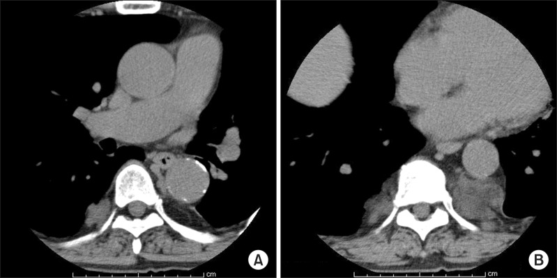
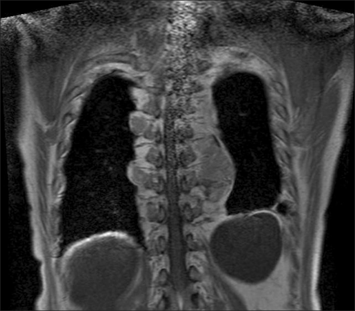
CT revealed three bilateral paravertebral thoracic tumors. Two of them were located on the right side of the vertebrae at the levels of Th-8 and Th-10. The tumors were 20 mm and 35 mm in diameter, respectively. The third tumor was located on the left side of the vertebrae, extending from Th-9 to Th-11, and was 75 mm in diameter. These tumors were heterogeneous. The CT density of the tumors ranged from -60.0 to +20.0 Hounsfield Units. There was no calcification in the tumors. Contrast medium was not used because of his poor renal function (Fig. 1). Magnetic resonance imaging revealed that the signal intensity of the tumors was mainly isointense with that of the bone marrow and partially isointense with that of the adipose tissue (Fig. 2). Differential diagnosis included neurogenic tumors, lymphomas, pleural mesotheliomas, and metastatic tumors of unknown origin, but none of them had any conclusive evidence.
Fig. 1.
(A and B) Thoracic computed tomography showed three bilateral paravertebral mediastinal tumors.
Fig. 2.
Magnetic resonance imaging showed that the signal intensity of the tumors was mainly isointense with that of the bone marrow and partially isointense with that of the adipose tissue.
Bilateral VATS with the three-port technique was performed. The tumors were soft and fragile and bled easily. The tumor on the left side adhered to its surroundings firmly and hence, took several hours to resect. The operation time was 5 hours and 27 minutes, and the blood loss was 327 mL.
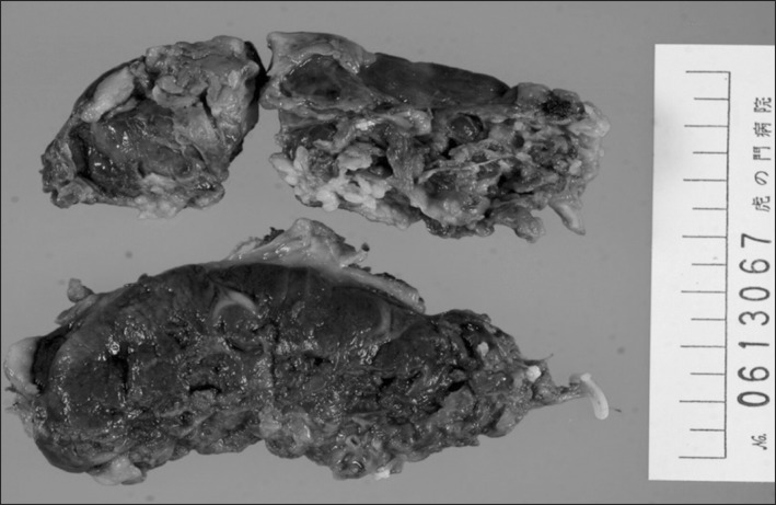
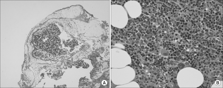
The enucleated tumors were brown and partially yellow, and measured 19×15×11 mm, 40×34×15 mm, and 75×45×30 mm, respectively (Fig. 3). Microscopic examination revealed mature adipose tissue with hematopoietic elements consistent with myelolipoma (Fig. 4). The surgical margins of the tumors were all negative. The patient's postoperative course was uncomplicated, and he was discharged on the fourth postoperative day. It has been four years since the operation, and there has been no sign of recurrence.
Fig. 3.
Macroscopic inspection of the tumor on the left side after resection showed that it was brown and partially yellow, and measured 75×45×30 mm.
Fig. 4.
(A and B) Histopathologic examination revealed mature adipose tissue with hematopoietic elements consistent with a myelolipoma (H&E, ×4 and ×200, respectively).
DISCUSSION
Myelolipomas are rare, benign tumors, and most of these tumors are detected in the adrenal glands as incidentalomas. Myelolipoma consists of mature adipose tissue and bone marrow cells. Symptoms caused by the tumor rarely occur; however, growth of the tumor may cause compression of the adjacent organs, resulting in several symptoms [1].
The proposed causes of myelolipoma include degenerative changes in hyperplastic tumor cells or adenomas of the adrenal glands, metaplasia in primary stem mesenchymal cells of the adrenal cortex, and displacement of differentiated bone marrow cells during embryogenesis [2]. Extra-adrenal myelolipoma is an extremely rare form of myelolipoma that can occur in the retroperitoneum, stomach, liver, or mediastinum. About half of all extra-adrenal myelolipomas are located in the presacral region [3]. In the thoracic area, they are frequently found in the mediastinum or the paravertebral thoracic region; however, sometimes they are located in the lung or the thoracic spine [4].
There are several distinctive imaging features of myelolipoma. It mostly occurs as a single, unilateral tumor and is often well-circumscribed and encapsulated. The ratios of bone marrow tissue and adipose tissue are different in each tumor. This reflects the heterogeneous nature of the tumors and explains the observation of varying CT densities. Calcification in the tumor is rare but has been reported [1].
The differential diagnosis includes neurogenic tumor, lymphoma, lymph node metastasis, malignant mesothelioma, and extramedullary hematopoietic tissue. Among them, differentiation between myelolipoma and extramedullary hematopoiesis is important. In cases of extramedullary hematopoiesis, there is often chronic anemia due to hematological diseases such as thalassemia or hereditary spherocytosis. Chronic anemia can be used to differentiate between myelolipoma and extramedullary hematopoiesis [5].
Most cases of myelolipoma are treated with surgical resection because myelolipoma is a rare disease and its preoperative diagnosis is difficult. However, there are some reports of successful diagnosis with CT-guided or endoscopic ultrasonography-guided biopsy [6,7]. There have been no reports of malignant myelolipoma. The decision for surgical resection is based on either increasing size or local symptoms such as chest pain, pleural effusion, or superior vena cava obstruction. Kenny et al. suggested that myelolipoma that is larger than 10 cm in diameter should be resected because the possibility of rupture and bleeding is higher with larger tumors [8].
Once there is a definitive diagnosis, and there are no signs or symptoms of increasing size, observation without any treatment can be a reasonable option. Surgical resection is the most common method of treatment, and a majority of resections are performed via thoracotomy. In this case, three bilateral myelolipomas in the posterior mediastinum were resected using bilateral VATS. To the best of our knowledge, this is the first case report of three bilateral mediastinal myelolipomas resected simultaneously using bilateral VATS.
Mediastinal myelolipoma is a rare tumor, but it should be considered when there is a differential diagnosis of mediastinal tumors. Most reports are cases of single and unilateral tumors; however, bilateral and multiple tumors can occur, although they are extremely rare. When surgery is necessary for diagnosis or treatment, VATS can be an alternative choice to thoracotomy.
Footnotes
No potential conflict of interest relevant to this article was reported.
References
- 1.Vaziri M, Sadeghipour A, Pazooki A, Shoolami LZ. Primary mediastinal myelolipoma. Ann Thorac Surg. 2008;85:1805–1806. doi: 10.1016/j.athoracsur.2007.11.023. [DOI] [PubMed] [Google Scholar]
- 2.Dieckmann KP, Hamm B, Pickartz H, Jonas D, Bauer HW. Adrenal myelolipoma: clinical, radiologic, and histologic features. Urology. 1987;29:1–8. doi: 10.1016/0090-4295(87)90587-5. [DOI] [PubMed] [Google Scholar]
- 3.Franiel T, Fleischer B, Raab BW, Fuzesi L. Bilateral thoracic extraadrenal myelolipoma. Eur J Cardiothorac Surg. 2004;26:1220–1222. doi: 10.1016/j.ejcts.2004.08.024. [DOI] [PubMed] [Google Scholar]
- 4.Omdal DG, Baird DE, Burton BS, Goodhue WW, Jr, Giddens EM. Myelolipoma of the thoracic spine. AJNR Am J Neuroradiol. 1997;18:977–979. [PMC free article] [PubMed] [Google Scholar]
- 5.Rosai J. Mediastinum. In: Rosai J, editor. Rosai and Ackerman's surgical pathology. 9th ed. St. Louis (MO): Mosby; 2004. p. 494. [Google Scholar]
- 6.Kawanami S, Watanabe H, Aoki T, et al. Mediastinal myelolipoma: CT and MRI appearances. Eur Radiol. 2000;10:691–693. doi: 10.1007/s003300050985. [DOI] [PubMed] [Google Scholar]
- 7.Rossi M, Ravizza D, Fiori G, et al. Thoracic myelolipoma diagnosed by endoscopic ultrasonography and fine-needle aspiration cytology. Endoscopy. 2007;39(Suppl 1):E114–E115. doi: 10.1055/s-2007-966147. [DOI] [PubMed] [Google Scholar]
- 8.Kenney PJ, Wagner BJ, Rao P, Heffess CS. Myelolipoma: CT and pathologic features. Radiology. 1998;208:87–95. doi: 10.1148/radiology.208.1.9646797. [DOI] [PubMed] [Google Scholar]


