Abstract

We have explored the isoelectronic replacement of the C=C double bond found at the core of many nonsteroidal estrogen ligands with a simple Schiff base (C=N). Di- and triaryl-substituted imine derivatives were conveniently prepared by the condensation of benzophenones with various anilines without the need for phenolic hydroxy protection. Most of these imines demonstrated high affinity for the estrogen receptors, which, in some cases exceeded that of estradiol. In cell-based assays, these imines profiled as ERα agonists but as ERβ antagonists, showing preferential reliance on the N-terminal activation function (AF1), which is more active in ERα. X-ray analysis revealed that the triaryl-imines distort the ligand-binding pocket in a new way: by controlling the separation of helices 3 and 11, which appears to alter the C-terminal AF2 surface that binds transcriptional coactivators. This work suggests that C=N for C=C substitution might be more widely considered as a general strategy for preparing drug analogues.
Introduction
There are many motivations for preparing analogues of pharmaceuticals (e.g., improving drug properties, reducing drug liabilities, seeking unclaimed intellectual property space, and simplifying or improving synthesis), and there are numerous approaches for the preparation of such analogues (e.g., substituting heteroatoms and replacing peripheral or structural core elements with sterically or electronically similar entities). We have sought to expand the chemical diversity of ligands for the estrogen receptor (ER) by replacing their internal scaffolding with various heterocycles and other structurally related elements, and in the process, we have obtained structurally novel compounds that are generally easy to prepare.1−7 Because estrogens, acting through their two receptors, ERα and ERβ, regulate a wide range of physiological and pathological processes and because various ER ligands can demonstrate marked tissue selectivity (based on ER subtype selectivity8,9 or selective engagement of coregulator proteins, i.e., SERM selectivity10,11), it is not surprising that in some cases our structurally novel estrogen analogues showed unusual patterns of estrogenic activity and selectivity.1,12
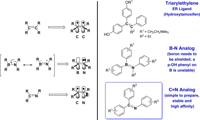
Many nonsteroidal estrogens are di- or triarylethylenes having a C=C double bond core, and in a recent publication, we examined the replacement of this C=C double bond with the isoelectronic and isostructural B—N bond (Figure 1, right, middle).13 To achieve hydrolytic stability, the electrophilicity of the boron center had to be sterically masked with a full array of flanking ortho methyl groups, and analogues with p-OH groups on the B-phenyl groups were unstable. Despite these restrictions, some of the anilino bis(2,6-dimethylphenyl)borane derivatives that we prepared were very stable and demonstrated reasonably high binding affinity and good cellular potency, being ERα agonists and ERβ antagonists.13 Those studies defined the structural determinants of stability and cellular bioactivity of a B—N for C=C substitution and provided a framework for further exploration of “elemental isomerism” for diversification of drug-like molecules.13
Figure 1.
Triarylethylene nonsteroidal estrogen as well as B—N and C=N double bond for C=C bond isoelectronic replacements.
Here, we have further explored the chemical diversification of ER ligands by another, simple isostructural and isoelectronic replacement, substituting a core C=C double bond with a simple Schiff base (C=N). As far as we are aware, there appears to be only limited and rather distant precedent for use of a C=N bond as a core element for estrogens.14,15 Furthermore, on the basis of a substructure search of the Merck Index, the C=N bond does not appear to be well-recognized as a surrogate for a C=C bond in the construction of bioactive molecules (except as a sub-element in 2-aryl-benzimidazoles, benzodiazepines, and other related heterocycles). As was the case earlier,13 di- and triarylethylene nonsteroidal estrogens offer a convenient C=C double bond core (Figure 1, right, top) for replacement by the isostructural C=N element (Figure 1, bottom, right). These Schiff base or imine-core ligands are very easy to synthesize, in most cases in one step (and much easier than their C=C double bond analogues), and to this end, we have prepared a series of triaryl-substituted (and some diaryl-substituted) Schiff base derivatives by a simple condensation that proceeds without phenolic hydroxy protection. Their binding affinities and cellular biological activities showed distinctive structure–activity relationships (SARs), and they profiled as potent ERα agonists and ERβ antagonists.
Results
Synthesis
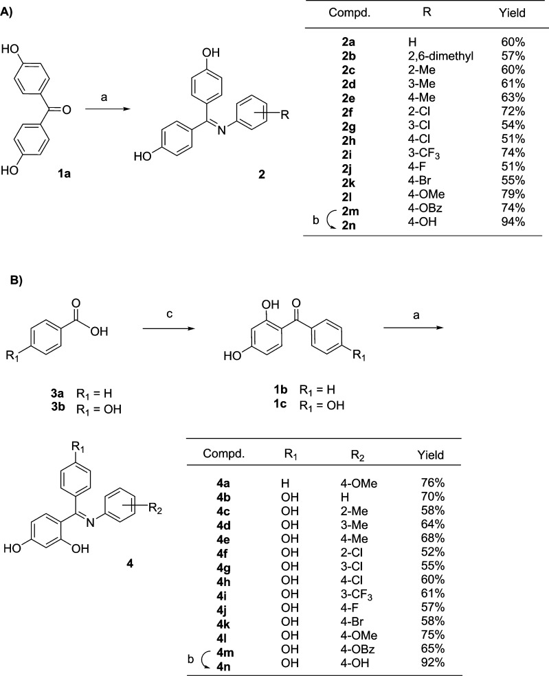
Representative members having an imine-core structure were prepared in three classes: 4,4′-dihydroxybenzophenone and 2,4,4′-trihydroxybenzophenone derivatives of various anilines and the corresponding diaryl imines. On the basis of previous work,16 we developed a HCl(g)-catalyzed condensation reaction without phenolic hydroxy protection for the synthesis of bisphenolic Schiff bases 2a–l in chlorobenzene with heating at 140–145 °C for 24 h (Scheme 1A). Although all yields were moderate, this improved method was superior to the phenol-protection strategy.17 We were unable to prepare triphenolic Schiff base 2n by the reaction of 4,4′-dihydroxybenzophenone 1c with 4-aminophenol or by demethylation of 2l. However, when 4,4′-dihydroxybenzophenone 1a was treated with 4-aminophenyl benzoate, it gave imine 2m in 74% yield, and deprotection with KOH proceeded at room temperature to give desired triphenolic product 2n in 94% yield. We also used this methodology for the synthesis of the corresponding phenolic Schiff bases 4 (Scheme 1B). Benzophenone derivatives 1b and 1c were prepared by a Friedel–Crafts acylation reaction and subsequent condensation with various anilines then afforded imines 4a–m in good yield. Product 4n was also prepared in 92% yield via a two-step procedure similar to that used for 2n. In all cases (4a–l), the (E) form of the imines was the only diastereoisomer formed, presumably because the intramolecular hydrogen bond, which can only form in the (E) isomer, contributes to the imine stability. These triaryl systems appeared to be hydrolytically stable indefinitely under aqueous conditions. By contrast, diaryl imines prepared from anilines and aldehydes or phenyl alkyl ketones displayed pronounced hydrolytic lability, undergoing substantial hydrolysis in aqueous MeOH at rt within 1–6 h. However, the diarylimines having an internal hydrogen bond between the imine nitrogen and an ortho phenolic OH group were considerably more stable.
Scheme 1. Synthesis of Triaryl-Substituted Schiff Base Analogues 2 and 4 with the Improved Method.
Reagents and conditions: (a) aniline derivatives (3 equiv), HCl(g), PhCl, 140–145 °C, 24 h; (b) KOH, MeOH, rt, 3 h; (c) resorcinol, AlCl3, sulfolane, 65–70 °C, 8 h.
Estrogen Receptor Binding Affinity Assays
The binding affinity of Schiff base analogues 2 and 4–6 for both ERα and ERβ was determined by a competitive radiometric assay, using methods that have been previously described, and are reported in Table 1.18 The affinities are represented here by relative binding affinity (RBA) values, where estradiol has an affinity of 100 (absolute affinities for estradiol: Kd 0.2 nM on ERα and 0.5 nM on ERβ).
Table 1. Relative Binding Affinities (RBAs) of Compounds 2 and 4–6 for Estrogen Receptor α and βa.
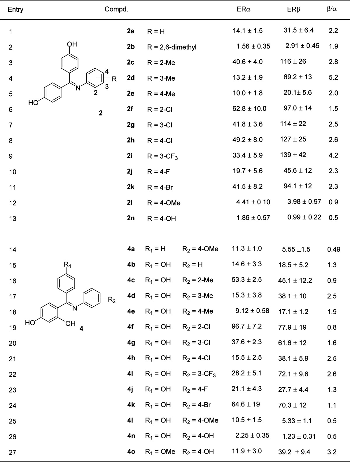
Determined by a competitive radiometric binding assay with [3H]estradiol; preparations of purified, full-length human ERα and ERβ (Invitrogen) were used; see the Experimental Section. Values are reported as the mean ± the range or SD of two or more independent experiments; the Kd for estradiol for ERα is 0.2 nM and for ERβ, 0.5 nM. Ki values for the reported compounds can be readily calculated using the formula Ki = (Kd[estradiol]/RBA)100.
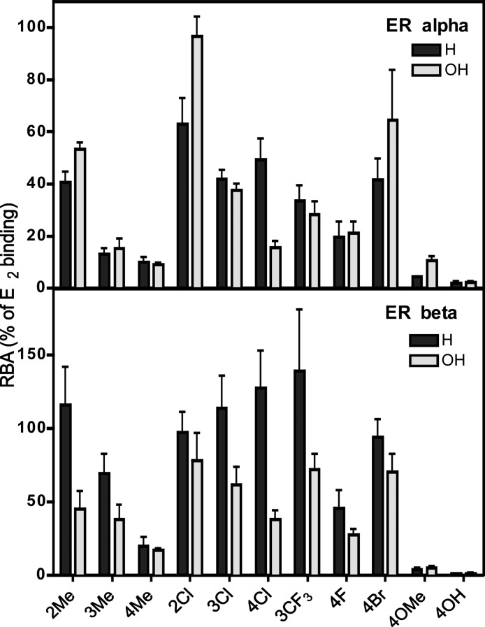
As a global observation, most of the imines are gratifyingly high-affinity ligands for both ERs, although the number and position of the hydroxyl group in the phenyl ring of the benzophenone moiety as well as the disposition and size of substituents on N-phenyl group have a marked influence on their binding affinity and selectivity. When assayed on the individual ER subtypes, ERα and ERβ, most compounds show only a modest binding-affinity preference for ERβ, at most 5-fold (compound 2d). Binding-affinity comparisons suggest that one of the p-hydroxy groups on the benzophenone moiety is playing the role of the phenolic hydroxyl of estradiol, which is well-known to be the dominant functional group ensuring high binding affinity.19 (This was confirmed by X-ray crystallography; see below.) Curiously, the presence of a para hydroxyl group in the distal N-phenyl group, which is essential for the high-affinity binding of a number of bisphenolic estrogens such as diethylstilbestrol and hexestrol as well as certain phenylindenes,20 actually has a markedly detrimental effect on binding, reducing affinity by about 7-fold (Table 1, compound 2a vs 2n and 4b vs 4n). Where studied, the second para hydroxy group in the second ring of the benzophenone unit has little effect on binding (Table 1, compound 4a vs 4l), although the methyl ether of this phenol is a good ERβ ligand (Table 1, 4o). Lastly, placement of a second hydroxyl group at the ortho position of the phenyl ring of the benzophenone moiety, which enables potential formation of an intramolecular hydrogen bond as in the series 4 compounds, caused, in most cases, a decrease in affinity for both ERα and ERβ; this was somewhat unexpected but can be rationalized from conformational changes noted in crystal structures (see Discussion and below). Parallel changes in the nature of the substituents on the N-phenyl group were made within the two major series, 2c–n and 4c–n, and in nearly every case, the binding affinities for both ERα and ERβ changed in a coordinated fashion, although with some bias for one or the other ER subtype. This is illustrated graphically in Figure 2.
Figure 2.
Graphical presentation of RBA values for imines 2c–n (black, H) and 4c–n (gray, OH)
In both series, the highest affinity analogues were those having somewhat bulky but nonpolar substituents, and where a substituent (Me or Cl) was placed at the C-2, -3, and -4 positions, there was a general decline in affinity with that progression of ring positions, although this was more evident in the ERα series than in the ERβ series. Among the various substituents, certain ones were preferred at each of the three positions, but these differed between the two series and with the two ER subtypes. Addition of a second methyl group, as in 2,6-dimethyl compound 2b, resulted in a dramatic, ca. 50-fold, reduction in affinity compared to 2-methyl analogue 2c. Electron-withdrawing substituents (halogens and CF3) were associated with higher affinity than alkyl (methyl) or electron-donating substituents (OH or OMe). Overall, these results illustrate that ER ligands having simple imine-core structures can be readily prepared but that high-affinity binding, as one might expect, requires an appropriate distribution of bulk, polarity, and functionality.
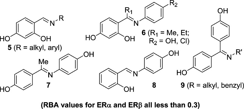
Previously, in connection with studies of dimethylgallium chelates that mimic nonsteroidal estrogens, we prepared a number of smaller imine-core systems (Figure 3), and although we published their synthesis and structures,16 we had, until now, not determined their ER-binding affinities. More recently, we also prepared a few additional diarylimines. In stark contrast to the triaryl systems, all of these monoaryl and diaryl imines have very low binding affinity (the data is given in Supporting InformationTable S1). The highest affinity compounds are benzophenone imines of alkyl or benzyl amines (9), which also have some ERβ-binding preference. Even on ERβ, however, these compounds have RBA values of less than 0.3%, and the affinities of the other diarylimines we prepared are typically more than 10-fold lower (see Supporting InformationTable S1). As noted earlier, some of these diaryl systems were also rather hydrolytically labile, which is not the case for all of the series 2 and 4 compounds.
Figure 3.
Other mono and diaryl imines (also see Supporting InformationTable S1).
Transcriptional Activity
We determined the effects of these imines on ER transcriptional activity using an ER-responsive luciferase reporter gene. Steroid-deprived HepG2 liver cells were transfected with a widely used 3×ERE-luciferase reporter and an ERα or ERβ expression plasmid for agonist activity (% efficacy) and potency (EC50) determinations. These cells were stimulated with increasing concentrations of 17β-estradiol (E2) or compounds 2a, 2c–k, 2n, 4b–k, and 4n. For antagonist mode assays (% efficacy and IC50), cells were stimulated with a combination of estradiol (10 nM) and an increasing concentration of the various compounds. The next day, luciferase activity was measured. HepG2 cells are particularly useful as a test system to distinguish the activity of estrogens through the two activation functions of the ERs, the N-terminal AF1, in the A/B domain, and AF2, which is in the ligand-binding domain.21 SERMs, such as tamoxifen, show tissue-selective agonist activity in some tissues, such as the uterus, and in the HepG2 cells via AF1 activity, but they work as antagonists in the breast through structural mechanisms that are not understood.
In general, these imines fully stimulated ER-mediated transcription in cells transfected with wild-type ERα, indicating that they are potent and highly efficacious ERα agonists (Table 2 and Figure 4A). In most cases, the number and position of the hydroxyl groups in the phenyl ring as well as the disposition and size of substituents on the N-phenyl group had no obvious effect on ERα-mediated transcription. However, compared to E2, none of the imines fully stimulated ER-mediated transcription in cells transfected with an ER construct that lacks AF1 because of the deletion of the N-terminal AB domain (Figure 4B), indicating that these imines do not fully induce AF2-mediated ER activity but also rely substantially on the AF1-mediated activity of ER to drive transcription.
Table 2. Effects of Imines on the Transcriptional Activities of Estrogen Receptor α and β.
| agonist
modea |
antagonist modea |
|||||||
|---|---|---|---|---|---|---|---|---|
| ERα |
ERβ |
ERα |
ERβ |
|||||
| EC50 (nM) | eff (% E2) | EC50 (nM) | eff (% E2) | IC50 (nM) | eff (% E2) | IC50 (nM) | eff (% E2) | |
| 2a | 3 | 110 ± 5 | 34 ± 12 | 126 ± 4 | 13 | 17 ± 3 | ||
| 2c | 7 | 103 ± 6 | 8 ± 3 | 149 ± 6 | 6 | 27 ± 9 | ||
| 2d | 5 | 109 ± 3 | 13 ± 9 | 93 ± 5 | 41 | 14 ± 0 | ||
| 2e | nd | nd | 7 ± 9 | nd | 45 | 10 ± 2 | ||
| 2f | 1 | 104 ± 3 | 37 ± 9 | 83 ± 9 | 19 | 30 ± 3 | ||
| 2g | 10 | 102 ± 3 | 10 ± 3 | 109 ± 5 | 16 | 11 ± 2 | ||
| 2h | 2 | 114 ± 4 | 9 ± 3 | 138 ± 8 | 9 | 6 ± 2 | ||
| 2i | 23 | 89 ± 3 | 3 ± 2 | 104 ± 6 | 24 | 11 ± 5 | ||
| 2j | 2 | 114 ± 4 | 10 ± 3 | 113 ± 4 | 7 | 8 ± 2 | ||
| 2k | 1 | 105 ± 2 | 11 ± 1 | 128 ± 10 | 6 | 4 ± 3 | ||
| 2n | 14 | 95 ± 2 | 332 | 16 ± 8 | 111 ± 4 | 300 | 33 ± 9 | |
| 4b | 7 | 102 ± 2 | 628 | 25 ± 8 | 134 ± 34 | 6 | 23 ± 12 | |
| 4c | 111 ± 4 | 2 | 31 ± 3 | 100 ± 5 | 16 | 29 ± 4 | ||
| 4d | 14 | 95 ± 2 | 9 ± 2 | 101 ± 2 | 51 | 11 ± 3 | ||
| 4e | 16 | 103 ± 3 | 10 860 | 20 ± 9 | 122 ± 34 | 241 | 21 ± 9 | |
| 4f | 0.1 | 113 ± 4 | 54 ± 1 | 136 ± 13 | 6 | 33 ± 6 | ||
| 4g | 15 | 117 ± 6 | 15 ± 2 | 118 ± 23 | 67 | 15 ± 0 | ||
| 4h | 4 | 114 ± 3 | 385 | 19 ± 5 | 123 ± 18 | 180 | 15 ± 1 | |
| 4i | 57 | 100 ± 4 | 11 ± 6 | 95 ± 9 | 28 | 16 ± 2 | ||
| 4j | 10 | 108 ± 3 | 23 ± 8 | 124 ± 6 | 139 | 14 ± 2 | ||
| 4k | 9 | 113 ± 4 | 12 460 | 24 ± 2 | 111 ± 5 | 100 | 13 ± 6 | |
| 4l | 17 | 102 ± 3 | 276 | 38 ± 13 | 102 ± 6 | 20 ± 24 | ||
| 4n | 7 | 97 ± 3 | 51 ± 10 | 108 ± 4 | 417 | 25 ± 10 | ||
In the agonist mode, ERE-luciferase assays were performed with 12-point dose curves of the indicated ligands, whereas in the antagonist mode, this was done in the presence of 10 nM E2.
Figure 4.

Graphical comparison of the agonist-mode efficacies of matched compounds on full-length ERα (A), ERα with the N-terminal AF1 deleted (B), and ERβ (C)
ERβ exhibits negligible AF1-mediated activity compared to ERα, but the AF2-mediated activities of the ER subtypes are similar in reporter assays,22 suggesting that despite their similar binding affinities for the two ER subtypes (Figure 2) these imines would act as partial ERβ agonists. Consistent with this supposition, all of the Schiff bases tested failed to stimulate ER-mediated transcription fully in HepG2 cells transfected with ERβ (Table 2 and Figure 4C). In fact, many of these compounds acted as potent and nearly complete ERβ-selective antagonists, suppressing the E2-induced activity of ERβ in the low nanomolar range (Table 2) and thereby underscoring the AF1-dependent ER subtype-selective properties of these compounds.
The N-phenyl substituents appear to affect the potency of these compounds as ERβ antagonists; however, whether some of the obvious differences in IC50 reflect variation in RBA toward ERβ is unclear. For example, an overview of the compound 4 series, which in general had lower RBAs (Figure 2), suggests that they were also less potent than the compound 2 series (Table 2); yet, in most cases, they were more efficacious on ERβ (Figure 4C). Overall, our results suggest that the imines generally profile as potent, efficacious, subtype-selective ER ligands that depend to a large extent on AF1 to induce ER-mediated transcription fully, but they poorly stimulate the AF2-mediated activity of ERα or ERβ. These differences underlie their ERα-selective agonist and ERβ-selective antagonist properties.
Structural Basis for the ER Subtype-Selective Profile of Triaryl-Substituted Schiff Base Analogues
We obtained crystal structures of the ERα ligand-binding domain complexed with the 2-Cl-substituted analogues, compounds 2f and 4f, and compared these new structures to previously reported full agonist- and antagonist-bound ERα structures.23−25 Full agonists, such as E2, fit into the ERα ligand-binding pocket in an orientation that facilitates hydrogen bonding of the phenolic OH to the side chains of helix 3 residue Glu353, helix 6 residue Arg394, and, more variably, the D-ring 17β-OH to helix 11 residue His524 (Figure 5A). This binding orientation allows the switch helix, helix 12, to dock against helices 3 and 11, where it forms one side of the coactivator binding site on the surface that constitutes the functional core of AF2, that is, it is a coregulator binding site.23 In contrast, antagonists and SERMs, such as 4-hydroxytamoxifen, contain an agonist-like core but have a bulky side group that protrudes between helices 3 and 11 and directly displaces helix 12 from its active conformation. As result, helix 12 docks along helix 3, thereby occluding the AF2 surface.24,25
Figure 5.

Imines induce a suboptimal conformation of the ERα ligand-binding domain. (A, B) Active and inactive ERα ligand-binding domain conformations show a ∼1 Å difference in distance between helices 3 and 11. The crystal structures of 17β-estradiol (E2; PDB ID: 1GWR) and 4-hydroxytamoxifen (TAM; PDB ID: 3ERT) bound complexes are shown. (C, D) Crystal structures of the ERα ligand-binding domain in complex with compounds 2f and 4f show a TAM-like binding orientation and increased h3–h11 distance compared to E2. (E, F) Crystal structures of the imine-bound ERα complexes were superposed. Compared to compound 2f (white), the additional hydroxyl group of compound 4f (coral) leads to a subtle distortion of the ligand-binding orientation.
Within the ligand-binding pocket, compounds 2f and 4f mimicked the binding orientation of the 4-hydroxytamoxifen core without a protruding side chain (Figure 5C,D), consistent with their high binding affinities toward ER subtypes. In addition, the N-phenyl groups were accommodated between helices 8 and 11, contacting M421, H524, and L525. The additional hydroxyl group of compound 4f was also accommodated easily, as this ring was utilized as the A-ring mimetic that forms a hydrogen bond with Glu353 (Figure 5E); however, this hydroxyl substitution also led to a 0.8 Å rotation of the other phenyl ring that forms a hydrogen bond with Thr347 (Figure 5F). Therefore, the different binding orientations of these imines within the ligand-binding pocket account for the fact that many of the matched 2 and 4 series compounds have slightly different binding affinities (Figure 2) but similar ERα-mediated activity profiles (Figure 4).
The structures of the imines suggest that reduced AF2 activity derives from a shift in the distance between helices 3 and 11. The active helix 12 conformer docks across helices 3 and 11; therefore, shifting helices 3 and 11 apart would undoubtedly destabilize helix 12 and lead to a suboptimal ligand-binding domain conformation that would likely affect the binding of coregulators. In the full agonist-bound conformation of ERα, the distance between helices 3 and 11, measured from the α-carbon of Thr347 to the α-carbon of Leu525, is a remarkably consistent 9 Å (Figure 5A).23 ER ligands that are not full agonists, including SERMs, increase this distance in a ligand-dependent manner. When bound to 4-hydroxytamoxifen, this distance is increased by about 1 Å (Figure 5B).25 In contrast, compounds 2f and 4f increased this distance by 0.5 and 0.7 Å, respectively (Figure 5C,D), indicating that these compounds induce a suboptimal conformation of the ERα ligand-binding domain, which explains their inability to stimulate the AF2-mediated activity of ERα fully (Figure 4B). Furthermore, the ER subtypes have similar ligand-binding pockets; therefore, imines are likely to also distort the active ERβ conformation through a similar structural mechanism, which underlies their partial ERβ agonist/antagonist phenotype (Table 2 and Figure 4C).
Discussion
The estrogen receptors are remarkable in binding and responding to ligands of great structural diversity,8,26,27 and this eclectic acceptance of ligands offers an opportunity to investigate chemically novel structures as potential selective estrogen receptor modulators (SERMs)10,11 or ER subtype-selective ligands.8,9 For example, recently, simple acyclic amide,3 diphenylamine,28 monoaryl- or diaryl-substituted salicylaldoxime,29−33 and anthranylaldoxime34,35 derivatives have been reported as ER ligands. Thus, the development of new ER ligands remains an important issue in medicinal chemistry because novel functions of estrogen are still being found.36,37
In this article, we have explored diversification of ligands for the estrogen receptor by replacing the C=C double bond with a simple Schiff Base or imine (C=N) core structure. A series of triaryl-substituted Schiff base derivatives were conveniently prepared, generally in one step, by the condensation of benzophenones with various anilines without the need for phenolic hydroxy group protection. Many of these compounds have very high binding affinities for ERs, rivaling or exceeding that of estradiol (up to 97% RBA for ERα and 140% for ERβ). Some of the compounds also show significant affinity selectivity in favor of ERβ (4- to 5-fold), and in cell-based assays for transcriptional activity, they profiled as ERα agonists and ERβ antagonists, a form of ER-subtype selectivity that appears to be based on their preferential activity through AF1, the N-terminal activation function of the ERs that is more active in ERα than ERβ. In addition, an unusual distortion of the ligand-binding domain, revealed by X-ray analysis, suggests that the function of AF2 is not fully engaged and highlights the diverse ways in which ligands can regulate the conformation, and ultimately the activity, of the ERs.
Beyond our general interest in preparing ER ligands of unusual structure,1 two other factors motivated our investigation of imine-core systems for ER ligand design. Some time ago, in an attempt to prepare gallium chelates that might have good affinity for ER and could be used in Ga-67- or Ga-68-labeled form for the imaging of ER in breast cancer using SPECT or PET, we prepared some similar imines, notably, those having the 2-hydroxyl group on the carbonyl component.16 Unfortunately, although we could prepare dimethylgallium complexes, engaging the oxygen and nitrogen of the o-hydroxyphenyl imine system as a bidentate chelate, and could even obtain their crystal structures, the aqueous stability of these complexes was very low.16 Nevertheless, at the time, we noted that a few of the imines had some ER binding affinity. (The complete binding data on these compounds is now given in Table S1.) We also returned to the imines when we prepared the B—N core compounds as part of our attempt to make the C=C and C=N systems that were isoelectronic with the hydrolytically stable B—N compounds; however, the one C=N analogue of these more sterically encumbered systems that we were able to prepare lacked the critical phenolic hydroxyl group and had other substituents that precluded high-affinity binding.13 By contrast, in this investigation, we were gratified by how easy it was to prepare a large series of triaryl-substituted imines and how many of them had high binding affinity for both ERα and ERβ.
Tolerance of Polar Groups in the Ligand Interior
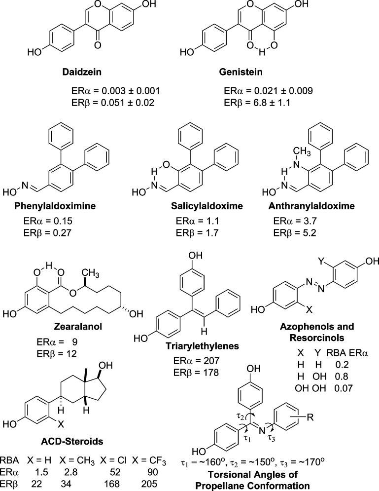
Because the interior of the ER ligand binding pockets is lined strictly with hydrophobic residues (except for the phenol–hydrogen bonding glutamate and arginine residues and one threonine),24 we expected that compounds in series 2 would have lower affinity compared those in the series 4: the isolated lone pair on the imine nitrogen in 2 was expected to exact a large desolvation energy penalty, whereas in the series 4 imines, the intramolecular hydrogen bond would internally solvate the imine nitrogen, thereby muting the impact of moving this polar function from water into the binding pocket. Relevant to this issue is the higher affinity of genistein than daidzein for both ERα and ERβ: the ketone group in genistein is shielded by an intramolecular hydrogen bond, whereas in daidzein, it is an isolated function (Figure 6).38 A similar intramolecular hydrogen-bonded system is present in the resorcylic acid lactone series and macrolides with high affinity for the ERs exemplified by zeralanol39 as well as in the salicylaldoximine and anthranylaldoxime systems developed by Minutolo, where an intermolecular hydrogen bond completes the formation of a pseudocycle that is thought to mimic the phenolic ring of estradiol and is important for high affinity binding (Figure 6).34 (In addition, ER ligands with polar, hydrogen-bonding cores, such as imidazoles and pyridazines, bind much less well than their less polar analogues, pyrazoles and pyrazines, respectively.1)
Figure 6.
Structure and relative binding affinities (RBAs, estradiol = 100) of various nonsteroidal and seco-steroidal estrogens as well as torsional angles of the triarylimine propellane conformation.
Despite these precedents for the importance of intramolecular hydrogen bonding to shield a polar element in the interior of ER ligands, in nearly every case, the imines of series 2, all of which lack this hydrogen bond, have comparable or higher affinity than the corresponding members in series 4 (Figure 2). This was particularly evident in their ERβ-binding affinities, where the average ratio of RBA (series 2)/RBA (series 4) is 1.88 ± 0.70, whereas it is only 1.16 ± 0.78 for ERα.
A reexamination of the crystal structures for compounds 2f and 4f shows that at the imine side of the ligands there is ample space between the ligands and the pocket residues, with no evidence for interaction of the imine lone pair in compound 2f with any elements of the protein. Also, the additional hydroxyl group in compound 4f does not engage in hydrogen bonding with the protein and is nicely accommodated without any obvious effects on the shape of the pocket at this side of the ligands. Perhaps the only factor contributing to the differences between the two series could be the increased twist angle of the N-phenyl ring noted in the X-ray structure of the series 4 compound 4f versus the series 2 compound 2f (Figure 5C–F), which appears to interfere with a productive interaction with the Thr347 residue. It is notable, as well, that in a number of ligand series, ERβ appears more tolerant to interior polar groups.8
Number of Substituents on the C=N Core
Although ligands having C=C or B—N core elements could be tetrasubstituted, the imine-core ligands, for valency reasons, can, at most, be trisubstituted. Nevertheless, triaryl ethylene ligands with a C=C core lacking a fourth substituent often have very good ER-binding affinity (Figure 6),41 as do our imines. By contrast, the mono and diaryl imines (Table S1) have uniformly low affinity, presumably because they are too small. There are a number of ER ligands in which a C=C core has been replaced by two nitrogens (i.e., an azo or N=N group), and these, for valence reasons, can be only disubstituted. These azophenol or azoresorcinol systems have low, but clearly measurable, ER-binding affinity (Figure 6) and are known to be estrogens, although of very low potency.42,43 In these molecules, as with the imines, there are ortho-substituted hydroxyl groups that can engage in intramolecular hydrogen bonds, but again, these hydrogen bonds reduce binding (Figure 6).
Effect of Phenyl Ring Substituents
In our imines, addition of a single ortho methyl group in the distal N-phenol ring improved binding considerably (Table 1, compound 2c vs 2a), whereas addition of a second ortho methyl group caused a precipitous drop in binding affinity (Table 1, compound 2b vs 2c). We encountered the beneficial effect of single ortho methyl substitution in A-CD estrogens (B-seco steroids lacking an intact B-ring), where addition of a methyl group (or other small substituent, Cl, CF3) ortho to the site of attachment of the phenol to the C-ring increased affinity (Figure 6).44 In these seco-estrogens, we interpreted the increase in binding resulting from this single ortho substitution as the dual effect of supplying bulk that was lost by deletion of the B-ring as well as twisting the aryl ring relative to the rest of the ligand, thereby increasing ligand volume, at least on one side. Similarly, in other ER ligands that we have explored, a single twist-inducing ortho substituent generally increased binding affinity.2,45,46
Substituents on the N-phenyl ring in our triarylimines are directed to different regions of the ER ligand-binding pocket than those in the B-secosteroids, and their enhancement of binding is most likely just due to increased hydrophobic bulk. There is a general trend, with binding affinity decreasing with the series C-2 > C-3 > C-4, which suggests that the ortho-disposed (C-2) groups might be causing an increased twist of the N-phenyl group. Simple MM2 energy minimization, however, shows that all of these triaryl imines adopt a propeller-like conformation; even the unsubstituted systems, 2a and 4b, show a coordinated twist of all three rings, giving dihedral angles of 160, 155, and 172° for τ1, τ2, and τ3, respectively (Figure 6). Notably, addition of the substituents shown in Table 1, even those at the C-2 position, changes these torsions by only a few degrees. Even 2,6-dimethyl substitution in 2c causes only a 10° change in τ3, suggesting that the marked drop in affinity results from a steric clash of the second methyl group in the ligand-binding pocket.
Structural Mechanisms for Ligand-Dependent Modulation of ER Activity
ER binds a diverse collection of ligands that modulate its activity through distinct structural mechanisms. Full agonists directly drive helix 12 to adopt an active conformation where it lies across helices 3 and 11, positioned to form one side of the AF2 coactivator-binding surface.24 In contrast, the bulky side chain of SERMs protrudes out of the pocket and directly displaces helix 12 from this active conformation.24,25 Yet, several other ligands disrupt the active conformation of helix 12 indirectly by distorting the C-terminal end of helix 11,7,45 thereby modulating ER activity. Here, we show that triaryl imine ligands modulate ER activity through a new type of indirect mechanism that involves a ligand-dependent increase in the distance separating helices 3 and 11 (Figure 5). Through this new mechanism, these imine ligands are able to modulate the AF2-mediated but not AF1-mediated activity of ER (Figure 4).
The ease with which these C=N imine analogues can be prepared, in some cases much more readily than their C=C double bond counterparts, and the high binding affinity that they have for the ERs suggest that this form of analogue development might be worth exploring more generally in drug discovery. Furthermore, the marked pattern of ER-subtype differential cellular efficacy, based on differential utilization of the two ER activation functions, displayed by the C=N analogues appears to be based on a novel mode of conformational control of the ER ligand-binding pocket that affects AF2 function, as revealed by our X-ray structural analyses. This new ligand design paradigm could also be more widely investigated for the development of estrogens having a novel spectrum of activities.
Experimental Section
Analytical Techniques and Instrumentation Used
Melting points were determined on a X-4 Beijing Tech melting point apparatus. 1H and 13C NMR spectra were recorded on a Bruker AV400 spectrometer (400 MHz, 1H NMR; 101 MHz, 13C NMR) at room temperature. NMR spectra were calibrated to the solvent signals of CDCl3 (δ 7.26 and 77.00 ppm), acetone-d6 (δ 2.05 and 29.84 ppm, 206.26 ppm), or DMSO-d6 (δ 2.50 and 39.43 ppm). The chemical shifts are provided in ppm, and the coupling constants, in Hz. The following abbreviations for multiplicities are used: s, singlet; d, doublet; dd, double doublet; t, triplet; dt, double triplet; q, quadruplet; m, multiplet; and br, broad. The purity of all compounds for biological testing was determined by HPLC analysis in two different solvent systems (normal and reversed phase), confirming >95% purity (see the Supporting Information).
Chemical Synthesis. General Procedure of the Improved Method for the Synthesis of Compounds 2a–m and 4a–m
A mixture of 1 (1 mmol) and aniline derivatives (3 mmol, 3 equiv) was dissolved in chlorobenzene (4 mL). Under a N2 atmosphere, the mixture was briefly exposed to HCl(g) and then heated at 140–145 °C for 24 h. The resulting solution was concentrated under reduced pressure, and the crude product was purified by silica gel chromatography (petroleum ether/ethyl acetate: Et3N = 4:1:0.5) to afford target molecules 2 and 4. Further purification was achieved by recrystallization (ethyl acetate/petroleum ether).
4,4′-((Phenylimino)methylene)diphenol (2a)
According to the general procedure of the improved method, 2a was obtained as a yellow solid (60% yield) and was further purified by recrystallization from ethyl acetate/petroleum ether (mp 273–275 °C). 1H NMR (400 MHz, acetone-d6) δ 8.83 (br s, 2H), 7.66–7.60 (m, 2H), 7.15–7.08 (m, 2H), 7.00–6.94 (m, 2H), 6.92–6.83 (m, 3H), 6.77–6.71 (m, 2H), 6.66 (dt, J = 8.4 Hz, 1.6 Hz, 2H). 13C NMR (101 MHz, acetone-d6) δ 167.97, 160.74, 158.37, 153.24, 132.58, 131.96, 131.94, 129.18, 128.51, 123.11, 121.78, 115.68, 115.49. HRMS (MALDI/DHB) calcd for C19H17NO2 (M + H)+m/z, 290.11811; found, 290.11756.
4,4′-(((2,6-Dimethylphenyl)imino)methylene)diphenol (2b)
According to the general procedure of the improved method, 2b was obtained as a yellow solid (58% yield) and was further purified by recrystallization from ethyl acetate/petroleum ether (mp 147–150 °C). 1H NMR (400 MHz, acetone-d6) δ 9.05 (s, 1H), 8.88 (s, 1H), 7.66 (d, J = 8.6 Hz, 2H), 6.98 (d, J = 8.5 Hz, 2H), 6.88 (dd, J = 14.6 Hz, 8.1 Hz, 4H), 6.75–6.66 (m, 3H), 1.99 (s, 6H). 13C NMR (101 MHz, acetone-d6) δ 167.02, 160.60, 158.84, 150.56, 132.56, 131.93, 131.06, 129.19, 128.35, 126.53, 122.89, 115.59, 115.28, 18.71. HRMS (MALDI/DHB) calcd for C21H20NO2(M + H)+m/z, 318.14941; found, 318.14886.
4,4′-((2-Tolylimino)methylene)diphenol (2c)
According to the general procedure of the improved method, 2c was obtained as a yellow solid (60% yield) and was further purified by recrystallization from ethyl acetate/petroleum ether (mp 126–130 °C). 1H NMR (400 MHz, acetone-d6) δ 8.94 (br s, 1H), 8.70 (br s, 1H), 7.65 (d, J = 8.7 Hz, 2H), 7.05 (d, J = 7.4 Hz, 1H), 6.97 (d, J = 8.5 Hz, 2H), 6.93–6.85 (m, 3H), 6.82–6.70 (m, 3H), 6.41 (d, J = 7.7 Hz, 1H), 2.12 (s, 3H). 13C NMR (101 MHz, acetone-d6) δ 167.34, 160.67, 158.45, 151.89, 132.53, 131.92, 131.41, 130.54, 128.83, 128.78, 126.62, 123.23, 120.57, 115.69, 115.45, 18.44. HRMS (MALDI/DHB) calcd for C20H19NO2 (M + H)+m/z, 304.13376; found, 304.13321.
4,4′-((3-Tolylimino)methylene)diphenol (2d)
According to the general procedure of the improved method, 2d was obtained as a yellow solid (61% yield) and was further purified by recrystallization from ethyl acetate/petroleum ether (mp 215–218 °C). 1H NMR (400 MHz, acetone-d6) δ 8.96 (br s, 1H), 8.71 (br s, 1H), 7.61 (d, J = 8.7 Hz, 2H), 6.98 (dd, J = 8.1 Hz, 6.2 Hz, 3H), 6.88 (d, J = 8.7 Hz, 2H), 6.74 (d, J = 8.5 Hz, 2H), 6.68 (d, J = 7.5 Hz, 1H), 6.53 (s, 1H), 6.43 (d, J = 7.9 Hz, 1H), 2.17 (s, 3H). 13C NMR (101 MHz, acetone-d6) δ 167.70, 160.65, 158.31, 153.17, 138.60, 132.66, 131.92, 131.90, 129.01, 128.59, 123.86, 122.50, 118.73, 115.65, 115.45, 21.45. HRMS (MALDI/DHB) calcd for C20H19NO2 (M + H)+m/z, 304.13376; found, 304.13321.
4,4′-((4-Tolylimino)methylene)diphenol (2e)
According to the general procedure of the improved method, 2e was obtained as a yellow solid (63% yield) and was further purified by recrystallization from ethyl acetate/petroleum ether (mp 128–131 °C). 1H NMR (400 MHz, acetone-d6) δ 8.85 (br s, 2H), 7.64–7.58 (m, 2H), 6.99–6.90 (m, 4H), 6.87 (d, J = 8.7 Hz, 2H), 6.75 (d, J = 8.5 Hz, 2H), 6.56 (d, J = 8.2 Hz, 2H), 2.19 (s, 3H). 13C NMR (101 MHz, acetone-d6) δ 167.78, 160.64, 158.31, 150.61, 132.74, 132.24, 131.90, 131.87, 129.74, 128.69, 121.83, 115.63, 115.51, 20.82. HRMS (MALDI/DHB) calcd for C20H19NO2 (M + H)+m/z, 304.13376; found, 304.13321.
4,4′-(((2-Chlorophenyl)imino)methylene)diphenol (2f)
According to the general procedure of the improved method, 2f was obtained as a yellow solid (72% yield) and was further purified by recrystallization from ethyl acetate/petroleum ether (mp 125–127 °C). 1H NMR (400 MHz, acetone-d6) δ 9.05 (br s, 1H), 8.77 (br s, 1H), 7.69–7.63 (m, 2H), 7.28 (dd, J = 8.0 Hz, 1.3 Hz, 1H), 7.07–7.00 (m, 3H), 6.93–6.85 (m, 3H), 6.78–6.72 (m, 2H), 6.62 (dd, J = 7.9 Hz, 1.5 Hz, 1H). 13C NMR (101 MHz, acetone-d6) δ 169.68, 161.14, 158.75, 150.72, 132.29, 131.82, 131.22, 129.96, 128.47, 127.89, 125.61, 124.26, 122.60, 115.77, 115.52. HRMS (MALDI/DHB) calcd for C19H16NO2Cl (M + H)+m/z, 324.07914; found, 324.07858.
4,4′-(((3-Chlorophenyl)imino)methylene)diphenol (2g)
According to the general procedure of the improved method, 2g was obtained as a yellow solid (54% yield) and was further purified by recrystallization from ethyl acetate/petroleum ether (mp 186–190 °C). 1H NMR (400 MHz, acetone-d6) δ 8.90 (br s, 2H), 7.63 (d, J = 8.7 Hz, 2H), 7.13 (t, J = 8.0 Hz, 1H), 7.01 (d, J = 8.5 Hz, 2H), 6.90 (dd, J = 5.5 Hz, 3.2 Hz, 3H), 6.78 (d, J = 8.5 Hz, 2H), 6.71 (t, J = 1.9 Hz, 1H), 6.61 (d, J = 8.0 Hz, 1H). 13C NMR (101 MHz, acetone-d6) δ 169.10, 161.04, 158.62, 154.85, 134.35, 132.18, 132.09, 121.90, 130.63, 128.03, 122.90, 121.66, 120.42, 115.74, 115.62. HRMS (MALDI/DHB) calcd for C19H16NO2Cl (M + H)+m/z, 324.07914; found, 324.07858.
4,4′-(((4-Chlorophenyl)imino)methylene)diphenol (2h)
According to the general procedure of the improved method, 2h was obtained as a yellow solid (51% yield) and was further purified by recrystallization from ethyl acetate/petroleum ether (mp 123–127 °C). 1H NMR (400 MHz, acetone-d6) δ 9.01 (br s, 1H), 8.75 (br s, 1H), 7.66–7.58 (m, 2H), 7.32–7.25 (m, 2H), 7.01–6.95 (m, 2H), 6.92–6.85 (m, 2H), 6.80–6.74 (m, 2H), 6.66–6.59 (m, 2H). 13C NMR (101 MHz, acetone-d6) δ 168.90, 161.01, 158.62, 152.12, 132.23, 132.10, 131.93, 129.16, 128.14, 127.86, 123.49, 115.76, 115.66. HRMS (MALDI/DHB) calcd for C19H16NO2Cl (M + H)+m/z, 324.07914; found, 324.07858.
4,4′-(((3-(Trifluoromethyl)phenyl)imino)methylene)diphenol (2i)
According to the general procedure of the improved method, 2i was obtained as a yellow solid (74% yield) and was further purified by recrystallization from ethyl acetate/petroleum ether (mp 212–215 °C). 1H NMR (400 MHz, acetone-d6) δ 9.02 (br s, 1H), 8.86 (br s, 1H), 7.67 (t, J = 11.2 Hz, 2H), 7.34 (t, J = 7.8 Hz, 1H), 7.20 (d, J = 7.7 Hz, 1H), 7.05–6.87 (m, 6H), 6.77 (d, J = 8.4 Hz, 2H). 13C NMR (101 MHz, acetone-d6) δ 169.64, 161.17, 158.67, 153.94, 132.24, 131.98, 131.91, 131.21, 130.90, 130.14, 128.65 (d, J = 270 Hz), 127.88, 125.63, 119.15 (d, J = 100 Hz), 115.79, 115.65. HRMS (MALDI/DHB) calcd for C20H16NO2F3 (M + H)+m/z, 358.10549; found, 358.10494.
4,4′-(((4-Fluorophenyl)imino)methylene)diphenol (2j)
According to the general procedure of the improved method, 2j was obtained as a yellow solid (51% yield) and was further purified by recrystallization from ethyl acetate/petroleum ether (mp 267–271 °C). 1H NMR (400 MHz, acetone-d6) δ 8.97 (br s, 1H), 8.76 (br s, 1H), 7.65–7.59 (m, 2H), 7.00–6.94 (m, 2H), 6.93–6.85 (m, 4H), 6.80–6.74 (m, 2H), 6.71–6.64 (m, 2H). 13C NMR (101 MHz, acetone-d6) δ 168.80, 160.86, 158.48, 159.53 (d, J = 237 Hz), 149.52, 132.44, 131.99, 131.93, 128.34, 123.32, (d, J = 8 Hz), 115.70, 115.63, (d, J = 22 Hz), 115.61. HRMS (MALDI/DHB) calcd for C19H16NO2F (M + H)+m/z, 308.10869; found, 308.10813.
4,4′-(((4-Bromophenyl)imino)methylene)diphenol (2k)
According to the general procedure of the improved method, 2k was obtained as a yellow solid (55% yield) and was further purified by recrystallization from ethyl acetate/petroleum ether (mp 127–130 °C). 1H NMR (400 MHz, acetone-d6) δ 9.03 (br s, 1H), 8.77 (br s, 1H), 7.66–7.58 (m, 2H), 7.17–7.11 (m, 2H), 7.01–6.95 (m, 2H), 6.93–6.85 (m, 2H), 6.80–6.74 (m, 2H), 6.71–6.64 (m, 2H). 13C NMR (101 MHz, acetone-d6) δ 168.85, 160.95, 158.60, 152.54, 132.23, 132.14, 132.13, 131.95, 128.11, 123.94, 115.75, 115.66, 115.58. HRMS (MALDI/DHB) calcd for C19H16NO2Br (M + H)+m/z, 368.02862; found, 368.02807.
4,4′-(((4-Methoxyphenyl)imino)methylene)diphenol (2l)
According to the general procedure of the improved method, 2l was obtained as a yellow solid (79% yield) and was further purified by recrystallization from ethyl acetate/petroleum ether (mp 117–119 °C). 1H NMR (400 MHz, acetone-d6) δ 7.60 (d, J = 8.7 Hz, 2H), 6.95 (dd, J = 8.7 Hz, 2.4 Hz, 3H), 6.86 (d, J = 8.7 Hz, 2H), 6.76 (d, J = 8.5 Hz, 2H), 6.69 (d, J = 8.8 Hz, 2H), 6.61 (d, J = 8.8 Hz, 2H), 3.68 (s, 3H). 13C NMR (101 MHz, acetone-d6) δ 167.76, 160.61, 158.34, 156.40, 146.14, 133.02, 131.94, 131.82, 128.78, 123.23, 115.76, 115.59, 114.45, 55.51. HRMS (MALDI/DHB) calcd for C20H18NO3 (M + H)+m/z, 320.12867; found, 320.12812.
4,4′-(((4-Hydroxyphenyl)imino)methylene)diphenol (2n)
According to the general procedure of the improved method, 2m was obtained as a yellow solid (74% yield). Then, 2m was dissolved in THF and MeOH, a solution of KOH (1 equiv) in MeOH was added to the stirred mixture for 3h, the mixture was concentrated under reduced pressure, and the residue was isolated by silica gel chromatography (petroleum ether/ethyl acetate = 2:1, including 0.5% Et3N) to afford the desired product 2n (94% yield), which was further purified by recrystallization from ethyl acetate/petroleum ether (mp 297–301 °C). 1H NMR (400 MHz, acetone-d6) δ 7.46 (d, J = 8.7 Hz, 2H), 6.85–6.79 (m, 2H), 6.73 (d, J = 8.7 Hz, 2H), 6.64 (t, J = 8.1 Hz, 2H), 6.51–6.44 (m, 2H), 6.42–6.36 (m, 2H). 13C NMR (101 MHz, acetone-d6) δ167.29, 160.52, 158.30, 153.94, 145.10, 132.99, 131.95, 131.73, 128.93, 123.34, 116.04, 115.82, 115.59. HRMS (MALDI/DHB) calcd for C19H17NO3 (M + H)+m/z, 306.11302; found, 306.11247.
4-(((4-Methoxyphenyl)imino)(phenyl)methyl)benzene-1,3-diol (4a)
According to the general procedure of the improved method, 4a was obtained as a yellow solid (76% yield) and was further purified by recrystallization from ethyl acetate/petroleum ether (mp 253–257 °C). 1H NMR (400 MHz, acetone-d6) δ 15.03 (s, 1H), 9.26 (s, 1H), 7.39 (dd, J = 5.1 Hz, 1.6 Hz, 3H), 7.28–7.19 (m, 2H), 6.82 (d, J = 8.8 Hz, 1H), 6.77–6.67 (m, 4H), 6.43 (d, J = 2.2 Hz, 1H), 6.28 (dd, J = 8.8 Hz, 2.2 Hz, 1H), 3.69 (s, 3H). 13C NMR (101 MHz, acetone-d6) δ 173.34, 165.44, 162.81, 157.50, 141.22, 134.55, 132.51, 132.15, 129.65, 129.52, 129.09, 124.60, 114.53, 107.53, 104.00, 55.54. HRMS (MALDI/DHB) calcd for C20H18NO3 (M + H)+m/z, 320.12867; found, 320.12812.
4-((4-Hydroxyphenyl)(phenylimino)methyl)benzene-1,3-diol (4b)
According to the general procedure of the improved method, 4c was obtained as a yellow solid (70% yield) and was further purified by recrystallization from ethyl acetate/petroleum ether (mp 249–252 °C). 1H NMR (400 MHz, acetone-d6) δ 14.82 (s, 1H), 9.15 (s, 1H), 8.81 (s, 1H), 7.18 (t, J = 7.8 Hz, 2H), 7.06 (d, J = 8.5 Hz, 2H), 7.02–6.95 (m, 2H), 6.82 (d, J = 8.4 Hz, 4H), 6.43 (d, J = 2.2 Hz, 1H), 6.31 (dd, J = 8.8 Hz, 2.3 Hz, 1H). 13C NMR (101 MHz, acetone-d6) δ 174.22, 166.08, 163.10, 158.77, 148.44, 134.86, 131.38, 129.31, 126.42, 124.78, 123.50, 115.77, 114.19, 107.47, 104.02. HRMS (MALDI/DHB) calcd for C19H17NO3 (M + H)+m/z, 306.11302; found, 306.11247.
4-((4-Hydroxyphenyl)(2-tolylimino)methyl)benzene-1,3-diol (4c)
According to the general procedure of the improved method, 4c was obtained as a yellow solid (58% yield) and was further purified by recrystallization from ethyl acetate/petroleum ether (mp 249–252 °C). 1H NMR (400 MHz, acetone-d6) δ 14.86 (s, 1H), 9.12 (s, 1H), 8.77 (s, 1H), 7.11 (d, J = 7.3 Hz, 1H), 7.04 (d, J = 8.3 Hz, 2H), 6.99 (d, J = 8.8 Hz, 1H), 6.95–6.84 (m, 2H), 6.79 (d, J = 8.5 Hz, 2H), 6.56 (d, J = 7.4 Hz, 1H), 6.43 (s, 1H), 6.31 (d, J = 8.7 Hz, 1H), 2.23 (s, 3H). 13C NMR (101 MHz, acetone-d6) δ 174.09, 166.18, 162.98, 158.74, 147.41, 134.87, 130.92, 130.77, 130.37, 126.72, 126.65, 124.96, 122.98, 115.67, 114.10, 107.39, 104.03, 18.58. HRMS (MALDI/DHB) calcd for C20H19NO3 (M + H)+m/z, 320.12867; found, 320.12812.
4-((4-Hydroxyphenyl)(3-tolylimino)methyl)benzene-1,3-diol (4d)
According to the general procedure of the improved method, 4d was obtained as a yellow solid (64% yield) and was further purified by recrystallization from ethyl acetate/petroleum ether (mp 239–243 °C). 1H NMR (400 MHz, acetone-d6) δ 14.88 (s, 1H), 9.15 (s, 1H), 8.82 (s, 1H), 7.07–7.00 (m, 3H), 6.97 (d, J = 8.8 Hz, 1H), 6.80 (t, J = 9.3 Hz, 3H), 6.68 (s, 1H), 6.56 (d, J = 7.9 Hz, 1H), 6.42 (s, 1H), 6.30 (d, J = 8.8 Hz, 1H), 2.20 (s, 3H). 13C NMR (101 MHz, acetone-d6) δ 173.98, 166.17, 163.09, 158.78, 148.23, 138.93, 134.82, 131.33, 129,12, 126.44, 125.55, 124.23, 120.50, 115.74, 114.18, 107.45, 104.02, 21.34. HRMS (MALDI/DHB) calcd for C20H19NO3 (M + H)+m/z, 320.12867; found, 320.12812.
4-((4-Hydroxyphenyl)(4-tolylimino)methyl)benzene-1,3-diol (4e)
According to the general procedure of the improved method, 4e was obtained as a yellow solid (68% yield) and was further purified by recrystallization from ethyl acetate/petroleum ether (mp 249–252 °C). 1H NMR (400 MHz, acetone-d6) δ 15.03 (s, 1H), 9.13 (s, 1H), 8.83 (s, 1H), 7.05 (d, J = 7.9 Hz, 2H), 6.97 (t, J = 8.0 Hz, 3H), 6.82 (d, J = 8.4 Hz, 2H), 6.70 (d, J = 7.7 Hz, 2H), 6.42 (s, 1H), 6.29 (d, J = 8.7 Hz, 1H), 2.20 (s, 3H). 13C NMR (101 MHz, acetone-d6) δ 173.88, 166.41, 163.02, 158.76, 145.50, 134.77, 134.27, 131.36, 129.90, 126.90, 123.49, 115.83, 114.23, 107.40, 104.05, 20.83. HRMS (MALDI/DHB) calcd for C20H19NO3 (M + H)+m/z, 320.12867; found, 320.12812.
4-((4-Hydroxyphenyl)(2-chloroimino)methyl)benzene-1,3-diol (4f)
According to the general procedure of the improved method, 4f was obtained as a yellow solid (52% yield) and was further purified by recrystallization from ethyl acetate/petroleum ether (mp 226–230 °C). 1H NMR (400 MHz, acetone-d6) δ 14.22 (s, 1H), 9.00 (br s, 2H), 7.35 (d, J = 8.0 Hz, 1H), 7.12–6.95 (m, 5H), 6.79 (dd, J = 16.4 Hz, 8.2 Hz, 3H), 6.47–6.43 (m, 1H), 6.37–6.30 (m, 1H). 13C NMR (101 MHz, acetone-d6) δ 175.65, 165.83, 163.47, 158.98, 146.45, 135.27, 130.72, 130.04, 127.95, 127.23, 126.50, 126.07, 124.81, 115.76, 113.88, 107.76, 104.02. HRMS (MALDI/DHB) calcd for C19H16NO3Cl (M + H)+m/z, 340.07405; found, 340.07350.
4-((4-Hydroxyphenyl)(3-chloroimino)methyl)benzene-1,3-diol (4g)
According to the general procedure of the improved method, 4g was obtained as a yellow solid (55% yield) and was further purified by recrystallization from ethyl acetate/petroleum ether (mp 209–211 °C). 1H NMR (400 MHz, acetone-d6) δ 14.74 (s, 1H), 9.20 (s, 1H), 8.86 (s, 1H), 7.05 (d, J = 8.3 Hz, 2H), 6.96 (dd, J = 18.6 Hz, 8.9 Hz, 3H), 6.87–6.79 (m, 4H), 6.43 (d, J = 2.0 Hz, 1H), 6.31 (dd, J = 8.8 Hz, 1.9 Hz, 1H). 13C NMR (101 MHz, acetone-d6) δ 174.82, 166.03, 163.12, 161.53, 158.82, 144.83, 134.96, 131.40, 126.30, 125.16, 125.09, 116.00, 115.87, 115.78, 114.16, 107.56, 103.99. HRMS (MALDI/DHB) calcd for C19H16NO3Cl (M + H)+m/z, 340.07405; found, 340.07350.
4-((4-Hydroxyphenyl)(4-chloroimino)methyl)benzene-1,3-diol (4h)
According to the general procedure of the improved method, 4h was obtained as a yellow solid (60% yield) and was further purified by recrystallization from ethyl acetate/petroleum ether (mp 255–257 °C). 1H NMR (400 MHz, acetone-d6) δ 14.50 (s, 1H), 9.25 (s, 1H), 8.90 (s, 1H), 7.20 (d, J = 8.5 Hz, 2H), 7.06 (d, J = 8.3 Hz, 2H), 6.99 (d, J = 8.8 Hz, 1H), 6.83 (dd, J = 8.5 Hz, 2.1 Hz, 4H), 6.43 (d, J = 2.0 Hz, 1H), 6.32 (dd, J = 8.8 Hz, 2.1 Hz, 1H). 13C NMR (101 MHz, acetone-d6) δ 174.91, 165.87, 163.20, 158.85, 147.55, 135.05, 131.40, 129.63, 129.30, 126.19, 125.19, 115.87, 114.12, 107.58, 103.95. HRMS (MALDI/DHB) calcd for C19H16NO3Cl (M + H)+m/z, 340.07405; found, 340.07350.
4-((4-Hydroxyphenyl)((3-(trifluoromethyl)phenyl)imino)methyl)benzene-1,3-diol (4i)
According to the general procedure of the improved method, 4i was obtained as a yellow solid (61% yield) and was further purified by recrystallization from ethyl acetate/petroleum ether (mp 238–242 °C). 1H NMR (400 MHz, acetone-d6) δ 14.28 (s, 1H), 9.26 (s, 1H), 8.86 (s, 1H), 7.40 (t, J = 7.8 Hz, 1H), 7.31 (d, J = 7.6 Hz, 1H), 7.17–7.05 (m, 4H), 7.00 (t, J = 8.4 Hz, 1H), 6.83 (d, J = 8.2 Hz, 2H), 6.45 (s, 1H), 6.34 (d, J = 8.6 Hz, 1H). 13C NMR (101 MHz, acetone-d6) δ 175.56, 165.78, 165.25, 163.38, 158.87, 135.30, 135.26, 131.41, 130.28, 127.38, 127.31, 126.38 (d, J = 269 Hz), 120.75 (d, J = 98 Hz), 115.88, 115.83, 107.78, 107.75, 103.96. HRMS (MALDI/DHB) calcd for C20H16NO3F3 (M + H)+m/z, 374.10041; found, 374.09985.
4-(((4-Fluorophenyl)imino)(4-hydroxyphenyl)methyl)benzene-1,3-diol (4j)
According to the general procedure of the improved method, 4j was obtained as a yellow solid (57% yield) and was further purified by recrystallization from ethyl acetate/petroleum ether (mp 241–245 °C). 1H NMR (400 MHz, acetone-d6) δ 14.75 (s, 1H), 9.19 (s, 1H), 8.85 (s, 1H), 7.05 (d, J = 8.6 Hz, 2H), 7.02–6.90 (m, 3H), 6.87–6.79 (m, 4H), 6.43 (d, J = 2.3 Hz, 1H), 6.31 (dd, J = 8.8 Hz, 2.4 Hz, 1H). 13C NMR (101 MHz, acetone-d6) δ 175.14, 165.23, 164.53 (d, J = 253 Hz), 158.85, 150.31, 135.21, 134.40, 131.38, 130.73, 124.61, 123.47, 122.15 (d, J = 8 Hz), 116.83 (d, J = 20 Hz), 107.72, 103.95. HRMS (MALDI/DHB) calcd for C19H16NO3F (M + H)+m/z, 324.10360; found, 324.10305.
4-(((4-Bromophenyl)imino)(4-hydroxyphenyl)methyl)benzene-1,3-diol (4k)
According to the general procedure of the improved method, 4k was obtained as a yellow solid (58% yield) and was further purified by recrystallization from ethyl acetate/petroleum ether (mp 279–282 °C). 1H NMR (400 MHz, acetone-d6) δ 14.45 (s, 1H), 9.18 (s, 1H), 8.82 (s, 1H), 7.34 (d, J = 8.6 Hz, 2H), 7.09–7.03 (m, 2H), 6.99 (d, J = 8.8 Hz, 1H), 6.83 (d, J = 8.4 Hz, 2H), 6.77 (dd, J = 8.7 Hz, 2.2 Hz, 2H), 6.43 (d, J = 2.2 Hz, 1H), 6.32 (dd, J = 8.8 Hz, 2.3 Hz, 1H). 13C NMR (101 MHz, acetone-d6) δ 174.83, 165.89, 163.26, 158.89, 147.97, 135.07, 132.28, 131.40, 126.21, 125.60, 117.45, 115.88, 114.13, 107.67, 104.00. HRMS (MALDI/DHB) calcd for C19H16NO3Br (M + H)+m/z, 384.02354; found, 384.02298.
4-((4-Hydroxyphenyl)((4-methoxyphenyl)imino)methyl)benzene-1,3-diol (4l)
According to the general procedure of the improved method, 4l was obtained as a yellow solid (75% yield) and was further purified by recrystallization from ethyl acetate/petroleum ether (mp 259–263 °C). 1H NMR (400 MHz, acetone-d6) δ 15.30 (s, 1H), 9.16 (s, 1H), 8.89 (s, 1H), 7.08–7.03 (m, 2H), 6.96 (d, J = 8.8 Hz, 1H), 6.87–6.83 (m, 2H), 6.74 (s, 4H), 6.41 (d, J = 2.4 Hz, 1H), 6.28 (dd, J = 8.8 Hz, 2.4 Hz, 1H), 3.70 (s, 3H). 13C NMR (101 MHz, acetone-d6) δ 173.49, 162.86, 158.73, 157.47, 141.06, 134.67, 131.37, 126.63, 124.78, 115.90, 114.56, 114.33, 110.90, 107.31, 104.00, 55.55. HRMS (MALDI/DHB) calcd for C20H19NO4 (M + H)+m/z, 336.12359; found, 336.12304.
4-((4-Hydroxyphenyl)((4-hydroxyphenyl)imino)methyl)benzene-1,3-diol (4n)
According to the general procedure of the improved method, 4m was obtained as a yellow solid (65% yield). Then, 4m was dissolved in THF and MeOH, a solution of KOH (1 equiv) in MeOH was added to the stirred mixture for 3 h, and the mixture was concentrated under reduced pressure, and the residue was isolated by silica gel chromatography (petroleum ether/ethyl acetate = 2:1, including 0.5% Et3N) to afford desired product 4n (92% yield), which was further purified by recrystallization from ethyl acetate/petroleum ether (mp 269–271 °C). 1H NMR (400 MHz, acetone-d6) δ 15.50 (s, 1H), 8.94 (s, 1H), 8.40 (s, 1H), 7.05 (d, J = 8.4 Hz, 2H), 6.96 (d, J = 8.8 Hz, 1H), 6.86 (d, J = 8.4 Hz, 2H), 6.65 (d, J = 8.7 Hz, 4H), 6.41 (d, J = 2.3 Hz, 1H), 6.27 (dd, J = 8.8 Hz, 2.3 Hz, 1H). 13C NMR (101 MHz, acetone-d6) δ 173.11, 162.84, 158.74, 144.22, 134.58, 132.02, 131.39, 129.65, 126.59, 124.95, 116.00, 115.93, 114.27, 107.23, 104.09. HRMS (MALDI/DHB) calcd for C19H17NO4 (M + H)+m/z, 322.10794; found, 322.10739.
Estrogen Receptor Binding Affinity Assays
Relative binding affinities were determined by a competitive radiometric binding assay, as previously described,47,48 using 2 nM [3H]estradiol as tracer ([2,4,6,7-3H]-estra-1,3,5(10)-triene-3,17β-diol, 70–115 Ci/mmol, PerkinElmer, Waltham, MA) and purified full-length human ERα and ERβ, which were purchased from PanVera/Invitrogen (Carlsbad, CA). Incubations were for 18–24 h at 0 °C. Hydroxyapatite (Bio-Rad, Hercules, CA) was used to absorb the receptor–ligand complexes, and free ligand was washed away. The binding affinities are expressed as relative binding affinity (RBA) values, with the RBA of estradiol set to 100%. The values given are the average ± range or SD of two to three independent determinations. Estradiol binds to ERα with a Kd of 0.2 nM and to ERβ with a Kd of 0.5 nM; these values were determined by Scatchard analysis using the binding assay protocol described previously.47
Gene Transcriptional Activity
Assays were performed as previously described.45 HepG2 cells cultured in Dulbecco’s minimum essential medium (DMEM) (Cellgro by Mediatech, Inc. Manassas, VA) supplemented with 10% fetal bovine serum (FBS) (Hyclone by Thermo Scientific, South Logan, UT), 1% nonessential amino acids (Cellgro), penicillin–streptomycin–neomycin antibiotic mixture, and Glutamax (Gibco by Invitrogen Corp. Carlsbad, CA) were maintained at 37 °C and 5% CO2. HepG2 cells were transfected with 10 μg of 3×ERE-luciferase reporter plus 1.6 μg of ERα or ERβ expression vector per 10 cm dish using FugeneHD reagent (Roche Applied Sciences, Indianapolis, IN). The next day, the cells were transferred to phenol red-free growth media supplemented with 10% charcoal-dextran sulfate-stripped FBS at a density of 20 000 cells/well, incubated in 384-well plates overnight at 37 °C with 5% CO2, and assayed in dose curves ranging from 10 μM to 100 pM for ERE luciferase assays in HepG2 cells. Luciferase activity was measured after 24 h using BriteLite reagent (PerkinElmer Inc., Shelton, CT) according to the manufacturer’s protocol. Raw data measured as relative light units were normalized for each plate using the average of DMSO-treated samples as 0% and the average of the top of the E2 curve as 100%.
X-ray Crystallography
As previously described,49 human ERα-Y537S ligand-binding domain was expressed in BL21(DE3) Escherichia coli cells, purified, mixed with SRC2 peptide, and crystallized at room temperature by the hanging-drop vapor-diffusion method. The ERα crystals obtained were then soaked in the different imine ligands. The X-ray diffraction data was scaled using HKL-2000 software.50 The crystal structures were solved via molecular replacement using the PHENIX software suite,51,52 with the protein components of PDB 2B1V as a starting model.53 The new structures were then completed upon ligand docking and extensive combinatorial refinement.54
Acknowledgments
This work was supported by grants from the NSFC (91017005, 81172935, and 81373255), the Key Project of the Ministry of Education (313040), the Scientific and Technological Innovative Research Team of Wuhan (2013070204020048), Hubei Province’s Outstanding Medical Academic Leader Program, and the Fundamental Research Funds for the Central Universities (201130602020001) as well as research support from the National Institutes of Health (PHS 5R37 DK015556 to J.A.K. and 5R01 DK077085 to K.W.N.). S.S. is supported by Frenchman’s Creek Women for Cancer Research. X-ray data reported in this study was collected at beamline 11-1, Stanford Synchrotron Radiation Lightsource, SLAC National Accelerator Laboratory.
Glossary
Abbreviations Used
- E2
estradiol
- ER
estrogen receptor
- AF1
transcriptional activation function 1
- AF2
transcriptional activation function 2
- SERMs
selective estrogen receptor modulators
- RBA
relative binding affinity
- HepG2
hepatoma-derived cancer cells
- ERE
estrogen response element
- DMEM
Dulbecco’s minimum essential medium
- FBS
fetal bovine serum
Supporting Information Available
ER-binding affinities of compounds 5–9, analytical methodology, summary of purities, and HPLC verification of the purity of the final compounds. This material is available free of charge via the Internet at http://pubs.acs.org.
The authors declare no competing financial interest.
Funding Statement
National Institutes of Health, United States
Supplementary Material
References
- Katzenellenbogen J. A. The 2010 Philip S. Portoghese Medicinal Chemistry Lectureship: Addressing the “core issue” in the design of estrogen receptor ligands. J. Med. Chem. 2011, 54, 5271–5282. [DOI] [PMC free article] [PubMed] [Google Scholar]
- Min J.; Wang P.; Srinivasan S.; Nwachukwu J. C.; Guo P.; Huang M.; Carlson K. E.; Katzenellenbogen J. A.; Nettles K. W.; Zhou H. B. Thiophene-core estrogen receptor ligands having superagonist activity. J. Med. Chem. 2013, 56, 3346–3366. [DOI] [PMC free article] [PubMed] [Google Scholar]
- Stauffer S. R.; Sun J.; Katzenellenbogen B. S.; Katzenellenbogen J. A. Acyclic amides as estrogen receptor ligands: Synthesis, binding, activity and receptor interaction. Bioorg. Med. Chem. 2000, 8, 1293–1316. [DOI] [PubMed] [Google Scholar]
- Fink B. E.; Mortensen D. S.; Stauffer S. R.; Aron Z. D.; Katzenellenbogen J. A. Novel structural templates for estrogen-receptor ligands and prospects for combinatorial synthesis of estrogens. Chem. Biol. 1999, 6, 205–219. [DOI] [PubMed] [Google Scholar]
- Ghosh U.; Ganessunker D.; Sattigeri V. J.; Carlson K. E.; Mortensen D. J.; Katzenellenbogen B. S.; Katzenellenbogen J. A. Estrogenic diazenes: Heterocyclic non-steroidal estrogens of unusual structure with selectivity for estrogen receptor subtypes. Bioorg. Med. Chem. 2003, 11, 629–657. [DOI] [PubMed] [Google Scholar]
- Mortensen D. S.; Rodriguez A. L.; Carlson K. E.; Sun J.; Katzenellenbogen B. S.; Katzenellenbogen J. A. Synthesis and biological evaluation of a novel series of furans: Ligands selective for estrogen receptor alpha. J. Med. Chem. 2001, 44, 3838–3848. [DOI] [PubMed] [Google Scholar]
- Zhu M.; Zhang C.; Nwachukwu J. C.; Srinivasan S.; Cavett V.; Zheng Y.; Carlson K. E.; Dong C.; Katzenellenbogen J. A.; Nettles K. W.; Zhou H. B. Bicyclic core estrogens as full antagonists: Synthesis, biological evaluation and structure-activity relationships of estrogen receptor ligands based on bridged oxabicyclic core arylsulfonamides. Org. Biomol. Chem. 2012, 10, 8692–8700. [DOI] [PMC free article] [PubMed] [Google Scholar]
- Minutolo F.; Macchia M.; Katzenellenbogen B. S.; Katzenellenbogen J. A. Estrogen receptor beta ligands: Recent advances and biomedical applications. Med. Res. Rev. 2011, 31, 364–442. [DOI] [PubMed] [Google Scholar]
- Nilsson S.; Koehler K. F.; Gustafsson J. A. Development of subtype-selective oestrogen receptor-based therapeutics. Nat. Rev. Drug Discovery 2011, 10, 778–792. [DOI] [PubMed] [Google Scholar]
- McDonnell D. P.; Wardell S. E. The molecular mechanisms underlying the pharmacological actions of ER modulators: Implications for new drug discovery in breast cancer. Curr. Opin. Pharmacol. 2010, 10, 620–628. [DOI] [PMC free article] [PubMed] [Google Scholar]
- Bryant H. U. Selective estrogen receptor modulators. Rev. Endocr. Metab. Disord. 2002, 3, 231–241. [DOI] [PubMed] [Google Scholar]
- Saijo K.; Collier J. G.; Li A. C.; Katzenellenbogen J. A.; Glass C. K. An ADIOL-ERbeta-CtBP transrepression pathway negatively regulates microglia-mediated inflammation. Cell 2011, 145, 584–595. [DOI] [PMC free article] [PubMed] [Google Scholar]
- Zhou H. B.; Nettles K. W.; Bruning J. B.; Kim Y.; Joachimiak A.; Sharma S.; Carlson K. E.; Stossi F.; Katzenellenbogen B. S.; Greene G. L.; Katzenellenbogen J. A. Elemental isomerism: A boron-nitrogen surrogate for a carbon-carbon double bond increases the chemical diversity of estrogen receptor ligands. Chem. Biol. 2007, 14, 659–669. [DOI] [PubMed] [Google Scholar]
- Nomura Y. Molecular structure and estrogenic activity of methylenimine derivatives. II. Nippon Kagaku Zasshi 1956, 77, 1072–1074. [Google Scholar]
- Nomura Y. Molecular structure and estrogenic activity of methylenimine derivatives. III. Nippon Kagaku Zasshi 1956, 77, 1074–1075. [Google Scholar]
- Chesnut R. W.; Cesati R. R.; Cutler C. S.; Pluth S. L.; Katzenellenbogen J. A. Four-coordinate dimethylgallium compounds vary in stability toward hydrolysis. Organometallics 1998, 17, 4889–4896. [Google Scholar]
- Takanashi K.; Yamamoto K. Divergent approach for synthesis and terminal modifications of dendritic polyphenylazomethines. Org. Lett. 2007, 9, 5151–5154. [DOI] [PubMed] [Google Scholar]
- Carlson K. E.; Choi I.; Gee A.; Katzenellenbogen B. S.; Katzenellenbogen J. A. Altered ligand binding properties and enhanced stability of a constitutively active estrogen receptor: Evidence that an open pocket conformation is required for ligand interaction. Biochemistry 1997, 36, 14897–14905. [DOI] [PubMed] [Google Scholar]
- Anstead G. M.; Carlson K. E.; Katzenellenbogen J. A. The estradiol pharmacophore: Ligand structure-estrogen receptor binding affinity relationships and a model for the receptor binding site. Steroids 1997, 62, 268–303. [DOI] [PubMed] [Google Scholar]
- Anstead G. M.; Wilson S. R.; Katzenellenbogen J. A. 2-Arylindenes and 2-arylindenones: Molecular structures and considerations in the binding orientation of unsymmetrical nonsteroidal ligands to the estrogen receptor. J. Med. Chem. 1989, 32, 2163–2171. [DOI] [PubMed] [Google Scholar]
- Gould J. C.; Leonard L. S.; Maness S. C.; Wagner B. L.; Conner K.; Zacharewski T.; Safe S.; McDonnell D. P.; Gaido K. W. Bisphenol A interacts with the estrogen receptor alpha in a distinct manner from estradiol. Mol. Cell. Endocrinol. 1998, 142, 203–214. [DOI] [PubMed] [Google Scholar]
- Cowley S. M.; Parker M. G. A comparison of transcriptional activation by ER alpha and ER beta. J. Steroid Biochem. Mol. Biol. 1999, 69, 165–175. [DOI] [PubMed] [Google Scholar]
- Warnmark A.; Treuter E.; Gustafsson J. A.; Hubbard R. E.; Brzozowski A. M.; Pike A. C. Interaction of transcriptional intermediary factor 2 nuclear receptor box peptides with the coactivator binding site of estrogen receptor alpha. J. Biol. Chem. 2002, 277, 21862–21868. [DOI] [PubMed] [Google Scholar]
- Brzozowski A. M.; Pike A. C.; Dauter Z.; Hubbard R. E.; Bonn T.; Engstrom O.; Ohman L.; Greene G. L.; Gustafsson J. A.; Carlquist M. Molecular basis of agonism and antagonism in the oestrogen receptor. Nature 1997, 389, 753–758. [DOI] [PubMed] [Google Scholar]
- Shiau A. K.; Barstad D.; Loria P. M.; Cheng L.; Kushner P. J.; Agard D. A.; Greene G. L. The structural basis of estrogen receptor/coactivator recognition and the antagonism of this interaction by tamoxifen. Cell 1998, 95, 927–937. [DOI] [PubMed] [Google Scholar]
- Katzenellenbogen J. A. The structural pervasiveness of estrogenic activity. Environ. Health Perspect. 1995, 103, 99–101. [DOI] [PMC free article] [PubMed] [Google Scholar]
- Katzenellenbogen J. A.; Muthyala R. S. Interactions of exogenous endocrine active substances with nuclear receptors. Pure Appl. Chem. 2003, 75, 1797–1817. [Google Scholar]
- Ohta K.; Chiba Y.; Ogawa T.; Endo Y. Promising core structure for nuclear receptor ligands: Design and synthesis of novel estrogen receptor ligands based on diphenylamine skeleton. Bioorg. Med. Chem. Lett. 2008, 18, 5050–5053. [DOI] [PubMed] [Google Scholar]
- Minutolo F.; Bertini S.; Papi C.; Carlson K. E.; Katzenellenbogen J. A.; Macchia M. Salicylaldoxime moiety as a phenolic “A-ring” substitute in estrogen receptor ligands. J. Med. Chem. 2001, 44, 4288–4291. [DOI] [PubMed] [Google Scholar]
- Minutolo F.; Antonello M.; Bertini S.; Rapposelli S.; Rossello A.; Sheng S. B.; Carlson K. E.; Katzenellenbogen J. A.; Macchia M. Synthesis, binding affinity, and transcriptional activity of hydroxy- and methoxy-substituted 3,4-diarylsalicylaldoximes on estrogen receptors alpha and beta. Bioorg. Med. Chem. 2003, 11, 1247–1257. [DOI] [PubMed] [Google Scholar]
- Minutolo F.; Bellini R.; Bertini S.; Carboni I.; Lapucci A.; Pistolesi L.; Prota G.; Rapposelli S.; Solati F.; Tuccinardi T.; Martinelli A.; Stossi F.; Carlson K. E.; Katzenellenbogen B. S.; Katzenellenbogen J. A.; Macchia M. Monoaryl-substituted salicylaldoximes as ligands for estrogen receptor β. J. Med. Chem. 2008, 51, 1344–1351. [DOI] [PubMed] [Google Scholar]
- Minutolo F.; Bertini S.; Granchi C.; Marchitiello T.; Prota G.; Rapposelli S.; Tuccinardi T.; Martinelli A.; Gunther J. R.; Carlson K. E.; Katzenellenbogen J. A.; Macchia M. Structural evolutions of salicylaldoximes as selective agonists for estrogen receptor β. J. Med. Chem. 2009, 52, 858–867. [DOI] [PubMed] [Google Scholar]
- Bertini S.; De Cupertinis A.; Granchi C.; Bargagli B.; Tuccinardi T.; Martinelli A.; Macchia M.; Gunther J. R.; Carlson K. E.; Katzenellenbogen J. A.; Minutolo F. Selective and potent agonists for estrogen receptor beta derived from molecular refinements of salicylaldoximes. Eur. J. Med. Chem. 2011, 46, 2453–2462. [DOI] [PMC free article] [PubMed] [Google Scholar]
- Minutolo F.; Antonello M.; Bertini S.; Ortore G.; Placanica G.; Rapposelli S.; Sheng S.; Carlson K. E.; Katzenellenbogen B. S.; Katzenellenbogen J. A.; Macchia M. Novel estrogen receptor ligands based on an anthranylaldoxime structure: Role of the phenol-type pseudocycle in the binding process. J. Med. Chem. 2003, 46, 4032–4042. [DOI] [PubMed] [Google Scholar]
- Tuccinardi T.; Bertini S.; Martinelli A.; Minutolo F.; Ortore G.; Placanica G.; Prota G.; Rapposelli S.; Carlson K. E.; Katzenellenbogen J. A.; Macchia M. Synthesis of anthranylaldoxime derivatives as estrogen receptor ligands and computational prediction of binding modes. J. Med. Chem. 2006, 49, 5001–5012. [DOI] [PubMed] [Google Scholar]
- van der Windt D. J.; Kok N. F.; Ijzermans J. Estrogen and tumors: For better or for worse?. Science 2007, 318, 1239–1240; author reply pp 1239–1240. [DOI] [PubMed] [Google Scholar]
- Prins G. S.; Korach K. S. The role of estrogens and estrogen receptors in normal prostate growth and disease. Steroids 2008, 73, 233–244. [DOI] [PMC free article] [PubMed] [Google Scholar]
- Muthyala R. S.; Ju Y. H.; Sheng S.; Williams L. D.; Doerge D. R.; Katzenellenbogen B. S.; Helferich W. G.; Katzenellenbogen J. A. Equol, a natural estrogenic metabolite from soy isoflavones: Convenient preparation and resolution of R- and S-equols and their differing binding and biological activity through estrogen receptors alpha and beta. Bioorg. Med. Chem. 2004, 12, 1559–1567. [DOI] [PubMed] [Google Scholar]
- Katzenellenbogen B. S.; Katzenellenbogen J. A.; Mordecai D. Zearalenones: Characterization of the estrogenic potencies and receptor interactions of a series of fungal beta-resorcylic acid lactones. Endocrinology 1979, 105, 33–40. [DOI] [PubMed] [Google Scholar]
- Lubczyk V.; Bachmann H.; Gust R. Investigations on estrogen receptor binding. The estrogenic, antiestrogenic, and cytotoxic properties of C2-alkyl-substituted 1,1-bis(4-hydroxyphenyl)-2-phenylethenes. J. Med. Chem. 2002, 45, 5358–5364. [DOI] [PubMed] [Google Scholar]
- Takahashi T. The estrogenic azo compounds. Nippon Kagaku Kaishi 1953, 74, 673–675. [Google Scholar]
- Urushibara Y.; Takahashi T. 2,2′,4,4′-Tetrahydroxyazobenzene a synthetic estrogen of a new type. Bull. Chem. Soc. Jpn. 1953, 26, 62–63. [Google Scholar]
- Wright J. S.; Shadnia H.; Anderson J. M.; Durst T.; Asim M.; El-Salfiti M.; Choueiri C.; Pratt M. A.; Ruddy S. C.; Lau R.; Carlson K. E.; Katzenellenbogen J. A.; O’Brien P. J.; Wan L. A-CD estrogens. I. Substituent effects, hormone potency, and receptor subtype selectivity in a new family of flexible estrogenic compounds. J. Med. Chem. 2011, 54, 433–448. [DOI] [PubMed] [Google Scholar]
- Zheng Y.; Zhu M.; Srinivasan S.; Nwachukwu J. C.; Cavett V.; Min J.; Carlson K. E.; Wang P.; Dong C.; Katzenellenbogen J. A.; Nettles K. W.; Zhou H. B. Development of selective estrogen receptor modulator (SERM)-like activity through an indirect mechanism of estrogen receptor antagonism: defining the binding mode of 7-oxabicyclo[2.2.1]hept-5-ene scaffold core ligands. ChemMedChem 2012, 7, 1094–1100. [DOI] [PMC free article] [PubMed] [Google Scholar]
- Wang P.; Min J.; Nwachukwu J. C.; Cavett V.; Carlson K. E.; Guo P.; Zhu M.; Zheng Y.; Dong C.; Katzenellenbogen J. A.; Nettles K. W.; Zhou H. B. Identification and structure-activity relationships of a novel series of estrogen receptor ligands based on 7-thiabicyclo[2.2.1]hept-2-ene-7-oxide. J. Med. Chem. 2012, 55, 2324–2341. [DOI] [PMC free article] [PubMed] [Google Scholar]
- Carlson K. E.; Choi I.; Gee A.; Katzenellenbogen B. S.; Katzenellenbogen J. A. Altered ligand binding properties and enhanced stability of a constitutively active estrogen receptor: Evidence that an open pocket conformation is required for ligand interaction. Biochemistry 1997, 36, 14897–14905. [DOI] [PubMed] [Google Scholar]
- Katzenellenbogen J. A.; Johnson H. J. Jr.; Myers H. N. Photoaffinity labels for estrogen binding proteins of rat uterus. Biochemistry 1973, 12, 4085–4092. [DOI] [PubMed] [Google Scholar]
- Nettles K. W.; Bruning J. B.; Gil G.; Nowak J.; Sharma S. K.; Hahm J. B.; Kulp K.; Hochberg R. B.; Zhou H.; Katzenellenbogen J. A.; Katzenellenbogen B. S.; Kim Y.; Joachmiak A.; Greene G. L. NFkappaB selectivity of estrogen receptor ligands revealed by comparative crystallographic analyses. Nat. Chem. Biol. 2008, 4, 241–247. [DOI] [PMC free article] [PubMed] [Google Scholar]
- Otwinowski Z.; Minor W.. Processing of X-ray Diffraction Data Collected in Oscillation Mode; Academic Press: New York, 1997; pp 307–326. [DOI] [PubMed] [Google Scholar]
- Adams P. D.; Afonine P. V.; Bunkoczi G.; Chen V. B.; Davis I. W.; Echols N.; Headd J. J.; Hung L. W.; Kapral G. J.; Grosse-Kunstleve R. W.; McCoy A. J.; Moriarty N. W.; Oeffner R.; Read R. J.; Richardson D. C.; Richardson J. S.; Terwilliger T. C.; Zwart P. H. PHENIX: A comprehensive Python-based system for macromolecular structure solution. Acta Crystallogr., Sect. D: Biol. Crystallogr. 2010, 66, 213–221. [DOI] [PMC free article] [PubMed] [Google Scholar]
- Adams P. D.; Afonine P. V.; Bunkoczi G.; Chen V. B.; Echols N.; Headd J. J.; Hung L. W.; Jain S.; Kapral G. J.; Grosse Kunstleve R. W.; McCoy A. J.; Moriarty N. W.; Oeffner R. D.; Read R. J.; Richardson D. C.; Richardson J. S.; Terwilliger T. C.; Zwart P. H. The Phenix software for automated determination of macromolecular structures. Methods 2011, 55, 94–106. [DOI] [PMC free article] [PubMed] [Google Scholar]
- Hsieh R. W.; Rajan S. S.; Sharma S. K.; Guo Y.; DeSombre E. R.; Mrksich M.; Greene G. L. Identification of ligands with bicyclic scaffolds provides insights into mechanisms of estrogen receptor subtype selectivity. J. Biol. Chem. 2006, 281, 17909–17919. [DOI] [PubMed] [Google Scholar]
- Nwachukwu J. C.; Southern M. R.; Kiefer J. R.; Afonine P. V.; Adams P. D.; Terwilliger T. C.; Nettles K. W. Improved crystallographic structures using extensive combinatorial refinement. Structure 2013, 21, 1923–1930. [DOI] [PMC free article] [PubMed] [Google Scholar]
Associated Data
This section collects any data citations, data availability statements, or supplementary materials included in this article.


