Transmigration of leukocytes through venular walls into the inflamed tissues plays a central role in innate immune responses against infection or injury. Transendothelial migration (TEM) was widely believed to be the final step in the process of leukocyte emigration into inflamed tissues. Leukocyte TEM is driven by large-scale, complicated molecular interactions that allow leukocyte TEM to occur with minimal disruption to the complex and delicate structures of vessel walls.1, 2 However, the molecular mechanisms underlying the leukocyte TEM have not been fully understood.3 It has been presumed that an active leukocyte TEM may be regulated by many endothelial junctional molecules such as platelet/endothelial cell adhesion molecule 1, intercellular adhesion molecules 1 and 2, junctional adhesion molecule A (JAM-A), JAM-B, JAM-C and endothelial cell-selective adhesion molecule,1, 2, 3 which are expressed at high levels at junctions between adjacent endothelial cells (ECs). However, most studies evaluating the roles of endothelial junctional molecule were done using in vitro systems. Recently, Woodfin and colleagues4 have demonstrated that JAM-C expressed at EC junctions is a critical regulator of the polarized migration of neutrophils in vivo (Figure 1).
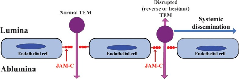
Figure 1.
rTEM of neutrophils. TEM of neutrophils, which is preferentially regulated by JAM-C expressed on junctional endothelia cells, occurred primarily through the paracellular route rather than the transendothelia route upon receiving inflammatory stimulation. Paracellular TEM of neutrophils can be divided into normal, hesitant and reverse TEM. Hesitant and reverse paracellular TEM were together named the disrupted polarized paracellular TEM by the authors; rTEM is the most severe form of disrupted polarized paracellular TEM. Importantly, neutrophil rTEM in which leukocytes migrate in an abluminal-to-luminal direction may contribute to the dissemination of inflammation to the second organ. JAM, junctional adhesion molecule; rTEM, reverse transendothelial migration; TEM, transendothelial migration.
To analyze leukocyte TEM in vivo, Woodfin et al4 built up a four-dimensional imaging system with a relatively high spatial and temporal resolution. With such a powerful imaging system in hand, they found that inflammation mainly triggered paracellular TEM, which could then be divided into normal and disrupted polarized TEM (Figure 1). They also found that disrupted TEM happened mostly under the conditions in which there was lower functional expression of JAM-C at EC junctions. More specifically, they showed that ischemia–reperfusion (I–R), an inflammatory insult that led to significantly lower expression of JAM-C by ECs, was associated with substantial disrupted polarized TEM. Thus, this work provides good evidence to show that EC JAM-C is one of the key regulators mediating the luminal-to-abluminal neutrophil paracellular TEM in vivo.
Until now, one of the main obstacles to directly visualize the migration of leukocytes with high temporal and spatial resolution was the efficient, clear labeling of the EC junction, as the traditional protocol of intravenous administration of fluorescence-labeled monoclonal antibody does not result in a reproducible labeling of ECs for tracking the movement of leukocytes in vivo, Woodfin et al4 have utilized Alexa Fluor555-conjugated monoclonal antibody 390 to platelet/endothelial cell adhesion molecule 1 to label the EC borders. The beauty of this labeling protocol is that it does not inhibit leukocyte transmigration but may result in a strong and reliable labeling of EC borders in cremasteric venules of mice. To elucidate how neutrophils undergo TEM in inflammatory conditions, the authors used proinflammatory cytokine IL-1β, chemotactic formylated tripeptide fMLP, or I–R injury to stimulate the cremaster muscles and mimic the effects of physiological and pathological results on leukocyte TEM. They found that TEM induced by these three stimuli occurred predominantly via the paracellular route rather than the transendothelia route. Notably, their findings suggest that ECs that receive inflammatory stimulation may redistribute their junctional molecules in a way that favors paracellular TEM; however, this is not an altogether surprising result as several studies2, 3, 5 have demonstrated that the paracellular route might be a preferential choice for the TEM of leukocytes.
One of the key findings in this work is that the paracellular TEM of leukocytes, which is triggered by inflammation, can be divided to three categories: (i) normal TEM, in which leukocytes migrate through ECs in a luminal-to-abluminal direction without pause; (ii) hesitant TEM, in which leukocytes show bidirectional movement in junctions with two to three oscillations in a luminal-to-abluminal direction before entering the sub-EC space; and (iii) reverse TEM (rTEM), in which leukocytes migrate in an abluminal-to-luminal direction before disengaging from the junction and crawling on the luminal surface. Because rTEM is a more severe form of hesitant TEM, they named these responses ‘disrupted polarized paracellular events' (Figure 1). It was previously thought that the transmigration of leukocytes through venules is divided into the following steps: capture, rolling, arrest, adhesion, crawling, and then paracellular or transcellular TEM3 However, very few studies, if any, have ever shown the normal, hesitant and reverse aspects of paracellular TEM of leukocytes.
While an understanding of the signaling processes that drive specific TEM of neutrophils, lymphocytes and monocytes may help identify new targets for potential therapeutic interventions, cell type-specific differences for the TEM remain to be discovered. To determine the association between inflammation and polarized paracellular events and examine the cell type-specific differences for the TEM, the authors analyzed the TEM of neutrophils and monocytes during I–R. Interestingly, they found that most neutrophils showed significant disrupted polarized TEM during I–R, whereas monocytes did not. Thus, they focused on neutrophils to study the detailed molecular mechanisms and pathophysiological roles of rTEM of neutrophils in inflammation.
Given the fact that inflammation during I–R is associated with the disrupted polarized TEM of neutrophils, the authors analyzed the expression of JAM-C during I–R to determine whether JAM-C was the true master mediator regulating the disrupted polarized neutrophil TEM during I–R. They found that I–R, not IL-1β, stimulation might induce a significantly lower expression of JAM-C at the EC junction. This finding has led to their hypothesis that EC JAM-C expression might mediate the polarized TEM of neutrophils. This hypothesis was indeed supported by their findings, which show that blocking of JAM-C at EC using monoclonal antibodies may trigger a much higher frequency of disrupted forms polarized paracellular TEM of neutrophils. It has previously been shown that JAM-C may not only mediate the migration of cells through EC junctions by providing an adhesive ligand for neutrophil Mac-1 but also regulate endothelia adherents junctions and barrier integrity.6, 7 This study therefore provides additional information to show that JAM-C may regulate the directionality of the migration of neutrophils through EC junctions in an abluminal-to-luminal direction.
The identification of rTEM, a severe form of disrupted polarized TEM mediated by JAM-C during I–R, prompted the most important question as to what the potential pathophysiological significance of disrupted polarized TEM of neutrophils was in the inflammatory response. The authors chose rTEM, the most extreme form of disrupted polarized TEM, to determine the role of disrupted polarized TEM in systemic dissemination of inflammation. They observed that neutrophils that had undergone rTEM were more responsive in terms of enhanced reactive oxygen species production, re-entered the circulation and were detected in a distant organ after local I–R injury. More importantly, they found that the presence of these cells was associated with tissue inflammation in a second organ (Figure 1). Thus, the important implication from this study is that neutrophils with rTEM potential might contribute to turning a local inflammatory response into a systemic multiorgan response.
Many biological questions remain in terms of understanding the rapid, complicated and systemic locomotion of leukocytes. Further studies will help us to understand how the immune system succeeds or fails in response to injury or infection.1, 2, 3, 5 Nevertheless, the direct visualization of neutrophils rTEM through high spatial and temporal resolution and the discovery of the correlation between rTEM and the systemic inflammatory response should enhance our understanding of the mechanisms underlying the innate immune response to infection or injury, and may shed new light on the way to discoveries of anti-inflammatory therapies.
References
- Dejana E. Endothelial cell–cell junctions: happy together. Nat Rev Mol Cell Biol. 2004;5:261–270. doi: 10.1038/nrm1357. [DOI] [PubMed] [Google Scholar]
- Ley K, Laudanna C, Cybulsky MI, Nourshargh S. Getting to the site of inflammation: the leukocyte adhesion cascade updated. Nat Rev Immunol. 2007;7:678–689. doi: 10.1038/nri2156. [DOI] [PubMed] [Google Scholar]
- Muller WA. Mechanisms of leukocyte transendothelial migration. Annu Rev Pathol. 2011;6:323–344. doi: 10.1146/annurev-pathol-011110-130224. [DOI] [PMC free article] [PubMed] [Google Scholar]
- Woodfin A, Voisin MB, Beyrau M, Colom B, Caille D, Diapouli FM, et al. The junctional adhesion molecule JAM-C regulates polarized transendothelial migration of neutrophils in vivo. Nat Immunol. 2011;12:761–769. doi: 10.1038/ni.2062. [DOI] [PMC free article] [PubMed] [Google Scholar]
- Nourshargh S, Hordijk PL, Sixt M. Breaching multiple barriers: leukocyte motility through venular walls and the interstitium. Nat Rev Mol Cell Biol. 2010;11:366–378. doi: 10.1038/nrm2889. [DOI] [PubMed] [Google Scholar]
- Santoso S, Sachs UJ, Kroll H, Linder M, Ruf A, Preissner KT, et al. The junctional adhesion molecule 3 (JAM-3) on human platelets is a counterreceptor for the leukocyte integrin Mac-1. J Exp Med. 2002;196:679–691. doi: 10.1084/jem.20020267. [DOI] [PMC free article] [PubMed] [Google Scholar]
- Chavakis T, Keiper T, Matz-Westphal R, Hersemeyer K, Sachs UJ, Nawroth PP, et al. The junctional adhesion molecule-C promotes neutrophil transendothelial migration in vitro and in vivo. J Biol Chem. 2004;279:55602–55608. doi: 10.1074/jbc.M404676200. [DOI] [PubMed] [Google Scholar]


