Abstract
AIM
To compare the anterior and posterior corneal parameters before and after collagen cross-linking therapy for keratoconus.
METHODS
Collagen cross-linking was performed in 31 eyes of 31 keratoconus patients (mean age 30.6±8.9y). Prior to treatment and an average 7mo after therapy, Scheimpflug analysis was performed using Pentacam HR. In addition to corneal thickness assessments, corneal radius, elevation, and aberrometric measurements were performed both on anterior and posterior corneal surfaces. Data obtained before and after surgery were statistically analyzed.
RESULTS
In terms of horizontal and vertical corneal radius, and central corneal thickness no deviations were observed an average 7mo after operation. Corneal higher order aberration showed no difference neither on anterior nor on posterior corneal surfaces. During follow-up period, no significant deviation was detected regarding elevation values obtained by measurement in mm units between the 3.0-8.0 mm-zones.
CONCLUSION
Corneal stabilization could be observed in terms of anterior and posterior corneal surfaces, elevation and higher order aberration values 7mo after collagen cross-linking therapy for keratoconus.
Keywords: corneal back surface, higher order aberration, elevation, collagen cross-linking, high resolution Pentacam
INTRODUCTION
The anterior and posterior surfaces jointly constitute corneal refractive power. However, corneal power is affected by the posterior corneal surface, which compensates for some 31% of anterior surface astigmatism and also highly affects corneal spherical aberration[1]-[5]. Previously, posterior corneal surfaces could be examined by Purkinje image photography, only with an accurate reading of central corneal thickness[3],[6]. With the introduction of OrbScan (Bausch & Lomb, Orbtek Inc., Salt Lake City, UT), and later Scheimpflug imaging with Pentacam (Oculus Optikgeräte GmbH, Wetzlar, Germany), more detailed information could be obtained of the posterior surface than ever before.
Corneal refractive surgeries cause a break in balance between anterior and posterior surfaces. Subsequent to corneal refractive operations, the investigation of posterior corneal changes is relevant, because our goal is to detect and prevent dreadful iatrogenic pathologic ectasia.
Our aim was to simultaneously assess anterior and posterior corneal changes (keratometric, higher order aberrations and elevation data) after collagen cross-linking therapy for keratoconus, using the high-resolution version of Pentacam.
SUBJECTS AND METHODS
Inclusion criteria for collagen cross-linking were the following: keratoconus diagnosed on the basis of the widely known criteria and the Collaborative Longitudinal Evaluation of Keratoconus Study[7]; cornea thicker than 400 µm documented by pachymetry; age 14-45y; a minimum of 1 row decline in visual acuity and/or keratometric values and corneal pachymetry in the past 3-6mo. Further conditions were: optically clear cornea and the lack of Vogt-striae or anterior stromal scarring. Exclusion criteria were dry eye syndrome encumbering the capture of Pentacam images, and pathologic eye conditions other than keratoconus.
Prior to operations, detailed ophthalmologic examinations were performed (keratorefractometry, visual acuity testing, slit-lamp examination, intraocular pressure measurement, fundus examination). Subsequently, three images were captured in all eyes preoperatively with the high-resolution version of Pentacam device (Pentacam HR, software version 1.17r139) using Scheimpflug imaging. We used the Pentacam system operating in a 25-image mode; 25 images were captured automatically within 2s. Imaging procedure was subsequently repeated in case of any capturing error occurred (blinking, lack of data, etc).
Collagen cross-linking was performed with the following details. After topical anesthesia with tetracaine hydrochloride, central corneal epithelium of 7 mm was removed, and 0.1 % riboflavin (in 20% dextrane solution) was instilled on the entire deepithelisated corneal surface 30min before irradiation, and every 2min thereafter. To keep eye moisture, physiological saline was instilled on the surface every 2min during treatment. UV-A irradiation was performed with emission at 370nm and a radiant energy of 3 mW/cm2 (=5.4 J/cm2) for 30min. Treatments were performed using the In-Pro CCL-Lix (Norderstadt, Germany) equipment. After treatment each patient was prescribed with steroid- and antibiotics eye drops and therapeutic contact lenses was given till the epithelial defect had closed. Therapeutic contact lenses were removed 5d after treatment. At an average of 7mo (range: 6.4-8.5mo) after cross-linking procedure, three images were captured again of the treated eyes using Pentacam HR.
Obtained data were calculated with the mean values of pre- and postoperative measurements. Horizontal and vertical corneal radius (Rh, Rv), central corneal thickness (CCT), total root mean square of higher- and lower order aberrations (RMS HOA, RMS LOA) up to 8th order and elevation data were measured. To assess anterior and posterior corneal shapes, elevation data were used by comparing given surface to reference surface (best-fit-sphere, BFS) in terms of micrometer. The reference BFS was determined by the central 8.0 mm-zone of the cornea (BSF setting was constant in Pentacam HR software during both preoperative and postoperative measurements). Positive elevation values were associated with ectatic deviations (forward protrusion of the posterior cornea). We used 8.0 mm pupil set in all measurements.
The method of operation was explained to all patients and informed consent was obtained. The research protocol adhered to the tenets of the Declaration of Helsinki (2008). The research protocol had been approved by the Institutional Ethic Comettee of the University of Debrecen.
Statistical analysis was performed with MedCalc 10.0. Descriptive statistical results were described as mean±standard deviation (SD). Wilcoxon paired sample test were carried out for comparison between groups or variables. A P value below 0.05 was considered statistically significant.
RESULTS
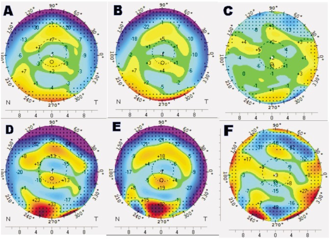
Thirty-one eyes of 31 keratoconus patients were examined (age: 30.6±8.9y; range: 17.1-45.2y). No significant, visible haze was observed in the postoperative period. Respect to horizontal and vertical corneal radius no difference was detected in both anterior and posterior surfaces. Compared to preoperative data, CCT also showed no difference at the end of the postoperative period (P=0.6). No significant changes were found in corneal HOA and LOA regarding the anterior and posterior surfaces (P=0.3-0.6). Seven months after surgery, no significant difference was detected in elevation data obtained between the 3.0-6.0 mm corneal zones (P=0.27-0.68) (Figure 1). Detailed data are shown in Table 1.
Figure 1. Anterior and posterior elevation maps of the cornea before and after collagen cross-linking (CXL) therapy obtained by Pentacam HR. We did not notice any significant changes during the examination period.
A: Elevation map of the anterior corneal surface before CXL therapy; B: Elevation map of the anterior corneal surface after CXL therapy; C: Difference elevation map of the anterior corneal surface after CXL therapy (after minus before); D: Elevation map of the posterior corneal surface before CXL therapy; E: Elevation map of the posterior corneal surface after CXL therapy; F: Difference elevation map of the posterior corneal surface after CXL therapy (after minus before).
Table 1. Anterior segment parameter changes after collagen cross-linking therapy measured by pentacam HR.
| Anterior segment parameter | Preoperative | Postoperative | P | |
| Rh front (mm) | 7.15±0.73 | 7.35±0.93 | 0.48 | |
| Rv front (mm) | 6.78±0.76 | 6.86±0.78 | 0.62 | |
| Rh back (mm) | 5.77±0.77 | 5.66±1.02 | 0.55 | |
| Rv back (mm) | 5.53±0.92 | 5.41±0.95 | 0.66 | |
| RMS HOA front (µm) | 2.22±1.63 | 2.04±1.46 | 0.65 | |
| RMS HOA back (µm) | 0.43±0.22 | 0.42±0.24 | 0.33 | |
| RMS HOA cornea (µm) | 1.87±1.5 | 1.68±1.28 | 0.77 | |
| RMS LOA front (µm) | 15.31±8.52 | 14.25±7.86 | 0.66 | |
| RMS LOA back (µm) | 3.34±1.51 | 3.4±1.5 | 0.32 | |
| RMS LOA cornea (µm) | 12.14±7.09 | 11.02±6.4 | 0.9 | |
| CCT (µm) | 441.7±67 | 444.3±56 | 0.7 | |
| anterior corneal elevation | zone 3.0 mm | 9.03±3.2 | 10.62±3.1 | 0.68 |
| zone 4.0 mm | 3.48±2.5 | 5.33±1.2 | 0.53 | |
| zone 5.0 mm | -1.61±0.9 | 0.3±0.25 | 0.35 | |
| zone 6.0 mm | -5.34±1.2 | -3.66±1.14 | 0.27 | |
| posterior corneal elevation | zone 3.0 mm | 19.34±5.6 | 23.57±3.9 | 0.51 |
| zone 4.0 mm | 6.97±3.6 | 10.3±2.9 | 0.45 | |
| zone 5.0 mm | -3.7±1.1 | -0.9±0.15 | 0.42 | |
| zone 6.0 mm | -10.9±1.5 | -8.28±2.32 | 0.45 | |
Rh: Horizontal radius of cornea; Rv: Vertical radius of cornea; RMS: Root mean square; HOA: Higher order aberration; LOA: Lower order aberration; CCT: Central corneal thickness.
DISCUSSION
Anterior and posterior corneal surfaces play an important role in determining the refractive power of the cornea. Posterior surface shape can change after keratorefractive surgeries performed on the anterior surface [8].
In 1999, Wang was the first to report a detailed analysis of the posterior corneal surface using scanning slit-beam topographer (Orbscan)[9]. His measurements were the first to prove that posterior corneal ectasia can be detected by Orbscan after LASIK treatments. Later on, further studies have been published observing corneal ectasia using Orbscan measurements[9]-[11]. Baek et al[10] described its possible explanations, namely that these deformations are affected by thinner corneas, higher intraocular pressure and higher myopia. Due to the known limitations of the device, the accuracy, the reliability and the validity of the results on posterior cornea using Orbscan have been questioned in many cases[12]-[14]. According to Nawa, true keratectasia occasionally exists after keratorefractive surgeries, but the result of posterior cornea measurements with Orbscan may be an artifact[13].
From 2005, the anterior segment of the eye could be analyzed by Pentacam; the first publication on posterior corneal surface measurement was issued in 2005; the first report analyzing posterior cornea deviations after keratorefractive surgery was published a year later[14],[15]. Based on Scheimpflug imaging, this method is suitable for measuring 25 000 true elevation points, and can detect posterior corneal elevations with mathematical reconstruction[15]. The repeatability of Pentacam in respect to the results of posterior and anterior surface measurements is good[16],[17]. At least three measurements are suggested to improve intersession repeatability, therefore, we performed three measurements in every case[17].
Investigations using Pentacam system found that, in PRK and LASIK treatments there is no posterior corneal surface displacement, though results contradicting this finding have been also published during LASIK treatments[9],[10],[15], [18]-[21]. Measuring the posterior corneal surface with Pentacam, Zhang and Wang[22] observed protrusion in the paracentral and midperipheral zones 1mo after LASIK, which decreased after 6mo, and also found deviation in the elevation in the center and paracentral zones 6mo after epi-LASIK treatment. In our study, ectatic progress was not observed at least 7mo after CXL therapy for keratoconus.
A decrease in corneal spherical aberrations can be observed with CSO EyeTop topographer after corneal cross-linking therapy, however, corneal coma showed no significant change[23]. Besides, a postoperative decrease in total HOA, total spherical aberration and total coma could be detected with Nidek OPD-Scan[23],[24]. According to the results of the Siena eye cross study, which was also using CSO EyeTop topographer, no anterior coma aberration reduction could be measured, however, there were no significant modifications of spherical aberration[25]. However, several studies using wavefront aberrometry reported no coma or spherical aberration deviations 1y after cross-linking treatment[26],[27]. Corneal aberrometric deviations measured with different devices after cross-linking show controversial results. Our results obtained with the high resolution version of Pentacam, showed no significant deviation in radius values, corneal HOA and LOA in anterior and posterior surfaces after collagen cross-linking therapy for keratoconus.
In summary, our study stated that anterior and posterior corneal surfaces show no significant deviations in respect to keratometry, corneal HOA, LOA or elevation compared to preoperative values in a follow up period of 7mo. Consequently, cross-linking treatment can stabilize not only refraction value, but also anterior and posterior corneas including corneal HOA.
Acknowledgments
Conflicts of Interest: Hassan Z, None; Modis L, None; Szalai E, None; Berta A, None; Nemeth G, None.
REFERENCES
- 1.Koch DD, Ali SF, Weikert MP, Shirayama M, Jenkins R, Wang L. Contribution of posterior corneal astigmatism to total corneal astigmatism. J Cataract Refract Surg. 2012;38(12):2080–2087. doi: 10.1016/j.jcrs.2012.08.036. [DOI] [PubMed] [Google Scholar]
- 2.Montalbán R, Piñero DP, Javaloy J, Alió JL. Scheimpflug photography-based clinical characterization of the correlation of the corneal shape between the anterior and posterior corneal surfaces in the normal human eye. J Cataract Refract Surg. 2012;38(11):1925–1933. doi: 10.1016/j.jcrs.2012.06.050. [DOI] [PubMed] [Google Scholar]
- 3.Garner LF, Owens H, Yap MK, Frith MJ, Kinnear RF. Radius of curvature of the posterior surface of the cornea. Optom Vis Sci. 1997;74(7):496–498. doi: 10.1097/00006324-199707000-00016. [DOI] [PubMed] [Google Scholar]
- 4.Dubbelman M, Sicam VA, van der Heijde GL. The shape of the anterior and posterior surface of the aging human cornea. Vision Res. 2006;46(6–7):993–1001. doi: 10.1016/j.visres.2005.09.021. [DOI] [PubMed] [Google Scholar]
- 5.Sicam VA, Dubbelman M, Van der Heijde RG. Spherical aberration of the anterior and posterior surfaces of the human cornea. J Opt Soc Am A Opt Image Sci Vis. 2006;23(3):544–549. doi: 10.1364/josaa.23.000544. [DOI] [PubMed] [Google Scholar]
- 6.Dunne MC, Royston JM, Barnes DA. Normal variations of the posterior corneal surface. Acta Ophthalmol. 1992;70(2):255–261. doi: 10.1111/j.1755-3768.1992.tb04133.x. [DOI] [PubMed] [Google Scholar]
- 7.Zadnik K, Barr JT, Gordon MO, Edrington TB. Biomicroscopic signs and disease severity in keratoconus. Collaborative Longitudinal Evaluation of Keratoconus (CLEK) Study Group. Cornea. 1996;15(2):139–146. doi: 10.1097/00003226-199603000-00006. [DOI] [PubMed] [Google Scholar]
- 8.Smadja D, Santhiago MR, Mello GR, Roberts CJ, Dupps WJ, Jr, Krueger RR. Response of the posterior corneal surface to myopic laser in situ keratomileusis with different ablation depths. J Cataract Refract Surg. 2012;38(7):1222–1231. doi: 10.1016/j.jcrs.2012.02.044. [DOI] [PubMed] [Google Scholar]
- 9.Wang Z, Chen J, Yang B. Posterior corneal surface topographic changes after laser in situ keratomileusis are related to residual corneal bed thickness. Ophthalmology. 1999;106(2):406–409. doi: 10.1016/S0161-6420(99)90083-0. [DOI] [PubMed] [Google Scholar]
- 10.Baek T, Lee K, Kagaya F, Tomidokoro A, Amano S, Oshika T. Factors affecting the forward shift of posterior corneal surface after laser in situ keratomileusis. Ophthalmology. 2001;108(2):317–320. doi: 10.1016/s0161-6420(00)00502-9. [DOI] [PubMed] [Google Scholar]
- 11.Seitz B, Langenbucher A, Torres F, Behrens A, Suárez E. Changes of posterior corneal astigmatism and tilt after myopic laser in situ keratomileusis. Cornea. 2002;21(5):441–446. doi: 10.1097/00003226-200207000-00001. [DOI] [PubMed] [Google Scholar]
- 12.Maldonado MJ, Nieto JC, Díez-Cuenca M, Piñero DP. Repeatability and reproducibility of posterior corneal curvature measurements by combined scanning-slit and placido-disc topography after LASIK. Ophthalmology. 2006;113(11):1918–1926. doi: 10.1016/j.ophtha.2006.05.053. [DOI] [PubMed] [Google Scholar]
- 13.Nawa Y, Masuda K, Ueda T, Hara Y, Uozato H. Evaluation of apparent ectasia of the posterior surface of the cornea after keratorefractive surgery. J Cataract Refract Surg. 2005;31(3):571–573. doi: 10.1016/j.jcrs.2004.05.050. [DOI] [PubMed] [Google Scholar]
- 14.Cairns G, McGhee CN. Orbscan computerized topography: attributes, applications, and limitations. J Cataract Refract Surg. 2005;31(1):205–220. doi: 10.1016/j.jcrs.2004.09.047. [DOI] [PubMed] [Google Scholar]
- 15.Ciolino JB, Belin MW. Changes in the posterior cornea after laser in situ keratomileusis and photorefractive keratectomy. J Cataract Refract Surg. 2006;32(9):1426–1431. doi: 10.1016/j.jcrs.2006.03.037. [DOI] [PubMed] [Google Scholar]
- 16.Kopacz D, Maciejewicz P, Kecik D. Pentacam--the new way for anterior eye segment imaging and mapping. Klin Oczna. 2005;107(10–12):728–731. [PubMed] [Google Scholar]
- 17.Chen D, Lam AK. Reliability and repeatability of the Pentacam on corneal curvatures. Clin Exp Optom. 2009;92(2):110–118. doi: 10.1111/j.1444-0938.2008.00336.x. [DOI] [PubMed] [Google Scholar]
- 18.Nishimura R, Negishi K, Saiki M, Arai H, Shimizu S, Toda I, Tsubota K. No forward shifting of posterior corneal surface in eyes undergoing LASIK. Ophthalmology. 2007;114(6):1104–1110. doi: 10.1016/j.ophtha.2006.09.014. [DOI] [PubMed] [Google Scholar]
- 19.Pérez-Escudero A, Dorronsoro C, Sawides L, Remón L, Merayo-Lloves J, Marcos S. Minor influence of myopic laser in situ keratomileusis on the posterior corneal surface. Invest Ophthalmol Vis Sci. 2009;50(9):4146–4154. doi: 10.1167/iovs.09-3411. [DOI] [PubMed] [Google Scholar]
- 20.Miyata K, Tokunaga T, Nakahara M, Ohtani S, Nejima R, Kiuchi T, Kaji Y, Oshika T. Residual bed thickness and corneal forward shift after laser in situ keratomileusis. J Cataract Refract Surg. 2004;30(5):1067–1072. doi: 10.1016/j.jcrs.2003.09.046. [DOI] [PubMed] [Google Scholar]
- 21.Twa MD, Roberts C, Mahmoud AM, Chang JS., Jr Response of the posterior corneal surface to laser in situ keratomileusis for myopia. J Cataract Refract Surg. 2005;31(1):61–71. doi: 10.1016/j.jcrs.2004.09.032. [DOI] [PubMed] [Google Scholar]
- 22.Zhang L, Wang Y. The shape of posterior corneal surface in normal, post-LASIK, and post-epi-LASIK eyes. Invest Ophthalmol Vis Sci. 2010;51(7):3468–3475. doi: 10.1167/iovs.09-4811. [DOI] [PubMed] [Google Scholar]
- 23.Vinciguerra P, Albè E, Trazza S, Rosetta P, Vinciguerra R, Seiler T, Epstein D. Refractive, topographic, tomographic, and aberrometric analysis of keratoconic eyes undergoing corneal cross-linking. Ophthalmology. 2009;116(3):369–378. doi: 10.1016/j.ophtha.2008.09.048. [DOI] [PubMed] [Google Scholar]
- 24.Vinciguerra P, Albè E, Trazza S, Seiler T, Epstein D. Intraoperative and postoperative effects of corneal collagen cross-linking on progressive keratoconus. Arch Ophthalmol. 2009;127(10):1258–1265. doi: 10.1001/archophthalmol.2009.205. [DOI] [PubMed] [Google Scholar]
- 25.Caporossi A, Mazzotta C, Baiocchi S, Caporossi T. Long-term results of riboflavin ultraviolet a corneal collagen cross-linking for keratoconus in Italy: the Siena eye cross study. Am J Ophthalmol. 2010;149(4):585–593. doi: 10.1016/j.ajo.2009.10.021. [DOI] [PubMed] [Google Scholar]
- 26.Vinciguerra P, Camesasca FI, Albè E, Trazza S. Corneal collagen cross-linking for ectasia after excimer laser refractive surgery: 1-year results. J Refract Surg. 2010;26(7):486–497. doi: 10.3928/1081597X-20090910-02. [DOI] [PubMed] [Google Scholar]
- 27.Doors M, Tahzib NG, Eggink FA, Berendschot TT, Webers CA, Nuijts RM. Use of anterior segment optical coherence tomography to study corneal changes after collagen cross-linking. Am J Ophthalmol. 2009;148(6):844–851. doi: 10.1016/j.ajo.2009.06.031. [DOI] [PubMed] [Google Scholar]


