Abstract
Objective
Impaired biomechanics and neuromuscular control have been suggested as probable links to female sex bias in the onset of patellofemoral pain syndrome. There are limited objective, clinical measures for assessment of impaired biomechanics and neuromuscular control. The primary objective of this investigation was to examine sex differences in vertical ground reaction force (vGRF) and force loading rate in young athletes performing maximum, repeated vertical single-leg hops (RVSHs). The authors hypothesised that females would demonstrate greater vGRF and force loading rate than males and show interlimb differences in force attenuation.
Design
Cross-sectional study.
Setting
Paediatric sports medicine clinic.
Participants
109 Healthy high school, soccer and basketball athletes.
Assessment of risk factors
Participants performed RVSHs for 15 seconds on a portable force plate with a sampling rate of 400 Hz (Accupower; AMTI, Watertown, Massachusetts, USA).
Main outcome measurements
Raw vGRF was filtered with a generalised cross-validation spline using a 50-Hz cutoff frequency and then normalised to potential energy. Force loading rate was calculated by dividing normalised vGRF by time-to-peak force. Group means were compared using analysis of variance.
Results
The females demonstrated significantly greater normalised vGRF (p<0.001) and force loading rate (p<0.001) during landing than their male counterparts. Neither sex demonstrated significant interlimb differences in force attenuation (p>0.05).
Conclusions
The female athletes may have altered force attenuation capability during RVSHs as identified by increased vGRF and force loading rate compared with the male athletes. Portable force plates may be potential tools to identify altered force attenuation in clinical settings.
Patellofemoral pain syndrome (PFPS) has been described as a peripatellar or retropatellar pain disorder of the knee.1,2 The syndrome is commonly experienced by young adult and adolescent athletes participating in running, jumping and cutting sports.3–6 Previous investigators reported that PFPS may account for 20% to 40% of all knee problems treated in sports medicine clinics.5 Other authors suggested a sex bias in the onset of PFPS, contending that adolescent females and young adult women are affected more than their male counterparts.7–9
In the conservative management of PFPS, sports medicine practitioners traditionally have based their clinical assessments on subjective measures such as patella alignment, patella excursion and patella compression tests.10,11 Commonly used static measures have been shown to have low reliability and poor association with functional abilities.12–14 Impaired biomechanics and neuromuscular control have been suggested as probable links to the pathomechanics of PFPS in young athletes.6,15–17 It is likely that altered force attenuation capabilities would predispose healthy female athletes to PFPS. However, there are limited objective, clinical measures for dynamic assessment of movement dysfunctions exemplified by athletes potentially at risk of PFPS.6,16
Measures of neuromuscular control have been quantified in biomechanics laboratories that use three-dimensional (3D) motion analysis.18,19 To promote advancement in conservative management of young athletes with PFPS, objective assessments that incorporate sports-specific mechanical loads must be made accessible to practitioners in clinical environments.20 In a previous investigation, Walsh and colleagues demonstrated that a portable force plate is both valid and reliable in measuring force and time data during a double-leg box-drop vertical jump.20 However, for more discriminating clinical assessments of interlimb and between-participant force loading differences, assessments of single-leg neuromuscular control is warranted.
Previous investigations that assessed single-leg neuromuscular control in biomechanics laboratories have used sports-specific tasks such as running, single-leg lateral landings, single-leg vertical landings, single-leg stride jumps and unanticipated cutting manoeuvres.21–25 The primary objective of this investigation was to determine whether sex differences would be observed in vertical ground reaction force (vGRF) and force loading rate in healthy, young athletes performing maximum, repeated vertical single-leg hops (RVSHs). The first hypothesis was that females would demonstrate greater vGRF and force loading rates than their male counterparts. The second hypothesis was that female athletes would exhibit interlimb differences in vGRF and loading rate.
METHODS
Participants
A total of 109 healthy, high school soccer and basketball athletes participated in the study. The participants consisted of 49 girls (mean (SD): age 15.6 (1.1) years; height 165.5 (6.1) cm; weight 60.6 (10.4) kg) and 60 boys (mean (SD): age 15.5 (1.1) years; height 175.6 (7.6) cm; weight 66.8 (11.1) kg). The participants were recruited from a county school system as part of a larger, longitudinal prospective investigation. All the participants read and signed the informed written consent approved by the Cincinnati Children’s Hospital Medical Center Institutional Review Board before participation. The athletes were included in the investigation if they participated in interscholastic soccer or basketball. They were excluded if they had any previous lower extremity orthopaedic surgeries, acute lower extremity injuries within the last 3 months, neurological problems or medical conditions that might affect postural stability.
Procedures
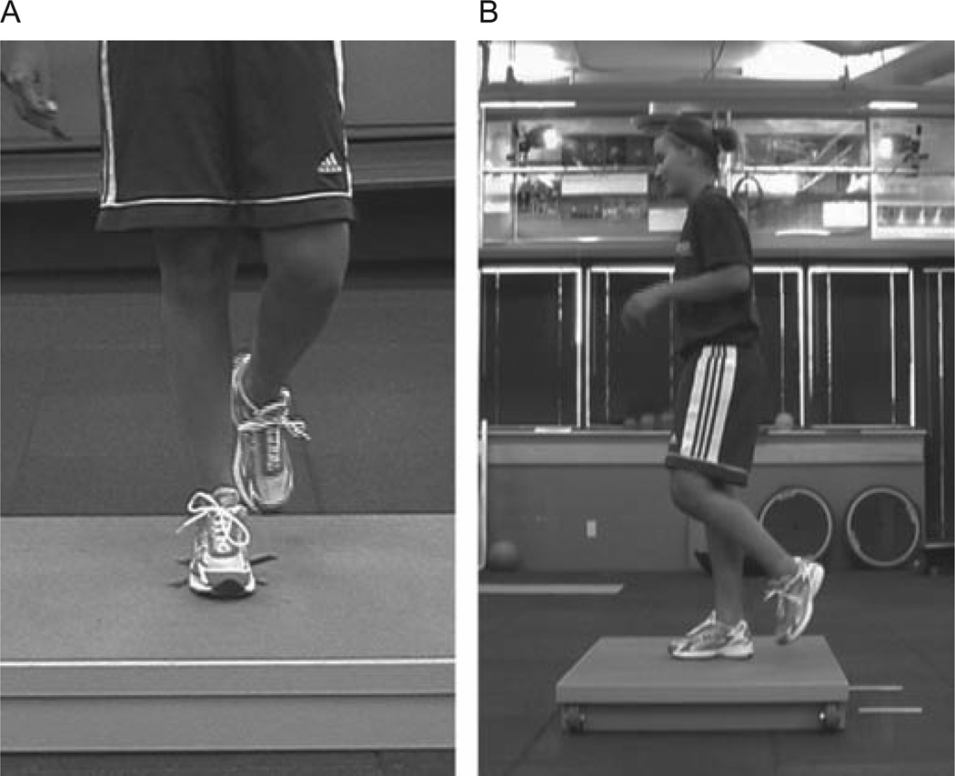
After the informed consent was obtained, height, weight and leg dominance were assessed. The investigators determined leg dominance by the leg the participants reported would be used to kick a ball as far as possible.26 vGRF was assessed using a portable force plate (Accupower; AMTI, Watertown, Massachusetts, USA) with dimensions of 76 × 102 × 12 cm (length × width × height) and sampled at 400 Hz as described by previous investigators.20 For RVSHs, the athletes were instructed to jump as high as they could, land under control, recover their balance and repeat vertical hopping for 15 seconds (fig 1). A counterbalanced testing order was used to eliminate any potential order effect, and each athlete was given three practice trials before data acquisition. One successful test trial was recorded for the dominant and non-dominant lower extremity. Faulty test trials were characterised by the athlete placing the contralateral foot onto the force plate and/ or landing outside the dimensions of the force plate. If a faulty trial was performed, the trial was stopped and then discarded. After full recovery, the athlete was instructed to repeat the test trial for that limb.
Figure 1.
A, Frontal plane view of single-leg hop starting position. B, Sagittal plane view of single-leg hop starting position.
Outcome measures
The primary dependent variables of this investigation included vGRF and force loading rate. The secondary dependent variable was potential energy (PE).25 PE was operationally defined as energy derived from the product of the mass of the participant (in kilograms), the gravitational acceleration of the earth (9.8 m/s2) and the single-leg jump height (in meters).25 Normalised vGRF was expressed as maximum vGRF (N) divided by PE (J), and normalised force loading rate was expressed as normalised vGRF (N/J) divided by the time-topeak landing force (in seconds).
Analysis
The data collected successfully from 109 participants (49 girls and 60 boys) were reduced and analysed. vGRF was analysed during the landing phase of stance. The participants were considered to be in stance phase when the vGRF exceeded a threshold of 10 N and ended when the force decreased below the 10 N threshold.26,27 Force peaks were selected from a time frame associated with the first 250 milliseconds of stance. Force peaks that exceeded 70% of the maximum vGRF peak were included in data analysis. Raw data was filtered with a generalised cross-validation spline using a 50 Hz cutoff frequency. vGRF was normalised to PE (mass × 9.8 m/s2 × height). Force loading rate was calculated by dividing the normalised vGRF by the time-to-peak force. Group means were compared using analysis of variance. The α level of 0.05 was established a priori to determine statistical significance. Statistical analyses were conducted in SPSS V.12.0 for Windows (SPSS Inc, Chicago, Illinois, USA).
RESULTS
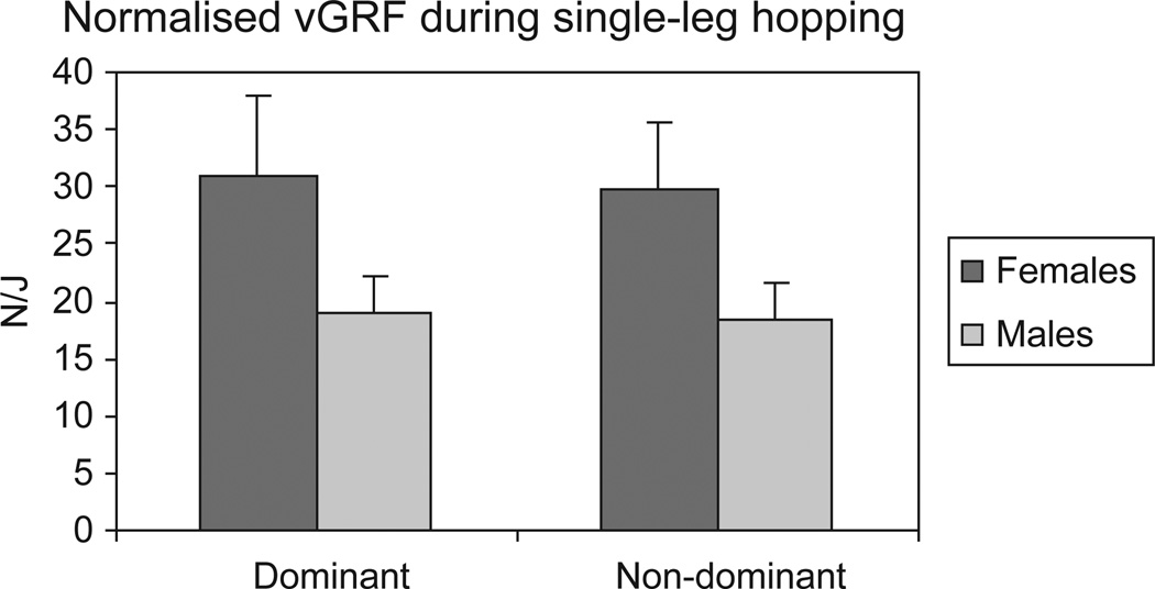
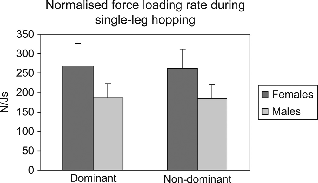
The within-session intraclass reliability of all the single-leg force production and force attenuation measures was ≥0.97 across the test trials, which indicated high test–retest reliability (p<0.05). Sex differences were observed in single-leg force production and force attenuation measures. The females demonstrated significantly greater normalised vGRF than the males on their dominant and non-dominant limbs (p<0.001; see fig 2). In addition, the females had significantly greater force loading rates than their male counterparts (p<0.001; fig. 3).
Figure 2.
Normalised vGRF during single-leg hopping.
Figure 3.
Normalised force loading rate during single-leg hopping.
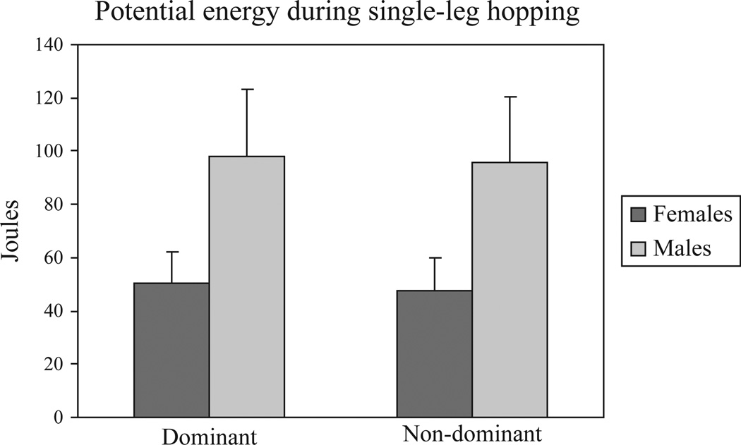
The male athletes demonstrated significantly greater PE than their female counterparts on both dominant and non-dominant limbs (p<0.001; fig 4). However, there were no significant interlimb differences in normalised vGRF (p=0.35) or force loading rates (p=0.46) for either sex.
Figure 4.
PE during single-leg hopping.
DISCUSSION
Conservative management of PFPS has long been considered an enigma for clinicians in sports medicine environments. The lack of reliable and valid measurements has perpetuated the use of anecdotal interventions among sports medicine practitioners. Several investigators have suggested that abnormal mechanical loading of the lower kinetic chain is associated with the onset of PFPS in recreational athletes.15,16,18,24
The primary objective of the current investigation was to determine whether sex differences exist in vGRF and force loading rates in healthy, young athletes performing RVSHs on a portable force plate. We hypothesised that females would demonstrate greater normalised vGRF and force loading rate than their male counterparts and would demonstrate interlimb differences in kinetic measures. This hypothesis was developed based on the available literature that suggests that adolescent girls and young adult women have a higher incidence of PFPS than their male counterparts.7–9 The results illustrated that females demonstrate significantly impaired force attenuation capability than their male counterparts during RVSHs. The females demonstrated greater landing forces than males despite the greater PE exhibited by the male participants. Contrary to the secondary hypothesis, the current investigation failed to demonstrate significant interlimb differences in kinetic measures for either sex.
It was expected that mass and vertical hop height would be greater in males compared with that in their female counterparts.28 Intuitively, an assumption of absolute Newtonian values of vGRF would be greater in male athletes compared with that in females. Use of a PE normalisation procedure that considers mass, acceleration due to gravity and vertical single-leg hop height presents a method to compare landing forces between sexes. Similar to previous investigations, it was discovered that females only exhibited 50% as much PE than males during a single-leg hop manoeuvre.29 Despite possessing significantly less PE, the female athletes demonstrated 1.5 times greater vGRF than males during the landing phase of an RVSH. These findings likely indicate that the neuromuscular strategies used by female athletes during landing from a single-leg hop may be inefficient in attenuating high-impact forces.
Previous investigations have used 3D motion analysis during double-leg landings to assess sex differences in force attenuation among young athletes.27,30,31 Kernozek and colleagues demonstrated that recreational female athletes exhibit greater normalised peak vGRF and posterior GRF than males during 60 cm drops from an adjustable hang bar.30 Salci and colleagues reported that female volleyball players applied significantly greater vGRF than males in simulated spike and block landings.31 It is probable that altered neuromuscular control observed in athletes participating in running and jumping sports may be contributory to the onset of PFPS. Specifically, high vGRF peaks during the landing phase of sports-specific manoeuvres may indicate the presence of high internal loads capable of causing injury to articular and periarticular tissues if not sufficiently attenuated by neuromuscular mechanisms.
In the current investigation, sex differences in force attenuation were assessed using a portable force plate designed for accessibility to practitioners in clinical environments. Similar to the current investigation, Hewett and colleagues used a large, portable force plate (Accupower; AMTI) to assess landing force and power production.27 The investigators reported that adolescent female athletes were unable to generate power and attenuate forces as effectively as their male counterparts during box-drop vertical jumps.27 They concluded that female athletes may exhibit potentially high-risk landing profiles when performing sports-specific manoeuvres during all stages of pubertal development.27 In an assessment of single-leg landing performance of college students, Wikstrom and colleagues found that females landed with significantly greater normalised vGRF than males.25 Force attenuation impairments exhibited by females during both double- and single-leg landings may indicate neuromuscular links to the pathomechanics of PFPS among young athletes.
Neuromuscular deficits assessed in adolescent athletes have been linked to decreased dynamic knee stability and increased risk for knee injury.26,24–32 Repetitive high-impact forces ineffectively attenuated during landings may compromise lower extremity articular structures and contribute to overuse injuries such as PFPS.35 Objective assessment of aberrant neuromuscular strategies that potentially lead to abnormal loading of the patellofemoral joint could establish a foundation for efficacious intervention programmes for young athletes. Development of new dynamic clinical tools is warranted for overall advancement in conservative management of athletes with PFPS. Using portable force plates to assess high-force landing profiles in healthy and rehabilitated athletes may prove beneficial in reducing anecdotal interventions by targeting pertinent modifiable factors.
A previous investigation conducted by Quatman and colleagues provided evidence for sex differences in force loading rates during a dynamic sports manoeuvre.36 Quatman and colleagues used 3D motion analysis to assess landing force and vertical jump performance in healthy pubertal and postpubertal athletes performing a box-drop vertical jump.36 Their findings showed that females had significantly greater force loading rates than their male counterparts. A similar sex difference in force loading rate was found using RVSHs in the current investigation. It has been suggested that excessive mechanical loading experienced by human structures such as peripatellar soft tissues can be attained by an abnormal magnitude of stress, normal stress in an abnormal direction or normal stress over an abnormal duration of time.37 It is plausible that the pathomechanics of PFPS may include repetitive, abnormal load distribution about the synovium, infrapatellar fat pad, retinacula and subchondral bone. Dye and Vaupel suggested that altered load shifting produces changes in the mechanical and chemical environments of the knee. Subsequently, these changes may trigger nocioceptive activity that leads to patellofemoral pain in young athletes.38
Other investigators have reported results contrary to the findings of the current investigation.28,39 Swartz and colleagues failed to find sex disparities in kinetic variables in prepubescent and postpubescent athletes.28 It is plausible that the differences in kinetic findings between Swartz and colleagues and the current investigators can be explained by assessment of differences in testing procedures and data reduction. Swartz and colleagues instructed participants to jump for a suspended target set at only 50% of their maximum vertical jump using a single-leg landing strategy.28 In the current investigation, kinetic variables were assessed while participants performed maximum effort hops. Objective testing that does not incorporate maximum effort levels may not be discriminative for assessment of force attenuation deficits during the landing phase of vertical hopping.
Decker and colleagues assessed sex differences in lower extremity kinematics, kinetics and energy absorption during drop landings in recreational athletes.39 They found no significant difference in vGRF during the landing phase. It is possible that the conflicting results observed between Decker and colleagues and the current investigation can be partially explained by participant recruitment differences between the investigations. Decker and colleagues assessed adult participants with a mean age of 27.3 years compared with adolescent athletes with a mean age of 15.5 years assessed in the current investigation.39 Previous investigators have suggested that male and females diverge in neuromuscular performance and injury rate after peak height velocity during maturation.40 Increases in skeletal long bone length and body mass potentially create greater joint forces at the knee and greater challenges to neuromuscular control during dynamic tasks.36,40 There is evidence that females exhibit decreased neuromuscular control at the knee from early to late puberty while males exhibit relatively increased neuromuscular control from early to late puberty.36,41 Sex differences in force attenuation may be apparent in the current investigation because of the effects of a sensitive period for neuromuscular development in the recruited adolescent participants.
Limitations to the current investigation include the lack of verification for maximum hop effort during RVSHs. This limitation was partially minimised by continuous encouragement provided to the participants during the testing and further minimised by the reduction and the analysis of force peaks that achieved at least 70% of the maximum vGRF peak in the 15-second trial. In addition, no clear criterion standard for normalisation of vGRF currently exists. Previous investigations assessing vGRF of participants during landings have generally used participant mass in normalisation procedures.21,23,39,42,43 Because of sex differences in vertical jump height and the variability of jump height between repetitions, the current investigation selected PE as the preferred normalisation factor for sex comparisons.
CONCLUSIONS
The results of this investigation indicate that healthy female athletes may have altered force attenuation capability as evidenced by increased vGRF and force loading rate compared with their male counterparts during single-leg landing. Decreased force attenuation may contribute to the onset of repetitive use injuries such as PFPS in young athletes participating in running and jumping sports. Portable force plates may be a potential tool to identify altered force attenuation in young athletes in clinical settings. Athletes identified with high-force landing profiles through accessible screening tools should be selected for specific neuromuscular training to reduce neuromuscular control deficits and to decrease potential risks for the onset of PFPS. Future investigations should combine portable force plates and 2D motion analysis systems for identification of kinetic and kinematic deficits in young athletes with PFPS during clinical preparticipation assessments.
Acknowledgements
The authors acknowledge the funding support from the National Institutes of Health grants 5R01-AR049735-04 and 5R01-AR049735-05 (TEH). The authors thank Randy Poe and all of the Boone County School Board members for cooperating in the development of this study.
Funding This study was funded by the National Institutes of Health.
Footnotes
Competing interest None.
Patient consent Obtained.
Ethics approval This study was conducted with the approval of the institutional review board committee.
Provenance and peer review Not commissioned; externally peer reviewed.
REFERENCES
- 1.Salsich GB, Brechter JH, Farwell D, et al. The effects of patellar taping on knee kinetics, kinematics, and vastus lateralis muscle activity during stair ambulation in individuals with patellofemoral pain. J Orthop Sports Phys Ther. 2002;32:3–10. doi: 10.2519/jospt.2002.32.1.3. [DOI] [PubMed] [Google Scholar]
- 2.Witvrouw E, Lysens R, Bellemans J, et al. Which factors predict outcome in the treatment program of anterior knee pain? Scand J Med Sci Sports. 2002;12:40–46. doi: 10.1034/j.1600-0838.2002.120108.x. [DOI] [PubMed] [Google Scholar]
- 3.Heintjes E, Berger M, Bierma-Zeinstra S, et al. Exercise therapy for patellofemoral pain syndrome. Hobken: John Wiley & Sons, Ltd; 2005. [Google Scholar]
- 4.Louden JK, Gajewski B, Goist-Foley HL, et al. The effectiveness of exercise in treating patellofemoral-pain syndrome. J Sport Rehab. 2004;13:323–342. [Google Scholar]
- 5.Natri A, Kannus P, Järvinen M. Which factors predict the long-term outcome in chronic patellofemoral pain syndrome? A 7-yr prospective follow-up study. Med Sci Sports Exerc. 1998;30:1572–1577. doi: 10.1097/00005768-199811000-00003. [DOI] [PubMed] [Google Scholar]
- 6.Witvrouw E, Lysens R, Bellemans J, et al. Intrinsic risk factors for the development of anterior knee pain in an athletic population. A two-year prospective study. Am J Sports Med. 2000;28:480–489. doi: 10.1177/03635465000280040701. [DOI] [PubMed] [Google Scholar]
- 7.Fulkerson JP. Diagnosis and treatment of patients with patellofemoral pain. Am J Sports Med. 2002;30:447–456. doi: 10.1177/03635465020300032501. [DOI] [PubMed] [Google Scholar]
- 8.Fulkerson JP, Arendt EA. Anterior knee pain in females. Clin Orthop Relat Res. 2000:69–73. doi: 10.1097/00003086-200003000-00009. [DOI] [PubMed] [Google Scholar]
- 9.Robinson RL, Nee RJ. Analysis of hip strength in females seeking physical therapy treatment for unilateral patellofemoral pain syndrome. J Orthop Sports Phys Ther. 2007;37:232–238. doi: 10.2519/jospt.2007.2439. [DOI] [PubMed] [Google Scholar]
- 10.Fredericson M, Yoon K. Physical examination and patellofemoral pain syndrome. Am J Phys Med Rehabil. 2006;85:234–243. doi: 10.1097/01.phm.0000200390.67408.f0. [DOI] [PubMed] [Google Scholar]
- 11.Wilson T. The measurement of patellar alignment in patellofemoral pain syndrome: are we confusing assumptions with evidence? J Orthop Sports Phys Ther. 2007;37:330–341. doi: 10.2519/jospt.2007.2281. [DOI] [PubMed] [Google Scholar]
- 12.Kannus P, Niittymäki S. Which factors predict outcome in the nonoperative treatment of patellofemoral pain syndrome? A prospective follow-up study. Med Sci Sports Exerc. 1994;26:289–296. [PubMed] [Google Scholar]
- 13.Loudon JK, Wiesner D, Goist-Foley HL, et al. Intrarater reliability of functional performance tests for subjects with patellofemoral pain syndrome. J Athl Train. 2002;37:256–261. [PMC free article] [PubMed] [Google Scholar]
- 14.Piva SR, Fitzgerald K, Irrgang JJ, et al. Reliability of measures of impairments associated with patellofemoral pain syndrome. BMC Musculoskelet Disord. 2006;7:33. doi: 10.1186/1471-2474-7-33. [DOI] [PMC free article] [PubMed] [Google Scholar]
- 15.Duffey MJ, Martin DF, Cannon DW, et al. Etiologic factors associated with anterior knee pain in distance runners. Med Sci Sports Exerc. 2000;32:1825–1832. doi: 10.1097/00005768-200011000-00003. [DOI] [PubMed] [Google Scholar]
- 16.Messier SP, Davis SE, Curl WW, et al. Etiologic factors associated with patellofemoral pain in runners. Med Sci Sports Exerc. 1991;23:1008–1015. [PubMed] [Google Scholar]
- 17.Powers CM. The influence of altered lower-extremity kinematics on patellofemoral joint dysfunction: a theoretical perspective. J Orthop Sports Phys Ther. 2003;33:639–646. doi: 10.2519/jospt.2003.33.11.639. [DOI] [PubMed] [Google Scholar]
- 18.Powers CM, Heino JG, Rao S, et al. The influence of patellofemoral pain on lower limb loading during gait. Clin Biomech (Bristol, Avon) 1999;14:722–728. doi: 10.1016/s0268-0033(99)00019-4. [DOI] [PubMed] [Google Scholar]
- 19.Powers CM, Maffucci R, Hampton S. Rearfoot posture in subjects with patellofemoral pain. J Orthop Sports Phys Ther. 1995;22:155–160. doi: 10.2519/jospt.1995.22.4.155. [DOI] [PubMed] [Google Scholar]
- 20.Walsh MS, Ford KR, Bangen KJ, et al. The validation of a portable force plate for measuring force-time data during jumping and landing tasks. J Strength Cond Res. 2006;20:730–734. doi: 10.1519/R-18225.1. [DOI] [PubMed] [Google Scholar]
- 21.Coventry E, O’Connor KM, Hart BA, et al. The effect of lower extremity fatigue on shock attenuation during single-leg landing. Clin Biomech (Bristol, Avon) 2006;21:1090–1097. doi: 10.1016/j.clinbiomech.2006.07.004. [DOI] [PubMed] [Google Scholar]
- 22.Flynn TW, Soutas-Little RW. Patellofemoral joint compressive forces in forward and backward running. J Orthop Sports Phys Ther. 1995;21:277–282. doi: 10.2519/jospt.1995.21.5.277. [DOI] [PubMed] [Google Scholar]
- 23.Madigan ML, Pidcoe PE. Changes in landing biomechanics during a fatiguing landing activity. J Electromyogr Kinesiol. 2003;13:491–498. doi: 10.1016/s1050-6411(03)00037-3. [DOI] [PubMed] [Google Scholar]
- 24.Stefanyshyn DJ, Stergiou P, Lun VM, et al. Knee angular impulse as a predictor of patellofemoral pain in runners. Am J Sports Med. 2006;34:1844–1851. doi: 10.1177/0363546506288753. [DOI] [PubMed] [Google Scholar]
- 25.Wikstrom EA, Tillman MD, Kline KJ, et al. Gender and limb differences in dynamic postural stability during landing. Clin J Sport Med. 2006;16:311–315. doi: 10.1097/00042752-200607000-00005. [DOI] [PubMed] [Google Scholar]
- 26.Ford KR, Myer GD, Smith RL, et al. A comparison of dynamic coronal plane excursion between matched male and female athletes when performing single leg landings. Clin Biomech (Bristol, Avon) 2006;21:33–40. doi: 10.1016/j.clinbiomech.2005.08.010. [DOI] [PubMed] [Google Scholar]
- 27.Hewett TE, Ford KR, Myer GD, et al. Gender differences in hip adduction motion and torque during a single-leg agility maneuver. J Orthop Res. 2006;24:416–421. doi: 10.1002/jor.20056. [DOI] [PubMed] [Google Scholar]
- 28.Swartz EE, Decoster LC, Russell PJ, et al. Effects of developmental stage and sex on lower extremity kinematics and vertical ground reaction forces during landing. J Athl Train. 2005;40:9–14. [PMC free article] [PubMed] [Google Scholar]
- 29.Wikstrom EA, Tillman MD, Smith AN, et al. A new force-plate technology measure of dynamic postural stability: the dynamic postural stability index. J Athl Train. 2005;40:305–309. [PMC free article] [PubMed] [Google Scholar]
- 30.Kernozek TW, Torry MR, Van Hoof H, et al. Gender differences in frontal and sagittal plane biomechanics during drop landings. Med Sci Sports Exerc. 2005;37:1003–1012. discussion 1013. [PubMed] [Google Scholar]
- 31.Salci Y, Kentel BB, Heycan C, et al. Comparison of landing maneuvers between male and female college volleyball players. Clin Biomech. 2004;19:622–628. doi: 10.1016/j.clinbiomech.2004.03.006. [DOI] [PubMed] [Google Scholar]
- 32.Ford KR, Myer GD, Hewett TE. Valgus knee motion during landing in high school female and male basketball players. Med Sci Sports Exerc. 2003;35:1745–1750. doi: 10.1249/01.MSS.0000089346.85744.D9. [DOI] [PubMed] [Google Scholar]
- 33.Ford KR, Myer GD, Toms HE, et al. Gender differences in the kinematics of unanticipated cutting in young athletes. Med Sci Sports Exerc. 2005;37:124–129. [PubMed] [Google Scholar]
- 34.Hewett TE, Myer GD, Ford KR, et al. Biomechanical measures of neuromuscular control and valgus loading of the knee predict anterior cruciate ligament injury risk in female athletes: a prospective study. Am J Sports Med. 2005;33:492–501. doi: 10.1177/0363546504269591. [DOI] [PubMed] [Google Scholar]
- 35.Zhang SN, Bates BT, Dufek JS. Contributions of lower extremity joints to energy dissipation during landings. Med Sci Sports Exerc. 1998:812–819. doi: 10.1097/00005768-200004000-00014. [DOI] [PubMed] [Google Scholar]
- 36.Quatman CE, Ford KR, Myer GD, et al. Maturation leads to gender differences in landing force and vertical jump performance: a longitudinal study. Am J Sports Med. 2006;34:806–813. doi: 10.1177/0363546505281916. [DOI] [PubMed] [Google Scholar]
- 37.Norkin CC, Levangie PK. Joint structure and function: a comprehensive analysis. 2nd edn. Philadelphia: FA. Davis Company; 1998. [Google Scholar]
- 38.Dye SF, Vaupel GL. The pathophysiology of patellofemoral pain. Sports Med Arthrosc Rev. 1994;2:203–210. [Google Scholar]
- 39.Decker MJ, Torry MR, Wyland DJ, et al. Gender differences in lower extremity kinematics, kinetics and energy absorption during landing. Clin Biomech (Bristol, Avon) 2003;18:662–669. doi: 10.1016/s0268-0033(03)00090-1. [DOI] [PubMed] [Google Scholar]
- 40.Hewett TE, Myer GD, Ford KR. Decrease in neuromuscular control about the knee with maturation in female athletes. J Bone Joint Surg Am. 2004;86-A:1601–1608. doi: 10.2106/00004623-200408000-00001. [DOI] [PubMed] [Google Scholar]
- 41.Hewett TE, Myer GD, Ford KR, et al. Preparticipation physical examination using a box drop vertical jump test in young athletes: the effects of puberty and sex. Clin J Sport Med. 2006;16:298–304. doi: 10.1097/00042752-200607000-00003. [DOI] [PubMed] [Google Scholar]
- 42.Pittenger VM, McCaw ST, Thomas DQ. Vertical ground reaction forces of children during one- and two-leg rope jumping. Res Q Exerc Sport. 2002;73:445–449. doi: 10.1080/02701367.2002.10609044. [DOI] [PubMed] [Google Scholar]
- 43.Zheng N, Barrentine SW. Biomechanics and motion analysis applied to sports. Phys Med Rehabil Clin N Am. 2000;11:309–322. [PubMed] [Google Scholar]


