Abstract
We developed a rational approach to identify a site in the vesicular stomatitis virus (VSV) glycoprotein (G) that is exposed on the protein surface and tolerant of foreign epitope insertion. The foreign epitope inserted was the six-amino-acid sequence ELDKWA, a sequence in a neutralizing epitope from human immunodeficiency virus type 1. This sequence was inserted into six sites within the VSV G protein (Indiana serotype). Four sites were selected based on hydrophilicity and high sequence variability identified by sequence comparison with other vesiculovirus G proteins. The site showing the highest variability was fully tolerant of the foreign peptide insertion. G protein containing the insertion at this site folded correctly, was transported normally to the cell surface, had normal membrane fusion activity, and could reconstitute fully infectious VSV. The virus was neutralized by the human 2F5 monoclonal antibody that binds the ELDKWA epitope. Additional studies showed that this site in G protein tolerated insertion of at least 16 amino acids while retaining full infectivity. The three other insertions in somewhat less variable sequences interfered with VSV G folding and transport to the cell surface. Two additional insertions were made in a conserved sequence adjacent to a glycosylation site and near the transmembrane domain. The former blocked G-protein transport, while the latter allowed transport to the cell surface but blocked membrane fusion activity of G protein. Identification of an insertion-tolerant site in VSV G could be important in future vaccine and targeting studies, and the general principle might also be useful in other systems.
Vesicular stomatitis virus (VSV) is a negative-strand RNA virus and is the prototype of the rhabdovirus family. VSV has an extremely broad tropism, possibly reflecting the use of the ubiquitous molecule phosphatidylserine in virus entry into cells (36). VSV infection is mediated by the binding and membrane fusion activity of the single transmembrane glycoprotein (G) (9, 29). Despite extensive use of VSV recombinants as vectors in vaccine studies (16, 28, 30, 34, 35) and VSV G for pseudotyping of other viruses (8, 39), there is relatively little information on the structure of G protein. VSV G is known to form trimers prior to transport from the endoplasmic reticulum (ER) (5, 18), but the three-dimensional structure of the molecule has not been determined. A G domain involved in membrane fusion has been identified (6, 10, 22, 42), although there is little information on how this domain functions. Because of the limited information on G-protein structure, it is difficult to predict exposed sites on the protein surface that might tolerate foreign epitope insertion.
The mature VSV G (serotype Indiana) has three domains: a 446-amino-acid ectodomain, a 20-amino-acid transmembrane domain, and a 29-amino-acid cytoplasmic tail. After VSV G-mediated binding of virus to cells, VSV is endocytosed. G protein then mediates membrane fusion at low pH to release the nucleocapsid from the endosome into the cytoplasm (9, 14, 29). The genome encased in nucleocapsid protein (N) is the template for transcription by the RNA-dependent RNA polymerase present in the virion (2, 33). Five mRNAs encoding the five structural proteins (N, P, M, G, and L) are synthesized by this polymerase. G protein is cotranslationally inserted into the membrane of the ER and glycosylated at two sites (33). G-protein monomers are assembled into trimers in the ER (5, 18) and are then transported to the Golgi bodies where the glycans are processed to the complex type (5). G is then transported to the plasma membrane where it assembles into budding virions.
VSV G forms a dense coat on the virus membrane. It has been suggested that the dense paracrystalline organization of G on the virus particle results in the strong T-cell-independent antibody response to G protein after VSV infection (1). Because of the vigorous immune response to VSV G, it might be an ideal platform on which to display foreign epitopes. However, an earlier study showed that insertion of even two- or three-amino-acid sequences at random sites in the G ectodomain interfered with folding and transport of the protein (22). Only two insertion sites in G, one within the signal sequence (which is removed from the protein cotranslationally), and one between the ectomembrane and transmembrane domains appeared to allow correct folding and transport of G protein. In this study, we undertook a rational approach to locate a permissive site within VSV G for foreign epitope display. We used sequence comparison of G proteins from five vesiculoviruses (the virus family which includes VSV) to locate potential epitope insertion sites that were variable in sequence and likely to be exposed on G protein.
We chose to insert the six-amino-acid sequence ELDKWA into each one of the six potential epitope display sites in VSV G. The ELDKWA sequence is within the epitope recognized by the human monoclonal antibody (MAb) 2F5 on the gp41 subunit of the human immunodeficiency virus type 1 (HIV-1) envelope protein. This B-cell epitope is present in 72% of HIV-1 isolates (26), with 82% containing the core LDKW sequence (7). Although HIV antibody escape mutants are generated rapidly, this epitope is relatively conserved, suggesting an important role in HIV envelope protein function. Thus, this epitope could be a good candidate for inclusion in an HIV-1 subunit vaccine. Previously, the ELDKWA epitope was inserted into a permissive site on the influenza virus hemagglutinin. Virus expressing this hemagglutinin-ELDKWA protein elicited HIV-neutralizing antibodies (25). However, antibodies neutralizing primary HIV isolates were not seen consistently (24). Because of previous failures to elicit an ELDKWA-specific antibody response that neutralizes primary HIV isolates, it was suspected that the 2F5 epitope (2F5e) was more complex than just the six-amino-acid ELDKWA sequence. Recent studies suggest that the 2F5 epitope may encompass as many as 16 amino acids with ELDKWA as the core sequence (27). Thus, the ability of G to tolerate this larger 16-amino-acid insertion was determined.
Neutralization of HIV by antibodies is the only method of achieving sterilizing protection to HIV. Thus, development of vectors displaying the 2F5 epitope could be important to HIV vaccine design. In addition, identification of permissive insertion sites in VSV G could allow insertions of specific targeting sequences. An insertion-tolerant site has been identified in the H protein of measles virus and has been used to modify virus tropism (13).
MATERIALS AND METHODS
Alignment of G proteins.
The Megalign program from the DNAstar software package was used to align the amino acid sequences of G proteins of the Indiana, New Jersey, Chandipura, Cocal, and Piry viruses (3, 4, 12, 23, 32). The Jotun Hein algorithm with default settings was employed.
Construction of G-ELDKWA and G-2F5e plasmids.
DNA encoding VSV G (32) was amplified by PCR using VENT polymerase (New England Biolabs), a forward primer that includes an MluI site (underlined) (5′-TAACAGAGAT CGATCTGTT TACGCGT-3′), and a reverse primer including SacI and NheI sites (underlined) (5′-GGATCCGAG CTCGCTAGCA GGATTTGAG TTACTTTCC-3′). The resulting fragment was digested with MluI and SacI and ligated between the MluI and SacI sites of a modified pBSSK+ vector.
The 18-nucleotide DNA sequence encoding the ELDKWA epitope was inserted into each of six sites using the QuikChange site-directed mutagenesis kit (Stratagene). Each insertion was made with complementary forward and reverse primers. The sequences of the six forward primers are listed in Table 1 with the nucleotides encoding ELDKWA in boldface type and underlined. Table 1 also shows the two amino acids in VSV G which are present on either side of the ELDKWA insertion. Complete nucleotide sequences of the G genes were verified.
TABLE 1.
ELDKWA insertion sites within VSV G protein
| VSV G amino acid position | VSV G sequence at insertion site | Oligonucleotide sequence (5′ to 3′)a |
|---|---|---|
| 52 | D-ELDKWA-L | GCTCAGATTTAAATTGGCATAATGACGAATTAGATAAGTGGGCTTTAATAGGCACAGCCATACAAGTC |
| 182 | T-ELDKWA-T | GCCCCACTGTCCATAACTCTACAGAATTAGATAAGTGGGCTACCTGGCATTCTGACTATAAGGTCT |
| 191 | K-ELDKWA-G | CATTCTGACTATAAGGTCAAAGAATTAGATAAGTGGGCTGGGCTATGTGATTCTAACCTCATTTCC |
| 266 | R-ELDKWA-F | GATAAGGATCTCTTTGCTGCAGCCAGAGAATTAGATAAGTGGGCTTTCCCTGAATGCCCAGAAGGGTCAAG |
| 366 | G-ELDKWA-T | CTCTCAAGAATGGTCGGAATGATCAGTGGAGAATTAGATAAGTGGGCTACTACCACAGAAAGGGAACTGTGGGATG |
| 464 | S-ELDKWA-S | GGTTGGTTCAGTAGTTGGAAAAGCGAATTAGATAAGTGGGCTTCTATTGCCTCTTTTTTCTTTATCATAGGG |
Nucleotides encoding ELDKWA are shown in boldface type and underlined.
To generate a full-length VSV clone encoding G with an ELDKWA insertion at position 191, the pBlueScript plasmid encoding 191 ELDKWA was digested with MluI and NheI. The pVSVXN2 plasmid that encodes the entire VSV genome (38) was digested with MluI and NheI to release the fragment coding for wild-type (wt) G, and the 191 ELDKWA insert was cloned in its place. The resulting plasmid was designated pVSV191ELDKWA.
To generate a VSV G containing the 16-amino-acid 2F5 epitope, 48 nucleotides were inserted into the VSV G gene using the QuikChange site-directed mutagenesis kit as described above, but using complementary forward and reverse primers (99-mers). The forward primer sequence with the nucleotides that encode the 2F5 epitope underlined was 5′-CTGGCATTCT GACTATAAGG TCAAAAATGA ACAAGAACTT CTCGAGCTTG ATAAGTGGGC TTCTCTTAAT TGGGGGCTAT GTGATTCTAA CCTCATTTC-3′. The gene was inserted into the VSV genome as described above for the smaller insertion mutants.
Metabolic labeling of G-ELDKWA proteins.
Baby hamster kidney cells (BHK-21) were maintained in Dulbecco's modified Eagle's medium (DMEM) supplemented with 5% fetal bovine serum (FBS). Cells on 6.0-cm-diameter plates were infected with vTF7-3 at a multiplicity of infection of 10. After 30 min, the pBSG-ELDKWA plasmids were transfected into the cells with a cationic liposome based on dimethyl-dioctadecyl ammonium bromide (31). After 4 h, the medium was removed, and cells were washed twice with prewarmed methionine-free DMEM. Methionine-free DMEM (2 ml) containing 200 μCi of [35S]methionine was added to each plate for 1 h at 37°C. To prepare extracts, the medium was removed, the cells were washed twice with phosphate-buffered saline and then lysed with 0.5 ml of detergent solution (1% Nonidet P-40, 0.4% deoxycholate, 50 mM Tris-HCl [pH 8], 62.5 mM EDTA) on ice for 5 min. Each cell lysate was collected and centrifuged for 2 min at 14,000 rpm (17,530 × g) in a microcentrifuge (Eppendorf).
Immunoprecipitation of G-ELDKWA proteins.
After the cells were lysed, the samples were split into thirds. To each sample, sodium dodecyl sulfate (SDS) was added to a final concentration of 0.2% for precipitation with rabbit polyclonal antibody to VSV or a final concentration of 0.1% for MAb I1 or I14 (20, 21). Samples were precleared by incubation with fixed Staphylococcus aureus bacteria for 30 min at 37°C (polyclonal antibody) or 4°C (MAbs). Bacteria were removed by centrifugation for 2 min at 14,000 rpm (17,530 × g) in a microcentrifuge (Eppendorf). Fixed S. aureus bacteria were then added, and the mixture was incubated for 30 min at 37°C for polyclonal antibody and for 2 h at 4°C for the MAbs. After S. aureus incubations, samples were centrifuged at 14,000 rpm (17,530 × g) in a microcentrifuge (Eppendorf) for 2 min. The pellets were washed three times with radioimmunoprecipitation assay buffer (1 M Tris [pH 7.4], 10% SDS, 1% deoxycholate, 1% Nonidet P-40, 3 M NaCl). Pellets were suspended in SDS-polyacrylamide gel electrophoresis (SDS-PAGE) sample buffer, boiled, and centrifuged. The supernatants were electrophoresed on an SDS-10% polyacrylamide gel.
Pulse-chase labeling and endoglycosidase H treatment.
The procedure for pulse-chase labeling used the same protocol as metabolic labeling with the following changes. Cells were labeled for 15 min with 100 μCi of [35S]methionine in DMEM. The cells were then washed with DMEM containing 10 mM methionine and incubated in the same medium for various times. After immunoprecipitation, the pellets were resuspended in 20 μl of 1% SDS-50 mM Tris-HCl (pH 6.8) and boiled for 3 min. The samples were then centrifuged for 2 min at 14,000 rpm (17,530 × g). The supernatant from each sample was divided in half, and 10 μl of 0.15 M sodium citrate (pH 5.5) was added. All samples were incubated overnight at 37°C with or without 500 U of endoglycosidase H (New England BioLabs). Samples were analyzed by SDS-PAGE and quantitated using PhosphorImager software.
Indirect immunofluorescence microscopy.
HeLa cells on coverslips in 35-mm-diameter dishes were infected with vTF7-3 (11) and transfected with each construct as described above. After 6 h, the cells were fixed in 3% paraformaldehyde. Cells were incubated with rabbit polyclonal anti-VSV antibody followed by Texas red-conjugated anti-rabbit antibody to detect VSV G on the cell surface. Cells were then washed, permeabilized with 1% Triton X-100 for 5 min, and then incubated with a guinea pig anti-VSV antibody and then with a fluorescein isothiocyanate (FITC)-conjugated anti-guinea pig antibody to detect intracellular VSV G.
Immunofluorescence to detect the ELDKWA or 2F5 epitope was performed with MAb 2F5 (AIDS Research and Reference Reagent Program, National Institutes of Health) followed by FITC-conjugated goat anti-human antibody.
Cells were examined by indirect immunofluorescence with a Nikon Microphot microscope FX with a 60× planapochromat objective and photographed with a SPOT digital camera.
Fusion assay.
Cells expressing VSV G protein were incubated for 1 min in 1 ml of prewarmed (37°C) fusion medium consisting of 1.85 mM NaH2PO4, 8.39 mM NaHPO4, 2.5 mM NaCl, 10 mM HEPES, and 10 mM 2-(N-morpholino)-ethanesulfonic acid (MES) adjusted to a final pH of 5.2 (9). The fusion medium was then replaced by DMEM with 5% FBS. After 1 h, this procedure was repeated. Syncytia were photographed after an additional 8-h incubation. Photographs were taken with a Nikon Diaphot microscope fitted with a Nikon COOLPIX950 digital camera.
Recovery of VSV Indiana 191 ELDKWA and VSV 2F5e.
Infectious recombinant VSVs were recovered as previously described (19, 37). The amounts of transfected plasmids were 10 μg of pVSV191ELDKWA or pVSV191-2F5e, 3 μg of pBS-N, 5 μg of pBS-P, and 2 μg of pBS-L. Recovery of viruses positive for VSV G and the ELDKWA or 2F5 epitope were identified initially using indirect immunofluorescence microscopy as described above.
Plaque reduction assay.
Antibody was serially diluted and incubated for 1 h at 37°C in DMEM with 100 PFU of either wt VSV or VSV with the ELDKWA sequence inserted. After incubation, samples were added to 75% confluent BHK cells in 35-mm-diameter dishes. The samples and cells were then incubated at 37°C for 30 min. Methylcellulose (1% methylcellulose in DMEM with 5% FBS) was then laid over the cells and mixture. Forty-eight hours later, the cells were fixed with ethanol containing 1% crystal violet, and plaques were counted.
RESULTS
Insertion of the HIV gp41 epitope ELDKWA into six sites within VSV G Indiana.
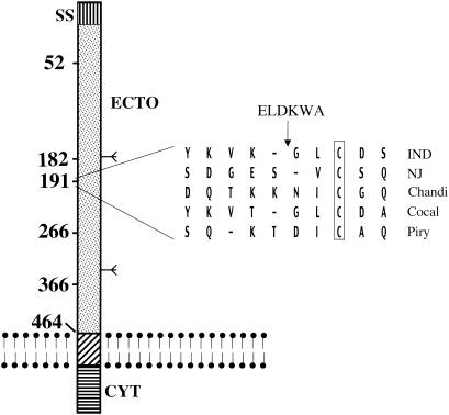
To locate sites within the VSV G ectodomain that might tolerate insertion of a foreign sequence while still allowing surface expression of the protein, we compared the amino acid sequences of G proteins from five vesiculoviruses. We looked for hydrophilic regions in which the sequences were variable in sequence and in length. Variability in sequence suggested that the region might not be critical for protein function. Hydrophilicity was important because polar or charged amino acids are likely to be exposed on the protein surface. Four sites following amino acid positions 52, 191, 266, and 366 fulfilled the criteria of sequence variation and hydrophilicity. The sequence alignment at the most variable of these sequences (position 191) is shown in Fig. 1. Note that only one amino acid, a cysteine, is conserved within the 10-amino-acid sequence around this position and that there is also variability in sequence length among the different G proteins. The overall sequence identity among the different G proteins is 30 to 50%.
FIG. 1.
Diagram of insertion sites in VSV G. VSV G is shown as a linear molecule inserted into the lipid bilayer. The relative positions of the six insertion sites are shown and numbered by the amino acid after which ELDKWA was inserted. The two N-linked glycosylation sites are indicated by the Ψ symbol. The computer-aligned (DNAstar; Megalign program) sequences of five serotypes of vesiculovirus G proteins around the permissive 191 site (Indiana [IND], New Jersey [NJ], Chandipura [Chandi], Cocal, and Piry) are shown. Gaps introduced into the sequences to maximize sequence alignment are indicated by dashes. The single conserved cysteine in the region is shown boxed. Abbreviations: SS, signal sequence; ECTO, ectodomain; CYT, cytoplasmic domain.
We chose the amino acid sequence ELDKWA for insertion at each of these sites. This sequence is part of a conserved epitope from HIV-1 gp41 (26) that is recognized by the human MAb 2F5. The sequence encoding ELDKWA was introduced using oligonucleotide-directed mutagenesis with complementary oligonucleotides that added the eighteen nucleotides encoding the six amino acids.
Two additional insertion sites were chosen for other reasons. The insertion at position 182 was made because it is near a glycosylation site and thus would likely be exposed on the surface of the protein. The insertion at position 464 (at the beginning of the transmembrane domain) was made because it was previously reported to tolerate a small insertion (22) and is also comparable to the membrane proximal position of ELDKWA in gp41. The relative positions of all insertions are shown on a linear diagram of the VSV G protein in Fig. 1. The proteins containing these insertions will be designated by the number of the amino acid in G after which the insertion begins (52, 182, 191, 266, 366, and 464).
Expression of proteins in BHK cells.
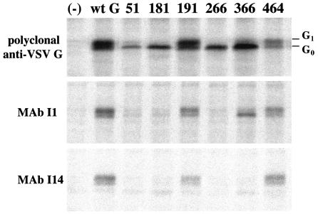
To determine whether insertion of the epitope in VSV G affected protein expression, each of the six proteins with insertions were initially expressed from pBlueScript plasmids using the vaccinia virus T7 system (vTF7-3) (11) and labeled with [35S]methionine. Proteins were then immunoprecipitated from cell lysates with either polyclonal or monoclonal anti-VSV G antibodies and subjected to SDS-PAGE (Fig. 2). VSV G has two N-linked glycans that are added cotranslationally in the ER and are further processed to the complex type in the Golgi, including the addition of sialic acid. The addition of sialic acid slows protein mobility, and the effect of this addition can be seen in the wt G, 191, and 464 proteins as an extra band labeled G1 (17). The appearance of the G1 band suggested initially that these two mutant proteins might be folding correctly and transported to the Golgi bodies. The other proteins 52, 182, 266, and 366 were expressed but showed little or no processing to the G1 form, indicating retention within the ER and suggesting misfolding.
FIG. 2.
Immunoprecipitation of G-ELDKWA mutants expressed from transfected cells. BHK cells were infected with vTF7-3 and then transfected with the indicated constructs or not transfected (−). After 4 h, proteins were labeled with [35S]methionine for 1 h, and cell lysates were immunoprecipitated with polyclonal anti-VSV G antibody or with VSV G MAb I1 or I14 as indicated. Proteins were then separated by SDS-PAGE. The positions of the unprocessed form (G0) and processed form (G1) of the glycoprotein are shown to the right of the gel.
Reactivity with VSV G MAbs.
We also used immunoprecipitation with the conformation-sensitive MAbs I1 and I14 to further assess the folding of each of the proteins (20, 21). The wt G, 191, and 464 proteins were immunoprecipitated by both antibodies, while the others were not (Fig. 2). Interestingly, the 366 protein is immunoprecipitated with I1 and but not with I14. Because the insertion at position 366 is near the I14 epitope (41), the insertion may affect antibody recognition locally or through effects of folding. Because I1 still recognizes this protein, it is likely that this protein folds correctly in other regions.
Rate of acquisition of endoglycosidase H resistance.
The rate of N-glycan processing of each protein was assessed to determine whether the insertions affected the rate of transport from the ER to the Golgi. Because VSV G proteins fold and form trimers in the ER before moving to the Golgi (5), the rate at which they acquire processed glycans in the Golgi bodies can be used to determine whether the insertions affect kinetics of folding. Endoglycosidase H removes the two N-linked glycans added to the G protein in the ER but cannot remove the glycans after they have been processed to the complex type in the Golgi.
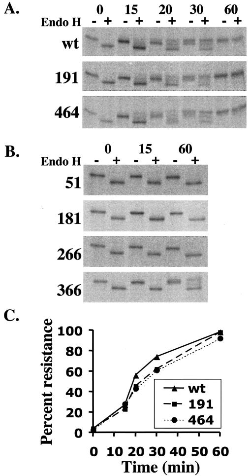
To measure the rate of processing, cells expressing the G proteins were pulsed for 15 min with [35S]methionine and chased for 15, 20, 30 or 60 min (Fig. 3) prior to lysis. Cell lysates were immunoprecipitated with polyclonal antibody to VSV G, and samples were split and either digested or not digested with endoglycosidase H. Endoglycosidase H-treated samples were electrophoresed adjacent to their nontreated counterparts on SDS-polyacrylamide gels. VSV wt G and the two mutants that were processed to the G1 form were analyzed in detail (Fig. 3A). These data are quantitated and show nearly identical kinetics of processing of wt G, 191, and 464 (Fig. 3C). The 52, 182, and 266 proteins showed little or no processing to endoglycosidase H resistance after 60 min (Fig. 3B). However, the 366 protein showed approximately 30% resistance to endoglycosidase H at 60 min, indicating that at least a fraction of the protein was able to fold and move to the Golgi.
FIG. 3.
Acquisition of endoglycosidase H resistance by wt and mutant proteins. BHK cells transfected with plasmids encoding wt or mutant G proteins were labeled with [35S]methionine and then incubated in chase medium with nonradioactive methionine for the indicated number of minutes (A and B). Cell lysates were immunoprecipitated with polyclonal anti-VSV G, and samples were incubated in the presence (+) or absence (−) of endoglycosidase H (Endo H). The kinetics of acquisition of endoglycosidase H resistance of the processed glycoproteins (A) were quantitated using ImageQuant software (C). The percentage of resistance is expressed as the percent of processed G protein relative to the total G protein detected.
Expression of mutant G proteins analyzed by indirect immunofluorescence.
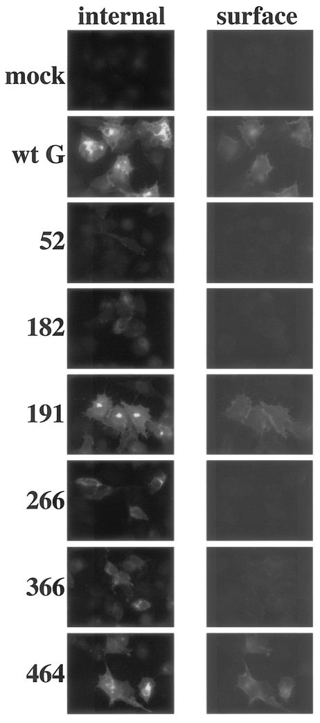
In order to assess further the expression and transport of the mutant proteins within the cell, indirect immunofluorescence microscopy was performed. HeLa cells expressing the proteins were fixed and incubated with a rabbit polyclonal anti-VSV G serum and then with a Texas red-conjugated anti-rabbit secondary antibody. The same cells were permeabilized and incubated first with guinea pig anti-VSV G serum and then with a FITC-conjugated anti-guinea pig secondary antibody. This method allows the same cell to be assayed for both internal and surface-expressed VSV G (Fig. 4). The wt G, 191, and 464 proteins show both internal (FITC) signal and surface (Texas red) signal. Thus, these proteins are clearly exported through the exocytic pathway to the cell surface. The 182, 266, and 366 proteins were detected only internally (FITC), and the 52 protein was barely visible even internally possibly because of degradation. We could detect a low level of signal from the 366 protein at the cell surface, although it does not appear clearly in the photograph.
FIG. 4.
Indirect immunofluorescence to detect intracellular or cell surface G proteins. HeLa cells were infected with vTF7-3 and transfected with the indicated mutants. After 6 h, cells were fixed and stained with rabbit polyclonal anti-VSV serum and then with a Texas red-conjugated anti-rabbit secondary antibody to detect surface G expression. The samples were then permeabilized and incubated with guinea pig anti-VSV G serum and then with a FITC-conjugated anti-guinea pig secondary antibody to show internal G expression. Cells were visualized by indirect immunofluorescence with a Nikon Microphot FX microscope and photographed with a SPOT digital camera.
Recognition of G-protein mutants by the human MAb 2F5.
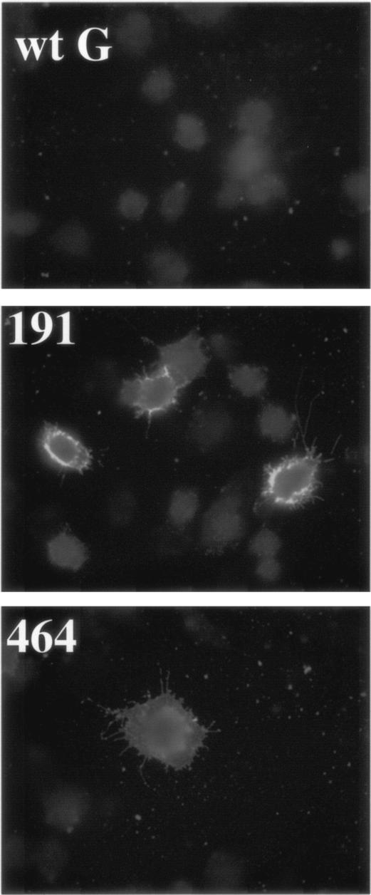
To ascertain whether the two ELDKWA G proteins expressed on the surface also present the ELDKWA epitope on the surface of the protein, immunofluorescence using the human MAb 2F5 was performed. MAb 2F5, which recognizes the ELDKWA epitope, has been shown neutralize a broad range of HIV-1 isolates (40). BHK cells expressing the wt G, 191, and 464 proteins were fixed and incubated with the human MAb 2F5 (Fig. 5). We readily detected the expression of the 2F5 epitope on cells expressing either the 191 or 464 mutant but never on cells expressing wt G.
FIG. 5.
Detection of G-ELDKWA mutants by indirect immunofluorescence with MAb 2F5. BHK cells were infected with vTF7-3 and transfected with plasmids encoding the indicated proteins. After 6 h, cells were fixed and incubated with the human MAb 2F5 and then with a FITC-conjugated anti-human secondary antibody. Cells were photographed as described in the legend to Fig. 4.
Fusion activity of 191 G-ELDKWA
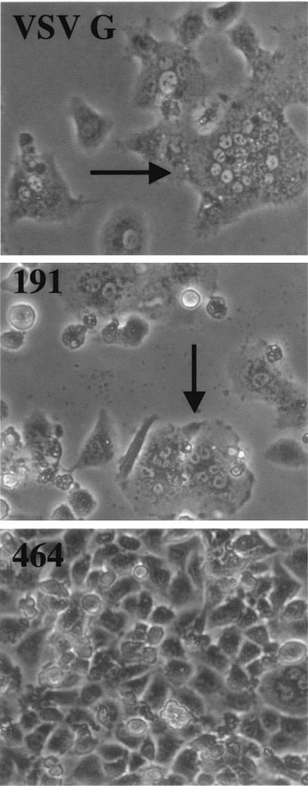
Although expression and processing of the 191 and 464 proteins appeared comparable to those of wt G, we wanted to assess function of these G proteins. During infection, VSV G mediates virus binding after which endocytosis of the virion occurs. The endocytic vesicle is acidified, triggering a conformational change in the G-protein trimer that mediates fusion of the viral envelope with the vesicle (14). Expressing G on the cell surface and reducing the pH of the medium to 5.5 result in cell-cell fusion and syncytial formation, thus mimicking the natural fusion event (9, 29). We used this assay in BHK cells expressing wt G and the 191 and 464 proteins. Only wt G and the 191 mutant showed normal fusion activity as evidenced by the formation of large syncytia (Fig. 6).
FIG. 6.
Fusion activity of 191 and 464 G mutants. BHK cells were infected with vTF7-3 and then transfected with plasmid encoding either wild-type G, 191, or 464 protein. After 6 h, cells were incubated for 1 min in fusion medium at pH 5.2 and again 1 h later as described in Materials and Methods. Large syncytia (multinucleated cells) are indicated (arrows).
Generation of replication-competent VSV expressing the ELDKWA epitope in its G protein.
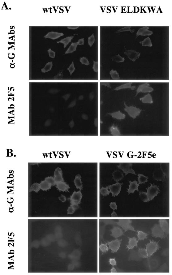
To determine whether the 191 protein was fully functional, we replaced the VSV G coding sequence in the VSV infectious clone with that of G 191 insertion. Infectious virus was recovered by standard procedures (37). To confirm that this virus expressed the ELDKWA sequence in G, BHK cells were infected with either VSV 191 ELDKWA or with wt VSV. Six hours later, cells were fixed and labeled for both G expression with polyclonal anti-G antibodies and for ELDKWA exposure with the 2F5 antibody (Fig. 7A). Cells infected with wt VSV reacted solely with the anti-G antibodies, while cells infected with VSV 191 ELDKWA reacted both with anti-G antibodies and with 2F5. Thus, even with the insertion, the 191 protein is fully functional in virus replication.
FIG. 7.
Indirect immunofluorescence microscopy of cells infected with VSV, VSV 191 G-ELDKWA, or VSV G-2F5e. BHK cells were infected with wt VSV or VSV 191 G-ELDKWA (A), or cells were infected with wt VSV or VSV G-2F5e (B). Cells were fixed and then incubated with mouse MAbs I1 and I14 to VSV G (α-G MAbs) and with the human MAb 2F5 as indicated. Cells were then incubated with a Texas red-conjugated anti-mouse antibody and a FITC-conjugated anti-human antibody. Cells were photographed as described in the legend to Fig. 4.
Neutralization of VSV 191 ELDKWA by 2F5 antibody.
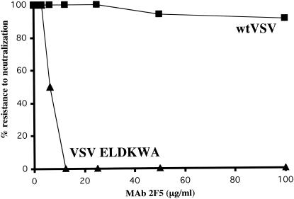
To determine whether MAb 2F5 was able to neutralize the infectivity of VSV 191 ELDKWA, a standard plaque reduction assay was performed. Virus (100 PFU) was incubated with various amounts of MAb 2F5 and then plated on BHK cells. VSV with the 191 protein was neutralized, while wt VSV was not (Fig. 8).
FIG. 8.
Neutralization of VSV-ELDKWA by MAb 2F5. Approximately 100 PFU of wt VSV or VSV-ELDKWA were incubated with the indicated concentrations of MAb 2F5 for 1 h at 37°C before addition to BHK cells. Cells were then overlaid with DMEM containing methylcellulose. After 48 h, cells were stained with crystal violet, and plaques were counted. Results are expressed as a percentage of the titer obtained with no antibody.
A replication-competent VSV expressing the 16-amino-acid 2F5 epitope in G protein.
We next determined whether VSV would tolerate the insertion of the entire 16-amino-acid 2F5 epitope (27). The VSV G gene in the infectious clone was replaced with a G gene containing the 2F5 epitope sequence NEQELLELDKWASLWN at position 191. We recovered infectious virus and showed that cells infected with the recombinant expressed the 2F5 epitope on the cell surface (Fig. 7B).
DISCUSSION
We have located a permissive insertion site in the VSV G protein using a rational approach of sequence comparisons of different vesiculovirus G proteins. One of the four insertions chosen on the basis of high sequence variability and hydrophilicity allowed normal G-protein transport and function after insertion of the six-amino-acid sequence ELDKWA. Interestingly, this site was located within the most variable region in terms of length and sequence among the G proteins compared. Normal function of the mutant protein was ultimately determined by the recovery of viable virus containing this glycoprotein. Additionally, the ELDKWA sequence was exposed on the surface of the glycoprotein as shown by immunofluorescence using MAb 2F5 and the ability of MAb 2F5 to neutralize VSV carrying the ELDKWA epitope in its G protein. The same site also tolerated a larger insertion of 16 amino acids. Similar sequence comparisons should allow prediction of permissive insertion sites in other viral glycoproteins. Identification of permissive sites could be useful for epitope insertion and for insertion of sequences that could alter viral vector targeting.
Insertions at the variable but nonpermissive sites in G (positions 52, 266, and 366) appeared to interfere with G-protein folding at least partially. These regions are likely critical for folding of the G monomer or subsequent assembly of trimers.
We chose the 182 site because it is close to a glycosylation site and thus should be exposed on the protein surface. However, this site is highly conserved in sequence and apparently critical to protein folding, since the protein with this insertion was retained within the ER and did not react with the conformation-sensitive G MAbs.
Position 464 at the junction of the ectodomain and the transmembrane domain was the only site previously reported to accept a small (two-amino-acid) insertion without interfering with G transport and fusion activity. We found that insertion of the ELKDWA sequence at this position did allow transport to the cell surface but also abolished membrane fusion activity. Apparently, the extra length, sequence composition, or both interfered with the fusion process. Because this site is between the transmembrane domain and a stalk sequence known to be involved in membrane fusion (15), the insertion is likely preventing conformational changes required for fusion.
Because recent studies have suggested that the 2F5 epitope may encompass as many as 16 amino acids (27), we also tested the ability of VSV G to tolerate and express this longer epitope (G-2F5e) at position 191. We were able to recover infectious virus with a G protein containing the longer epitope, it grew to normal titers, and cells infected with it expressed the 2F5 epitope in G protein on the cell surface.
These viruses are currently being tested in animal models for induction of neutralizing antibody to HIV-1. VSV recombinants carrying the 2F5 epitope do elicit HIV-1 neutralizing antibodies in mice, but only when the mice were given booster doses of a VSV recombinant carrying the same HIV epitope in the VSV New Jersey G protein. Future studies will address the breadth of the neutralizing antibody response generated by the 6- and 16-amino-acid epitope constructs, as well as the types of antibody (e.g., serum versus mucosal) generated in mouse and monkey models.
Acknowledgments
We thank the members of the Rose lab for critical reading of this manuscript. The human MAb 2F5 was isolated by Hermann Katinger and obtained through the NIH AIDS Research and Reference Reagent Program, Division of AIDS, NIAID, NIH.
This work was supported in part by grant AI40357 from the National Institutes of Health.
REFERENCES
- 1.Bachmann, M. F., H. Hengartner, and R. M. Zinkernagel. 1995. T helper cell-independent neutralizing B cell response against vesicular stomatitis virus: role of antigen patterns in B cell induction? Eur. J. Immunol. 25:3445-3451. [DOI] [PubMed] [Google Scholar]
- 2.Baltimore, D., A. S. Huang, and M. Stampfer. 1970. Ribonucleic acid synthesis of vesicular stomatitis virus. II. An RNA polymerase in the virion. Proc. Natl. Acad. Sci. USA 66:572-576. [DOI] [PMC free article] [PubMed] [Google Scholar]
- 3.Bhella, R. S., S. T. Nichol, E. Wanas, and H. P. Ghosh. 1998. Structure, expression and phylogenetic analysis of the glycoprotein gene of Cocal virus. Virus Res. 54:197-205. [DOI] [PubMed] [Google Scholar]
- 4.Brun, G., X. Bao, and L. Prevec. 1995. The relationship of Piry virus to other vesiculoviruses: a re-evaluation based on the glycoprotein gene sequence. Intervirology 38:274-282. [DOI] [PubMed] [Google Scholar]
- 5.Doms, R. W., D. S. Keller, A. Helenius, and W. E. Balch. 1987. Role for adenosine triphosphate in regulating the assembly and transport of vesicular stomatitis virus G protein trimers. J. Cell Biol. 105:1957-1969. [DOI] [PMC free article] [PubMed] [Google Scholar]
- 6.Durrer, P., Y. Gaudin, R. W. Ruigrok, R. Graf, and J. Brunner. 1995. Photolabeling identifies a putative fusion domain in the envelope glycoprotein of rabies and vesicular stomatitis viruses. J. Biol. Chem. 270:17575-17581. [DOI] [PubMed] [Google Scholar]
- 7.Eckhart, L., W. Raffelsberger, B. Ferko, A. Klima, M. Purtscher, H. Katinger, and F. Ruker. 1996. Immunogenic presentation of a conserved gp41 epitope of human immunodeficiency virus type 1 on recombinant surface antigen of hepatitis B virus. J. Gen. Virol. 77:2001-2008. [DOI] [PubMed] [Google Scholar]
- 8.Emi, N., T. Friedmann, and J. K. Yee. 1991. Pseudotype formation of murine leukemia virus with the G protein of vesicular stomatitis virus. J. Virol. 65:1202-1207. [DOI] [PMC free article] [PubMed] [Google Scholar]
- 9.Florkiewicz, R. Z., and J. K. Rose. 1984. A cell line expressing vesicular stomatitis virus glycoprotein fuses at low pH. Science 225:721-723. [DOI] [PubMed] [Google Scholar]
- 10.Fredericksen, B. L., and M. A. Whitt. 1995. Vesicular stomatitis virus glycoprotein mutations that affect membrane fusion activity and abolish virus infectivity. J. Virol. 69:1435-1443. [DOI] [PMC free article] [PubMed] [Google Scholar]
- 11.Fuerst, T. R., P. L. Earl, and B. Moss. 1987. Use of a hybrid vaccinia virus-T7 RNA polymerase system for expression of target genes. Mol. Cell. Biol. 7:2538-2544. [DOI] [PMC free article] [PubMed] [Google Scholar]
- 12.Gallione, C. J., and J. K. Rose. 1983. Nucleotide sequence of a cDNA clone encoding the entire glycoprotein from the New Jersey serotype of vesicular stomatitis virus. J. Virol. 46:162-169. [DOI] [PMC free article] [PubMed] [Google Scholar]
- 13.Hammond, A. L., R. K. Plemper, J. Zhang, U. Schneider, S. J. Russell, and R. Cattaneo. 2001. Single-chain antibody displayed on a recombinant measles virus confers entry through the tumor-associated carcinoembryonic antigen. J. Virol. 75:2087-2096. [DOI] [PMC free article] [PubMed] [Google Scholar]
- 14.Helenius, A., and M. Marsh. 1982. Endocytosis of enveloped animal viruses. Ciba Found. Symp. 1982: 59-76. [DOI] [PubMed] [Google Scholar]
- 15.Jeetendra, E., C. S. Robison, L. M. Albritton, and M. A. Whitt. 2002. The membrane-proximal domain of vesicular stomatitis virus G protein functions as a membrane fusion potentiator and can induce hemifusion. J. Virol. 76:12300-12311. [DOI] [PMC free article] [PubMed] [Google Scholar]
- 16.Kahn, J. S., M. J. Schnell, L. Buonocore, and J. K. Rose. 1999. Recombinant vesicular stomatitis virus expressing respiratory syncytial virus (RSV) glycoproteins: RSV fusion protein can mediate infection and cell fusion. Virology 254:81-91. [DOI] [PubMed] [Google Scholar]
- 17.Katz, F. N., J. E. Rothman, D. M. Knipe, and H. F. Lodish. 1977. Membrane assembly: synthesis and intracellular processing of the vesicular stomatitis viral glycoprotein. J. Supramol. Struct. 7:353-370. [DOI] [PubMed] [Google Scholar]
- 18.Kreis, T. E., and H. F. Lodish. 1986. Oligomerization is essential for transport of vesicular stomatitis viral glycoprotein to the cell surface. Cell 46:929-937. [DOI] [PMC free article] [PubMed] [Google Scholar]
- 19.Lawson, N. D., E. A. Stillman, M. A. Whitt, and J. K. Rose. 1995. Recombinant vesicular stomatitis viruses from DNA. Proc. Natl. Acad. Sci. USA 92:4477-4481. [DOI] [PMC free article] [PubMed] [Google Scholar]
- 20.Lefrancois, L., and D. S. Lyles. 1982. The interaction of antibody with the major surface glycoprotein of vesicular stomatitis virus. I. Analysis of neutralizing epitopes with monoclonal antibodies. Virology 121:157-166. [PubMed] [Google Scholar]
- 21.Lefrancois, L., and D. S. Lyles. 1982. The interaction of antibody with the major surface glycoprotein of vesicular stomatitis virus. II. Monoclonal antibodies of nonneutralizing and cross-reactive epitopes of Indiana and New Jersey serotypes. Virology 121:168-174. [DOI] [PubMed] [Google Scholar]
- 22.Li, Y., C. Drone, E. Sat, and H. P. Ghosh. 1993. Mutational analysis of the vesicular stomatitis virus glycoprotein G for membrane fusion domains. J. Virol. 67:4070-4077. [DOI] [PMC free article] [PubMed] [Google Scholar]
- 23.Masters, P. S., R. S. Bhella, M. Butcher, B. Patel, H. P. Ghosh, and A. K. Banerjee. 1989. Structure and expression of the glycoprotein gene of Chandipura virus. Virology 171:285-290. [DOI] [PubMed] [Google Scholar]
- 24.Muster, T., B. Ferko, A. Klima, M. Purtscher, A. Trkola, P. Schulz, A. Grassauer, O. G. Engelhardt, A. Garcia-Sastre, P. Palese, and H. Katinger. 1995. Mucosal model of immunization against human immunodeficiency virus type 1 with a chimeric influenza virus. J. Virol. 69:6678-6686. [DOI] [PMC free article] [PubMed] [Google Scholar]
- 25.Muster, T., R. Guinea, A. Trkola, M. Purtscher, A. Klima, F. Steindl, P. Palese, and H. Katinger. 1994. Cross-neutralizing activity against divergent human immunodeficiency virus type 1 isolates induced by the gp41 sequence ELDKWAS. J. Virol. 68:4031-4034. [DOI] [PMC free article] [PubMed] [Google Scholar]
- 26.Muster, T., F. Steindl, M. Purtscher, A. Trkola, A. Klima, G. Himmler, F. Ruker, and H. Katinger. 1993. A conserved neutralizing epitope on gp41 of human immunodeficiency virus type 1. J. Virol. 67:6642-6647. [DOI] [PMC free article] [PubMed] [Google Scholar]
- 27.Parker, C. E., L. J. Deterding, C. Hager-Braun, J. M. Binley, N. Schulke, H. Katinger, J. P. Moore, and K. B. Tomer. 2001. Fine definition of the epitope on the gp41 glycoprotein of human immunodeficiency virus type 1 for the neutralizing monoclonal antibody 2F5. J. Virol. 75:10906-10911. [DOI] [PMC free article] [PubMed] [Google Scholar]
- 28.Reuter, J. D., B. E. Vivas-Gonzalez, D. Gomez, J. H. Wilson, J. L. Brandsma, H. L. Greenstone, J. K. Rose, and A. Roberts. 2002. Intranasal vaccination with a recombinant vesicular stomatitis virus expressing cottontail rabbit papillomavirus L1 protein provides complete protection against papillomavirus-induced disease. J. Virol. 76:8900-8909. [DOI] [PMC free article] [PubMed] [Google Scholar]
- 29.Riedel, H., C. Kondor-Koch, and H. Garoff. 1984. Cell surface expression of fusogenic vesicular stomatitis virus G protein from cloned cDNA. EMBO J. 3:1477-1483. [DOI] [PMC free article] [PubMed] [Google Scholar]
- 30.Roberts, A., E. Kretzschmar, A. S. Perkins, J. Forman, R. Price, L. Buonocore, Y. Kawaoka, and J. K. Rose. 1998. Vaccination with a recombinant vesicular stomatitis virus expressing an influenza virus hemagglutinin provides complete protection from influenza virus challenge. J. Virol. 72:4704-4711. [DOI] [PMC free article] [PubMed] [Google Scholar]
- 31.Rose, J. K., L. Buonocore, and M. A. Whitt. 1991. A new cationic liposome reagent mediating nearly quantitative transfection of animal cells. BioTechniques 10:520-525. [PubMed] [Google Scholar]
- 32.Rose, J. K., and C. J. Gallione. 1981. Nucleotide sequences of the mRNAs encoding the vesicular stomatitis virus G and M proteins determined from cDNA clones containing the complete coding regions. J. Virol. 39:519-528. [DOI] [PMC free article] [PubMed] [Google Scholar]
- 33.Rose, J. K., and M. A. Whitt. 2001. Rhabdoviridae: the viruses and their replication, p. 1221-1277. In D. M. Knipe and P. M. Howley (ed.), Fields virology, 4th ed., vol. 1. Lippincott-Raven, New York, N.Y. [Google Scholar]
- 34.Rose, N. F., P. A. Marx, A. Luckay, D. F. Nixon, W. J. Moretto, S. M. Donahoe, D. Montefiori, A. Roberts, L. Buonocore, and J. K. Rose. 2001. An effective AIDS vaccine based on live attenuated vesicular stomatitis virus recombinants. Cell 106:539-549. [DOI] [PubMed] [Google Scholar]
- 35.Rose, N. F., A. Roberts, L. Buonocore, and J. K. Rose. 2000. Glycoprotein exchange vectors based on vesicular stomatitis virus allow effective boosting and generation of neutralizing antibodies to a primary isolate of human immunodeficiency virus type 1. J. Virol. 74:10903-10910. [DOI] [PMC free article] [PubMed] [Google Scholar]
- 36.Schlegel, R., T. S. Tralka, M. C. Willingham, and I. Pastan. 1983. Inhibition of VSV binding and infectivity by phosphatidylserine: is phosphatidylserine a VSV-binding site? Cell 32:639-646. [DOI] [PubMed] [Google Scholar]
- 37.Schnell, M. J., L. Buonocore, E. Kretzschmar, E. Johnson, and J. K. Rose. 1996. Foreign glycoproteins expressed from recombinant vesicular stomatitis viruses are incorporated efficiently into virus particles. Proc. Natl. Acad. Sci. USA 93:11359-11365. [DOI] [PMC free article] [PubMed] [Google Scholar]
- 38.Schnell, M. J., L. Buonocore, M. A. Whitt, and J. K. Rose. 1996. The minimal conserved transcription stop-start signal promotes stable expression of a foreign gene in vesicular stomatitis virus. J. Virol. 70:2318-2323. [DOI] [PMC free article] [PubMed] [Google Scholar]
- 39.Schnitzer, T. J., R. A. Weiss, and J. Zavada. 1977. Pseudotypes of vesicular stomatitis virus with the envelope properties of mammalian and primate retroviruses. J. Virol. 23:449-454. [DOI] [PMC free article] [PubMed] [Google Scholar]
- 40.Trkola, A., A. B. Pomales, H. Yuan, B. Korber, P. J. Maddon, G. P. Allaway, H. Katinger, C. F. Barbas III, D. R. Burton, D. D. Ho, and J. P. Moore. 1995. Cross-clade neutralization of primary isolates of human immunodeficiency virus type 1 by human monoclonal antibodies and tetrameric CD4-IgG. J. Virol. 69:6609-6617. [DOI] [PMC free article] [PubMed] [Google Scholar]
- 41.Vandepol, S. B., L. Lefrancois, and J. J. Holland. 1986. Sequences of the major antibody binding epitopes of the Indiana serotype of vesicular stomatitis virus. Virology 148:312-325. [DOI] [PubMed] [Google Scholar]
- 42.Whitt, M. A., P. Zagouras, B. Crise, and J. K. Rose. 1990. A fusion-defective mutant of the vesicular stomatitis virus glycoprotein. J. Virol. 64:4907-4913. [DOI] [PMC free article] [PubMed] [Google Scholar]


