Abstract
The hemagglutinin-neuraminidase (HN) protein of Newcastle disease virus (NDV) is an important determinant of its virulence. We investigated the role of each of the four functional N-linked glycosylation sites (G1 to G4) of the HN glycoprotein of NDV on its pathogenicity. The N-linked glycosylation sites G1 to G4 at residues 119, 341, 433, and 481, respectively, of a moderately pathogenic NDV strain Beaudette C (BC) were eliminated individually by site-directed mutagenesis on a full-length cDNA clone of BC. A double mutant (G12) was also created by eliminating the first and second glycosylation sites at residues 119 and 341, respectively. Infectious virus was recovered from each of the cDNA clones of the HN glycoprotein mutants, employing a reverse genetics technique. There was a greater delay in the replication of G4 and G12 mutant viruses than in the parental virus. Loss of glycosylation does not affect the receptor recognition by HN glycoprotein of NDV. The neuraminidase activity of G4 and G12 mutant viruses and the fusogenicity of the G4 mutant virus were significantly lower than those of the parental virus. The fusogenicity of the double mutant virus (G12) was significantly higher than that of the parental virus. Cell surface expression of the G4 virus HN was significantly lower than that of the parental virus. The antigenic reactivities of the mutants to a panel of monoclonal antibodies against the HN protein indicated that removal of glycosylation from the HN protein increased (G1, G3, and G12) or decreased (G2 and G4) the formation of antigenic sites, depending on their location. In standard tests to assess virulence in chickens, all of the glycosylation mutants were less virulent than the parental BC virus, but the G4 and G12 mutants were the least virulent.
Newcastle disease virus (NDV) is a member of the family Paramyxoviridae and has been assigned to the genus Avulavirus in the subfamily Paramyxovirinae (9, 26). It causes a serious respiratory and neurological disease in all species of birds and is an economically important infectious agent, causing significant losses to the poultry industry. Newcastle disease varies in the degree of severity, ranging from an inapparent infection to 100% mortality, depending on the virus strain into lentogenic (mildly pathogenic), mesogenic (moderately pathogenic), and velogenic (highly pathogenic) pathotypes (2).
NDV contains a single-stranded, negative-sense, nonsegmented RNA genome. The genomic RNA is 15,186 nucleotides in length (21, 34). The genomic RNA contains six genes that encode at least seven proteins (33, 44). The envelope of NDV contains two glycoproteins, the hemagglutinin-neuraminidase (HN) and fusion (F) proteins. The F glycoprotein mediates fusion of the viral envelope with cellular membranes (7). The HN glycoprotein of NDV is a multifunctional protein. It recognizes sialic acid-containing receptors on cell surfaces, it promotes the fusion activity of F protein of NDV, thereby allowing the virus to penetrate the cell surface, and it acts as a neuraminidase (NA) by removing the sialic acid from progeny virus particles to prevent viral self-aggregation (23).
The HN glycoprotein is a type 2 homotetrameric integral membrane protein (40) which undergoes N-linked glycosylation (28, 30). This process occurs in the rough endoplasmic reticulum of host cells when an N-linked carbohydrate attaches covalently as a core oligosaccharide side chain to asparagines on the nascent polypeptide chain in response to the consensus sequence motif NXT (Asn-X-Thr) or NXS (Asn-X-Ser), where X is any amino acid except aspartic acid or proline. The structure of the oligosaccharide side chain is then extensively modified as the protein moves through the membrane systems of the cell (20). N-linked glycosylation influences many properties of glycoproteins, including initiation and maintenance of folding of the proteins into their biologically active conformation, maintenance of protein stability and solubility, intracellular transport of the proteins to various subcellular compartments and the cell surface, and influencing the antigenicity and immunogenicity of the protein (32, 37).
The HN glycoprotein sequence of NDV strain Beaudette C (BC) contains six predicted sites for the addition of N-linked carbohydrates (residues 119, 341, 433, 481, 508, and 538) (28). A previous study has shown that four of these addition sites (G1, G2, G3, and G4 at residues 119, 341, 433, and 481, respectively) are used, whereas two addition sites (G5 and G6 at residues 508 and 538, respectively) are not used (28). The same study has shown that G1 and G2 play little role in maturation but modulate the biological activities of the protein, whereas G3 and G4 influence both folding and activity of the protein (28). In that study, the role of individual oligosaccharide chains in the activities of the HN glycoproteins was examined by using a plasmid transfection system. Thus, the role of each oligosaccharide chain in viral replication and pathogenesis could not be determined. In our present study, a reverse genetics system was used to generate recombinant viruses with mutations in the glycosylation sites of the HN protein. These mutations eliminated each of the four functional glycosylation sites individually (G1, G2, G3, and G4) and, in G1 and G2, in combination. This allowed the determination of the role played by the individual glycans in the context of viral replication and pathogenesis. Our results showed that elimination of the residue at G4 and combined elimination of residues at G1 and G2 significantly attenuated the pathogenicity of the viruses. It also indicated that G4 played an important role in the transport of the HN protein. Interestingly, elimination of G1 and G2 significantly increased fusion promotion activities of the viruses, which does not correlate with pathogenicity in chickens. This study provides a useful tool for studying the impact of glycosylation of the HN protein of NDV on virus pathogenicity and may be able to provide insights for designing better recombinant attenuated NDV vaccines.
MATERIALS AND METHODS
Cells and viruses.
The chicken embryo fibroblast cell line (DF1) was obtained from Douglas Foster (University of Minnesota) and grown in Dulbecco's modified Eagle's medium containing 10% fetal bovine serum. HEp-2 cells were grown in Eagle's minimal essential medium containing 10% fetal bovine serum. A moderately pathogenic (mesogenic) NDV strain BC and other recombinant viruses generated from BC virus for this study were grown in 9-day-old embryonated specific-pathogen-free (SPF) chicken eggs. The modified vaccinia virus Ankara recombinant that expresses the T7 RNA polymerase (a generous gift of Bernard Moss, National Institutes of Health) was grown in primary chicken fibroblast (CEF) cells.
Introduction of HN mutations into full-length NDV cDNA.
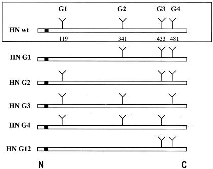
The construction of plasmid pNDVfl carrying the full-length cDNA of the NDV strain BC has been described previously (22). To introduce mutations into the HN gene of pNDVfl, an AgeI-MluI fragment containing the HN gene was excised from pNDVfl and subcloned into plasmid pGEM-7Z (+) (Promega, Madison, Wis.). Using appropriate oligomers, site-directed mutagenesis (6) was performed on the cDNA clone of the HN gene to generate a panel of HN mutants, as shown in Fig. 1. Mutants were named with a G (for glycosylation site) and the number of the functional glycosylation site mutated (1 to 4). The primers used for generating the cDNA clones are as follows: clone G1, 5′-6766CAGAACAGCGGGTGGGGGGCACCT6789-3′ (forward) and 5′-6765CGCAGCTCCATTAATCTGATAAGAGAG6739-3′ (reverse); clone G2, 5′-7432CAGGACACATGCCCAGATGAG7452-3′ (forward) and 5′-7431GTATCGCTTGTATATTACATATTTCCC7405-3′ (reverse); clone G3, 5′-7708CAGAAAACAGCCACTCTTCATAGTCCC7734-3′ (forward) and 5′-7707GCTGACTGTCATAGGATATAATAACGC7681-3′ (reverse); clone G4, 5′-7852CAGCACACCTTGCGAGGGGTA7872-3′ (forward) and 5′-7851CCTATAGAAGATTAGGGGATATGG7828-3′ (reverse). Letters in boldface type represent the mutated nucleotides, which changed the corresponding amino acids. To eliminate each glycosylation site, the asparagine residue in the NXT/NXS sequence motif was changed to glutamine. A double HN mutant, G12, was also created by eliminating glycosylation sites 1 and 2. This was done by mutating the second glycosylation site on the cDNA clone G1 with the primer specific for making the G2 cDNA clone. All mutant HN cDNAs were sequenced in their entirety to confirm the presence of the intentional mutations. The mutagenized AgeI-MluI fragments were then excised from pGEM 7Z (+) and inserted into the AgeI-MluI site of pNDVfl.
FIG. 1.
Schematic representation of the HN glycosylation mutants generated by site-directed mutagenesis. The HN wild-type (wt) protein is shown within the box at the top of the figure. Symbols: open bar, 571-amino-acid polypeptide chain; filled bar, N-terminal transmembrane anchor; Y, functional N-linked glycosylation site. The residue numbers indicate the Asn residues in the HN amino acid sequence. The designations of the carbohydrate chains (G1, G2, G3, and G4) are indicated above each site. HN glycosylation mutants are represented in a similar fashion, with the designated name shown at the left of each diagram. Wild-type HN has four functional Asn-linked glycosylation sites, represented by G1, G2, G3, and G4, present at amino acid residues 119, 341, 433, and 481, respectively. The HN G1 mutant has its glycosylation site G1 at residue 119 eliminated, mutant HN G2 has its G2 glycosylation site at residue 341 eliminated, mutant HN G3 has its G3 glycosylation site at residue 433 eliminated, mutant HN G4 has its G4 glycosylation site at residue 481 eliminated, and the double mutant HN G12 has two of its glycosylation sites at residues 119 and 341 eliminated. All glycosylation sites were eliminated by mutating the asparagine (Asn) residue at each site to glutamine (Glu) by site-directed mutagenesis.
Generation of recombinant viruses.
Transfection and recovery of recombinant NDV were performed by a reverse genetics technique described previously (22). Recovered viruses were designated rBC for the virus obtained from transfection with wild-type plasmid construct pNDVfl and rG1, rG2, rG3, rG4, and rG12 for the mutant viruses obtained from plasmid constructs G1, G2, G3, G4, and G12, respectively.
RT-PCR and sequence analysis of HN gene.
RNA was isolated from the recovered HN mutant viruses by using TRIzol reagent (Invitrogen). Reverse transcription (RT)-PCR was performed by using the Thermoscript RT-PCR kit (Invitrogen) with primers P1 (5′-6202GTGAACACAGATGAGGAACG6221-3′, positive sense) and P1R (5′-9369ATATCATTGGGGAGGAGGCCG9349-3′, negative sense) to amplify the HN gene. The amplified cDNA fragments were then sequenced with the Themosequenase kit (Amersham) to confirm the presence of the introduced mutations in the recovered viruses. The primer used to confirm the mutations were as follows: G1 (amino acid residue at position 119 on the HN gene), 5′-6623CATCTGCACTTGGTTCCAATC6643-3′; G2 (residue 341), 5′-7294TGGGTGGCCAACTACCCAGG7313-3′; G3 (residue 433) and G4 (residue 481), 5′-7660GGGTCATCATACTTCTCTCCCGCG7683-3′. For the mutation at G12 (residues 119 and 341), the same primers used previously for confirming the G1 and G2 mutations were used. The HN RNA amplified by RT-PCR of the recovered viruses was also sequenced entirely to ensure that the rest of the sequence of the protein remained unchanged.
Characterization of mutant viruses.
The growth rate of each recombinant virus was examined by multiple-cycle growth conditions. The DF1 cells were infected with each virus at a multiplicity of infection (MOI) of 0.01. A sample of the supernatant was harvested at 8-h intervals for a period of 56 h. The amount of virus in the supernatant was determined by plaque assay on DF1 cells. The size and morphology of the observed plaques were also examined.
The receptor recognition properties of the recombinant viruses were evaluated by their ability to adsorb guinea pig red blood cells (19). The NA activity of the recombinant viruses was determined by a fluorescence-based NA assay according to the procedures of Potier et al. (36). The NA activity of the recovered viruses was measured with the 4-methylumbelliferone released from the fluorogenic substrate, methylumbelliferone. Released 4-methylumbelliferone was quantified by fluorometric determination with an excitation wavelength of 360 nm and an emission wavelength of 450 nm. Readings from the substrate blanks were subtracted from the virus sample readings, and the mean values of duplicate readings were calculated.
The ability of each recombinant virus to form syncytia was determined according to the procedures described by Kohn (19). The syncytia obtained after infection with each virus at an MOI of 0.1 in Vero cells were quantitated, and the fusion index was calculated for each virus. The fusion index is the ratio of the total number of nuclei to the number of cells in which these nuclei are present (i.e., the mean number of nuclei per cell).
Radioimmunoprecipitation and analysis of stability of HN protein.
DF1 cells were infected with recombinant viruses at an MOI of 10. After 4 to 6 h of incubation at 37°C, the medium was removed and the cells were washed with phosphate-buffered saline (PBS) and starved in 5 ml of methionine-cysteine-free medium (Invitrogen) for 30 min. The cells were then pulse labeled for 2 h with medium containing 100 μCi of [35S]methionine-cysteine (1,000 Ci/mmol; Amersham) per ml. After labeling, cells were washed in PBS and lysed in cold radioimmunoprecipitation buffer (20 mM Tris [pH 7.5], 150 mM NaCl, 5 mM EDTA [pH 8.0], 0.5% sodium deoxycholate, 0.1% sodium dodecyl sulfate [SDS], 1.0% Triton X-100). The cell lysates were centrifuged to remove the nuclei. Immunoprecipitations were performed by adding anti-NDV-specific polyclonal antibody (prepared against the whole virus) to the cell lysates and incubating the mixtures for 3 h at 4°C. Immune complexes were precipitated by incubation with Staphylococcus aureus protein A for 30 min. Immune complexes were then solubilized by boiling in buffer H (1% SDS, 0.15 M Tris-HCl [pH 7.5]). Solubilized proteins were analyzed on SDS-12% polyacrylamide gels (SDS-polyacrylamide gel electrophoresis ). A portion of each solubilized protein was digested with endoglycosidase H (endo H) (Boehringer-Mannheim Corp). Briefly, endo H (1.5 mU) and sodium citrate buffer (0.05 M, final concentration), pH 5.5, were added to the immunoprecipitated proteins and incubated overnight at 37°C. The digestion was stopped by boiling, and the samples were analyzed by SDS-10% PAGE.
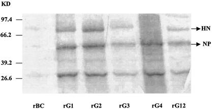
To assess the virion content of the HN proteins in the recombinant viruses, equal amounts of each purified virus sample were analyzed on an SDS-10% PAGE gel and visualized after staining with Coomassie blue. The concentrations of the viral proteins of each recombinant virus were standardized by using the Micro BSA protein assay reagent kit (Pierce). The HN protein of NDV has an approximate molecular mass of 74 kDa.
Cell surface expression of HN proteins.
DF1 cells at 70% confluence in six-well plates were inoculated with each virus at an MOI of 0.1 and incubated at 37°C. After 48 h, the cells were washed with PBS and fixed onto the plate with 3% paraformaldehyde in PBS for 30 min. After washing three times with PBS, the cells were permeabilized with either 0.1% Triton X-100 or only PBS for 30 min. Following three further washes with PBS, the cells were treated with a 1:500 dilution of primary antibody (HN-specific monoclonal antibody [MAb]) in PBS at room temperature for 30 min. The cells were then washed three times with PBS and incubated with affinity-purified, fluorescein-labeled goat anti-mouse immunoglobulin (Kirkegaard and Perry Laboratories) for 30 min. The cells were washed once again with PBS and visualized under a Nikon Eclipse TE 300 (Tokyo, Japan) epifluorescent microscope.
Flow cytometry.
DF1 cells were infected with each virus at an MOI of 1. After 16 h, the cells were washed once with calcium- and magnesium-free PBS containing 0.02% sodium azide (PBSA). The cells were lifted off the dish with PBS containing 50 mM EDTA. The cell suspensions were washed once with PBSA and then bound with a mixture of a cocktail of NDV HN-specific MAbs (24) at a dilution of 1:200 and incubated on ice for 30 min. The cells were then washed three times with PBSA and bound with a fluorescein isothiocyanate-conjugated goat anti-mouse immunoglobulin (Kirkegaard and Perry Laboratories) for 30 min on ice. Cells were then washed three times with PBSA. The fluorescence of 10,000 cells was measured with a FACSVantage flow cytometer (Becton Dickinson).
ELISA.
The reactivities of the HN-specific MAbs of NDV were tested in a standard enzyme-linked immunosorbent assay (ELISA). The MAbs used were 15C4, AVS, B79, and 10D11. The MAb 15C4 neutralizes and inhibits hemagglutination (HA) of all lentogenic, mesogenic, and velogenic NDV strains but not the pigeon paramyxovirus 1 strain. Antibody 10D11 also inhibits HA activity, but inhibition is more selective and is limited to the mesogenic and domestic, or indigenous, velogenic strains of NDV. MAb B79 reacts in all serologic assays with an antigenic site common to all serotype 1 avian paramyxoviruses. AVS is a paramyxovirus 1 lentogenic strain-specific MAb, which reacts with avirulent strains only, and was used as a negative control (24). Equal amounts of purified parental and mutant recombinant viruses were used as antigens. The concentrations of the viral proteins of each recombinant virus were standardized by using the Micro BSA protein assay reagent kit (Pierce). A 1:2,000 dilution of MAbs was used in the ELISA. Affinity-purified peroxidase-labeled goat anti-mouse immunoglobulin (Kirkegaard and Perry Laboratories) was used as the secondary antibody. The results of the ELISA were graded as + to ++++, depending on the reactivity of MAbs to the virus. Uninfected cell antigen was used as a negative control.
Pathogenicity studies.
The virulence of the recombinant viruses was determined by three in vivo tests: the mean death time (MDT), intracerebral pathogenicity index (ICPI), and intravenous pathogenicity index (IVPI) tests.
The MDT was determined to examine the pathogenicity of the recombinant viruses in embryonated chicken eggs as described previously (1). Briefly, a series of 10-fold dilutions of fresh, infective allantoic fluid were made in sterile PBS, and 0.1 ml of each dilution was inoculated into the allantoic cavity of each of five 9-day-old embryonated chicken eggs. The eggs were incubated at 37°C and examined four times daily for 7 days. The time that each embryo was first observed dead was recorded. The highest dilution that killed all embryos was considered the minimum lethal dose. The MDT was recorded as the time (in hours) for the minimum lethal dose to kill the embryos. The MDT has been used to classify NDV strains as velogenic (taking under 60 h to kill), mesogenic (taking between 60 and 90 h to kill), and lentogenic (taking more than 90 h to kill).
To examine the pathogenicity of recombinant viruses in vivo in chickens, the ICPI and IVPI tests were performed as described previously, with modifications (1). For ICPI, 103 PFU of each virus/bird was inoculated into groups of 10 1-day-old SPF chicks via the intracerebral route. The inoculation was done using a 27-guage needle attached to a 1-ml stepper syringe dispenser that was set to dispense 0.05 ml of inoculum per inoculation. The birds were inoculated by inserting the needle up to the hub into the right or left rear quadrant of the cranium. The birds were observed daily for 8 days, and at each observation, the birds were scored 0 if normal, 1 if sick, and 2 if dead. The ICPI value is the mean score per bird per observation. Highly virulent viruses give values approaching 2, and avirulent viruses give values approaching 0. For IVPI, 103 PFU of each virus/bird was inoculated intravenously into groups of 10 6-week-old chickens. The dispenser was set to dispense 0.1 ml of inoculum per inoculation. The birds were observed daily for a period of 10 days for clinical symptoms and mortality. They were scored 0 if normal, 1 if sick, 2 if paralyzed, and 3 if dead. The weighted sums were then added and divided by the total number of observations (10 birds × 10 days = 100 observations), which provided the mean score per bird per observation. Each experiment had mock-inoculated controls that received a similar volume of sterile PBS by the respective route. Highly virulent viruses give values approaching 3, and avirulent viruses give values approaching 0.
RESULTS
Generation of recombinant NDV containing HN N-linked glycosylation site mutations.
Previously, recovery of recombinant NDV from an infectious cDNA clone (pNDVfl) derived from a mesogenic strain of NDV, BC, was reported from our laboratory (22). In this study, the established reverse genetics system was used to determine the role of N-linked carbohydrates of the HN protein on the biological activities of NDV. To achieve this goal, the AgeI-MluI subclone containing the HN gene derived from the full-length clone of BC (pNDVfl) was mutated. Each of the four functional N-linked glycosylation sites (28) was altered by site-directed mutagenesis changing asparagine, the first amino acid residue of the acceptor site, to glutamine (Fig. 1). Substitutions were made by altering the first and third positions (underlined) of each asparagine codon to create a codon for glutamine (AAT or AAC to CAG). Glutamine was chosen because it is structurally similar to asparagine, differing by only a single methylene group. Moreover, a minimum of two nucleotide changes are necessary for the altered sequence to revert to any codon specifying asparagine, thereby reducing the likelihood of direct reversion of the targeted codon during virus replication in vivo. To examine the effect of the combined loss of two N-linked glycosylation sites, a double mutant was also created by eliminating the N-linked glycosylation sites 1 and 2 (G1 and G2, respectively). The mutant HN cDNA subclones were then inserted into the full-length cDNA of BC (pNDVfl), thus generating four single mutant cDNA clones (G1, G2, G3, and G4) and one double mutant cDNA clone (G12). To ensure the presence of the introduced mutations, the entire HN cDNA clone was sequenced by the dideoxy chain termination method.
Recombinant viruses expressing wild-type HN (rBC) and mutant proteins G1 (rG1), G2 (rG2), G3 (rG3), G4 (rG4), and G12 (rG12) were recovered by transfection of HEp-2 cells with full-length mutant HN cDNA clones and support plasmids and amplification of viruses in DF1 cells. Recovered viruses were subjected to RT-PCR, and the HN genes were sequenced in their entirety to confirm the presence of the introduced mutations. To determine the stability of each HN mutation, the recovered viruses were passaged five times in 9- to 11-day-old embryonated chicken eggs and the sequence of the HN gene was determined in viruses recovered at each passage level. These sequence analyses showed that the introduced HN mutations were unaltered, even after five egg passages (data not shown).
Growth and plaque morphology of mutant viruses.
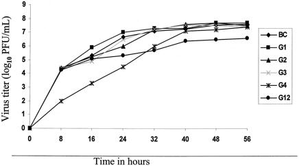
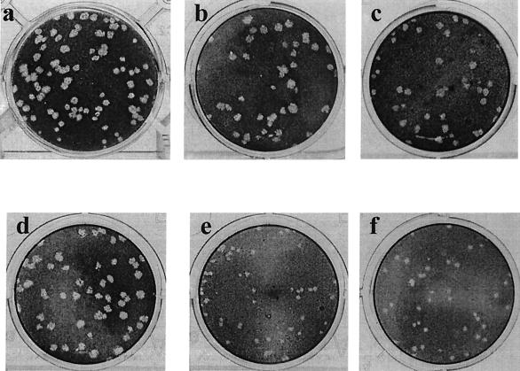
The efficiencies of replication of the parental (rBC) and mutant (rG1, rG2, rG3, rG4, and rG12) viruses were compared in a multistep growth cycle in DF1 cells (Fig. 2). The replication kinetics of the mutant viruses were comparable to that of the parental virus. Some of the mutant viruses (rG1, rG2, and rG3), however, had significantly higher titers than the parental virus at 32 and 40 h postinfection (PI) (P < 0.05), probably due to the rapid destruction of the cell sheet by the parental virus. Interestingly, the rG4 virus showed a delayed growth (16 h) and rG4 and rG12 viruses had lower virus yields (1.5 to 2.0 log U lower) than the parental and other mutant viruses until 40 h. However, after 40 h, the virus titer of rG4 reached a level comparable to that of the parental virus, but rG12 was at least 1.0 log U lower than that of the parental virus until 56 h. The virus titers of rG4 and rG12 at these time points were significantly different than the parental virus (P < 0.05). The virus yields had reached a plateau by 48 h PI. The parental rBC virus produced syncytia at 24 h PI, whereas the rG12 virus initiated syncytium formation at 48 h PI and rG4 virus induced syncytia only at 72 h PI. Most mutant viruses had plaque sizes similar to those of the parental virus, all measuring 3 to 5 mm in diameter, except the mutants rG4 and rG12, which had considerably smaller plaques (1 to 2 mm in diameter), as shown in Fig. 3.
FIG. 2.
Growth kinetics of viruses in tissue culture. Multicycle growth of parental and HN glycosylation mutant viruses in chicken embryo fibroblast (DF1) cells. Cells were infected with the indicated parental or chimeric virus at an MOI of 0.01. Samples were taken at 8-h intervals, and virus titers were determined by plaque assay. Values are averages from the results from three independent experiments.
FIG. 3.
Plaque morphology in DF1 cells by wild-type and HN glycosylation mutant viruses 4 days PI. Recovered viruses were titrated in duplicate in 12-well plates. Supernatant collected from virus-inoculated samples was serially diluted, and 100 μl of each serial dilution was added per well to confluent DF1 cells. After 60 min of adsorption, cells were overlaid with Dulbecco's modified Eagle's medium (containing 2% fetal bovine serum and 0.9% methylcellulose) and incubated at 37°C for 3 to 4 days. The cells were then fixed with ethanol and stained with crystal violet for observation of plaques. Plaque size and morphology are shown for rBC (a), rG1 (b), rG2 (c), rG3 (d), rG4 (e), and rG12 (f) viruses.
Analysis of the HN protein of glycosylation mutants by radioimmunoprecipitation assay.
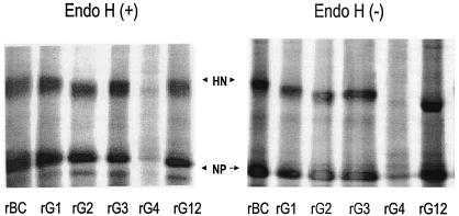
To confirm that the mutated glycosylation sites actually lead to the loss of carbohydrates in the HN protein, DF1 cells infected with recombinant NDVs expressing the mutant HN proteins were labeled with [35S]methionine-cysteine 4 to 6 h after infection. The labeled proteins were immunoprecipitated from cell lysates with NDV-specific polyclonal antibodies and analyzed by SDS-PAGE. One-half of the cell lysates were treated with endo H, and the other half was undigested. Both samples were subjected to SDS-PAGE analysis (Fig. 4). Our results showed that endo H-digested mutant HN proteins comigrated with the parental viruses on polyacrylamide gels. In the undigested samples, the HN proteins of single mutants (G1, G2, G3, and G4) migrated slightly faster on polyacrylamide gels than the parental HN protein while the HN protein with mutations in two sites (G12) migrated faster than the single mutant HN proteins. The protein missing site 1 migrated slower than proteins missing site 2, 3, or 4. On average, glycosylation of a single site is predicted to contribute approximately 2 to 3 kDa to the apparent molecular mass of a protein (20). These results corroborated with earlier findings with transfected HN mutant plasmids (28) that glycosylation sites at residues 119, 341, 433, and 481 are actually used in the HN protein of NDV. Further, the slightly slower migration of the G1 mutant than the other mutants, identified in this study with the BC strain and in an earlier study with the Australia-Victoria strain of NDV, may be due to a smaller carbohydrate linked to site 1 than those linked to other sites (28). Variation in the size of carbohydrate side chains has been observed in other glycoproteins (30, 42). None of the precipitated proteins, endo H-digested or undigested, including the parental HN protein, showed any partially resistant side chains reported earlier (28) in polyacrylamide gels. This may be due to the differences in the strain of virus employed and plasmid transfection versus whole virus infection methods employed. However, the amount of precipitated HN protein with rG4 mutant virus was extremely small (Fig. 4). Coomassie blue staining of purified virus run on SDS-10% polyacrylamide gels also showed extremely small amounts of HN protein of rG4 virus (Fig. 5). This suggests that mutation of the glycosylation site 4 (Asn-481) inhibited the maturation of HN glycoprotein of NDV. Pulse-chase experiments also indicated that the small amounts of rG4 HN protein precipitated were not due to instability of the protein, since all the mutant proteins were present at similar levels to that of the parental virus after a 2-h labeling, followed by a chase at 2, 6, and 12 h postlabeling (data not shown).
FIG. 4.
Immunoprecipitation of parental rBC and HN glycosylation mutants from infected cells and treatment with endo H. DF1 cells were infected with each of the viruses for 4 to 6 h, and proteins were labeled with [35S]methionine-cysteine for 2 h. Equal amounts of cell lysates were immunoprecipitated with a cocktail of NDV-specific polyclonal antibodies and S. aureus protein A and analyzed by SDS-PAGE. The immunoprecipitates were digested (+) or mock-digested (−) with endo H and then analyzed by SDS-PAGE. NP, nucleocapsid protein.
FIG. 5.
Expression of HN protein glycosylation mutants. Equal amounts of proteins from parental and mutant purified viruses were analyzed by SDS-PAGE. The migration and protein stability of the HN protein from each virus were examined after Coomassie blue staining. The HN protein of mutant rG4 virus was found to be degraded. A protein molecular mass marker was also run along with the purified viral samples to assess the viral proteins based on their molecular masses. The molecular mass of the HN protein of NDV is approximately 74 kDa. Three independent experiments with purified viral proteins confirmed the degradation of the HN glycoproteins of the rG4 mutant virus.
Reactivity of glycosylation mutant HN proteins with MAbs.
It is suggested that the formation of antigenic sites present only on the mature HN molecule is a measure of maturation of the protein (28). To examine the effect of the loss of carbohydrate on the formation of antigenic sites, the parental and mutant viruses were tested for reactivities to a panel of HN-specific MAbs (15C4, 10D11, B79, and AVS) by ELISA. Purified parental and mutant viruses were used as antigens. ELISA results showed that all mutant proteins reacted with most of the antibodies tested except the mutants rG2 and rG4 (Table 1). None of the mutants or the parental virus reacted to MAb AVS, which recognizes only avirulent strains. The mutant rG1, rG3, and rG12 viruses showed higher rates of reactivity than the other mutants with MAb B79, and the rG1 and rG3 viruses showed higher reactivity to MAb 15C4 than the parental rBC virus and other mutant viruses. The antibody binding sites are unknown for these MAbs, but they were shown to be neutralizing (24). Hence, enhanced recognition of HN by antibodies B79 and 15C4 by these viruses is important in the context of eliciting a neutralizing antibody response in the host. The rG4 and rG2 viruses were poorly reactive to the 15C4 and B79 MAbs and were totally unreactive to the MAb 10D11. Thus, loss of different carbohydrate side chains differentially influences the formation of antigenic epitopes. The presence of the G2 and G4 mutations eliminated the detection of a particular epitope recognized by the 10D11 MAb. The poor efficiency of binding to 15C4 and B79 antibodies by rG4 and rG2 viruses suggests that a significant population of these mutant proteins remains at least partially misfolded.
TABLE 1.
MAb reactivity, endo H sensitivity, and cell surface expression of HN glycosylation mutant viruses
| Virus | Reactivity to MAba:
|
endo H sensitivityb | Cell surface expressionc | |||
|---|---|---|---|---|---|---|
| 15C4 | 10D11 | B79 | AVS | |||
| rBC | +++ | + | + | − | S | Yes |
| rG1 | ++++ | + | +++ | − | S | Yes |
| rG2 | ++ | − | − | − | S | Yes |
| rG3 | ++++ | + | +++ | − | S | Yes |
| rG4 | ++ | − | + | − | S | Yes |
| rG12 | +++ | + | +++ | − | S | Yes |
The reactivities of the HN-specific MAbs to the HN protein of NDV were tested by an ELISA. The data are presented as the relative amounts of HN captured by each of the MAbs. ++++, A450, >2.0; +++, A450, >1.1; ++, A450, 0.9 to 1.0; +, A450, 0.5 to 0.8; −, A450, <0.4. Data are the results from three independent experiments.
S, carbohydrate side chains were sensitive to endo H digestion. Data are the results from three independent experiments.
Cell surface expression was analyzed by indirect immunofluorescence with an anti-HN MAb cocktail. Data are the results from three independent experiments.
Cell surface expression of glycosylation mutant HN proteins.
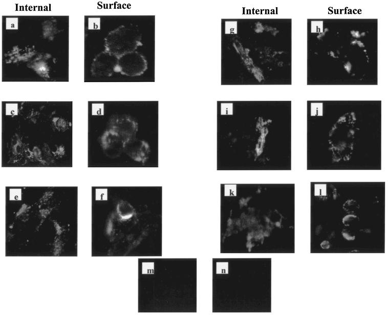
To study the transport of the mutant HN proteins synthesized in virus-infected cells from the cytoplasm to the cell surface, indirect immunofluorescence studies were carried out. As shown in Fig. 6, all mutant and parental viruses showed the presence of surface immunofluorescence in fixed cells and internal cytoplasmic immunofluorescence in fixed and permeabilized cells. Flow cytometry was used to quantitate these results. DF1 cells infected with each of these viruses were incubated with HN-specific MAb cocktail, and surface expression was determined by flow cytometry. The percentage of cells expressing the different mutated HN proteins was very similar except for rG4 virus, which had a significantly lower percentage (P < 0.05) of cells expressing the HN at the surface (Table 2). The mean fluorescence intensities ranged from 60 to 220% of the parental rBC virus. Cells infected with the rG4 virus also had a more decreased mean fluorescence intensity than that of the parental rBC virus. These results indicated that all glycosylation mutant HN proteins retained the ability to be transported to the cell surface except rG4 virus, which had decreased cell surface transport.
FIG. 6.
Detection of cell surface immunofluorescence of parental and mutant HN proteins. Virus-infected DF1 cells (0.1 MOI) were washed with PBS at 48 h PI and fixed with 3% paraformaldehyde in PBS for cell surface fluorescence (b, d, f, h, j, l, and n) or fixed and then permeabilized with 0.1% Triton X-100 in PBS for intracytoplasmic fluorescence (a, c, e, g, i, k, and m). HN proteins were stained with an anti-HN MAb cocktail, followed by affinity-purified fluorescein-labeled goat anti-mouse immunoglobulin (Kirkegaard and Perry Laboratories). (a and b) rBC; (c and d) rG1; (e and f) rG2; (g and h) rG3; (i and j) rG4; (k and l) rG12; (m and n) negative control. Cells were photographed at a magnification of ×200.
TABLE 2.
Cell surface expression of HN proteins of glycosylation mutants
| Virus | Efficiency of cell surface expressiona
|
|
|---|---|---|
| % Of positive cells ± SE | Relative mean fluorescence intensity | |
| rBC | 87 ± 3.1 | 1.00 |
| rG1 | 90 ± 7.0 | 1.60 |
| rG2 | 96 ± 1.5* | 2.20b |
| rG3 | 97 ± 0.6* | 2.00 |
| rG4 | 76 ± 1.3* | 0.60 |
| rG12 | 88 ± 5.7 | 1.00 |
| None (mock-infected cells) | 0.01 | |
Cell surface expression of HN was determined by flow cytometry. DF1 cells were infected with each virus at an MOI of 1. Surface expression of the HN proteins was assessed by flow cytometry at 18 h PI with a cocktail of NDV HN-specific MAbs followed by a fluorescein isothiocyanate-conjugated secondary antibody. Surface immunofluorescence was quantitated by fluorescence-activated cell sorter analysis with the FACSVantage flow cytometer (Becton Dickinson). Noninfected DF1 cells were used as negative controls. Values shown are averages of results from three independent experiments.
P < 0.05.
Biological activities of glycosylation mutant viruses.
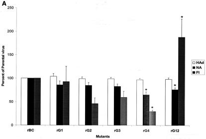
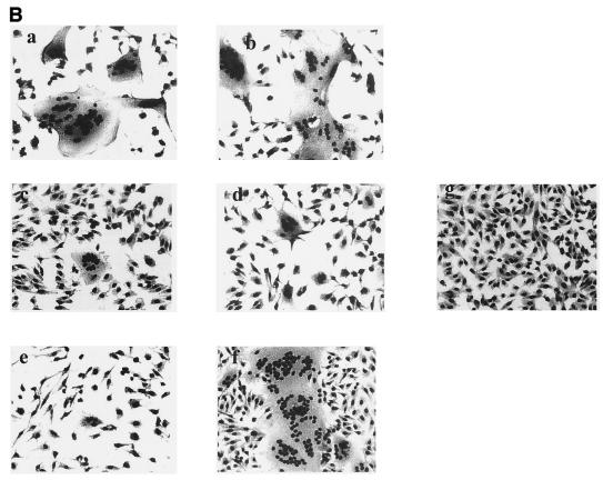
The results described above show that glycosylation at sites 2 and 4 affects the formation of a mature HN molecule. But the cell surface expression of virus missing site 2 was similar to that of the parental virus. However, proteins missing oligosaccharides at sites 1 and 3 singly or sites 1 and 2 in combination have no effect on antigenic site formation or cell surface expression. It is possible that the combined loss of two glycosylation sites alters the folding of the HN protein with better exposure of the antigenic epitopes. To explore the role of these carbohydrates in the biological activities of the HN protein, cells infected with the recombinant viruses were assayed for NA, hemadsorption (HAd), and fusion activities (Fig. 7A). Our results showed that the HAd values of all glycosylation mutant viruses were similar to that of parental virus, indicating that glycosylation probably does not play a major role in the attachment of NDV. The NA activity of mutants G1, G2, and G3 was also similar to that of the parental BC virus. Deletion of the G4 and G12 glycosylation sites, however, resulted in only 65 and 76% NA activity of the parental virus, respectively (P < 0.05). This was in contrast to earlier reported work, where the deletion of these sites resulted in a significant loss of NA activity. Loss of site G3 resulted in NA activity which was only 40% compared to the wild-type transfected HN, and loss of site G4 resulted in >80% loss of NA activity (28). It was also reported that there was a fourfold increase in the HAd activity of HN with deletion of sites 1 and 2. In our study, deletion of these sites did not have any deleterious effect on receptor recognition and the viruses with deleted sites 3 and 4 also recognized the receptor more efficiently than reported previously (28). This reinforces the view that there is no alternative to study the biological activities of individual glycoproteins in the context of viral infection. The fusion index of rG1 virus was similar to that of the parental rBC virus. The rG2 and rG3 viruses, however, had lower fusion indices numerically than the parental virus but were not considered significantly different (P > 0.05). Interestingly, the rG12 virus showed significantly (1.8-fold) higher fusogenicity (P < 0.05), and the fusion promotion activity of rG4 virus was approximately one-third of that of the parental virus (P < 0.05). The rG4 virus also showed reduced surface expression. The extent of fusion induced by parental and mutant viruses is shown in Fig. 7B.
FIG.7.
Biological activities of HN glycosylation mutants. The HAd, NA, and fusogenic activities of HN glycosylation mutants were examined. (A) HAd was measured as the percentage of the hemoglobin released compared to the parental rBC virus. The NA and fusion indices (FI) of the mutant viruses were also shown in comparison with those activities of the rBC virus. NA activity of purified viruses was measured by a fluorometric assay. The FI is the ratio of the total number of nuclei to the number of cells in which the nuclei were observed (i.e., the mean number of nuclei per cell). The averages of the results from three experiments are shown. *, P < 0.05. (B) Fusogenicity of mutant viruses in Vero cells in comparison to parental rBC virus. Infected cells were fixed at 48 h p.i. and stained with hematoxylin-eosin. The extent of fusion of the parental rBC (a) and the mutant viruses rG1, rG2, rG3, rG4, and rG12 (b, c, d, e, and f, respectively) in Vero cells was photographed on a Nikon Eclipse TE 300 microscope (magnification, ×168). An uninfected Vero cell monolayer (g) was included as a negative control for comparison.
Effect of loss of glycosylation from the HN protein on virulence.
Three internationally accepted tests were used to assess the degree of virulence of NDV isolates: the MDT test in 9- to 11-day-old embryonated SPF chicken eggs, the ICPI test in 1-day-old chicks, and the IVPI test in 6-week-old chickens. We examined the virulence of the parental and glycosylation mutant viruses by all three tests (Table 3). The MDT result showed a significant increase in the time required by rG4 virus (70 h) to kill the chicken embryos compared to the time required by the parental rBC virus (62 h), indicating a reduction in virulence of rG4 in vivo. The rG1 and rG3 viruses had MDT values of 60 h each, whereas rG2 and rG12 had MDT values of 64 h each. The ICPI values of all glycosylation mutants were lower than those of the parental virus (Table 3), indicating the loss of virulence in these mutants. All of the glycosylation mutants had lower IVPI values than that of the parental virus, indicating the loss of virulence. In aggregate, these results indicated that loss of an individual glycan(s) in the HN protein leads to reduction in virulence of NDV. Further, the loss of glycosylation at residue 481 (G4) or at residues 119 and 341 (G12) together greatly attenuated the virus.
TABLE 3.
HN glycosylation mutants of NDV and virus pathogenicity in vivo
| Virus | MDT (n) | ICPI | IVPI |
|---|---|---|---|
| rBC | 62 | 1.50 | 1.32 |
| rG1 | 60 | 1.12 | 0.37 |
| rG2 | 64 | 0.87 | 0.44 |
| rG3 | 60 | 1.07 | 0.75 |
| rG4 | 70 | 0.66 | 0.31 |
| rG12 | 64 | 0.76 | 0.67 |
DISCUSSION
N-linked carbohydrates play important roles for a variety of structural and functional activities of glycoproteins (32, 37, 46). Previous studies have shown that N-linked carbohydrates are important for folding, transport, and biological activities of the HN glycoprotein of NDV (10, 15, 28). We have characterized N-linked glycosylation mutants of NDV HN protein in terms of their biological activities and virulence. By using reverse genetics, we analyzed the role of individual N-linked oligosaccharides on the pathogenicity of NDV in its natural avian host.
It has been reported that only four of the six putative N-linked glycosylation sites in the NDV (Australia-Victoria strain) HN sequence are normally used for carbohydrate addition (28). Our results confirmed that the above-described four N-linked glycosylation sites are actually used in the context of infectious virus. Further, we have shown that all four glycosylation sites in the HN glycoprotein could be mutated individually without adversely affecting the recovery of replication-competent NDV. A double HN glycoprotein mutant virus lacking carbohydrates in sites 1 and 2 was also recovered. Similar to other virus systems (17, 38), conserved N-linked oligosaccharides of NDV HN protein are dispensable for viral replication.
The effect of substitution of single or combinations of several amino acid residues on HN protein expression and function has been reported for NDV (10, 15, 28), but none of the studies investigated the role of individual N-glycans in viral replication. We recovered viruses with individual mutations at glycosylation sites 1, 2, 3, and 4 and a combined mutation at glycosylation sites 1 and 2, and we tested the effects of these substitutions on viral replication in DF1 cells. Interestingly, viruses with mutations at site 4 (rG4) and sites 1 and 2 (rG12) had decreased growth rates and smaller plaque sizes than the other mutants or the parental virus. Each of the individual sites is dispensable for virus replication, although all sites appear to contribute to optimal replication efficiency. Glycosylation sites 1 to 4 are highly conserved in the HN protein of 13 different strains of NDV (39), which suggests that these sites are used and that all four side chains serve important biological functions (28). But conserved glycosylation sites in NDV are found to be largely dispensable for virus replication. Conserved N-linked glycans of human immunodeficiency virus type 1 gp41 envelope protein were found to be dispensable for viral replication (17). The delay in replication displayed by the G4 and G12 mutants may be the result of inefficient processing of the HN protein. At least for G4 virus, we demonstrated that the cell surface expression was compromised in comparison to the other mutants.
Antibody-selected NDV mutants with additional glycan chains on the HN protein decreased the ability of HN protein to complement the viral F protein in the promotion of cellular fusion (10). In our study, even though the in vitro growth rate of rG12 virus was lower, its fusogenicity was significantly higher than that of the parental virus. Its cell surface expression was similar to the parental virus. This result is in agreement with a previous study by McGinnes and Morrison (28), who demonstrated higher fusogenicity of G12 double mutant virus than that of the parental virus. These authors have suggested that increased fusion by rG12 may be due to the location of the first glycosylation site (residue 119) on the HN protein near the Cys-123 residue in the stalk region. This cysteine residue plays an important role in the disulfide linkage of the HN homodimers (27, 29, 35). Elimination of glycosylation at residue 119 may result in better covalent linkage by increasing the efficiency of participation of the adjacent cysteines in the covalent bond (28). But the NDV strain BC employed in this study has a tryptophan residue at position 123 instead of a cysteine residue. Therefore, in this strain, the increased fusogenicity might be a result of simultaneous elimination of carbohydrates at residues 119 and 341, leading to a better interaction of the HN mutant G12 with the F protein. Stem oligosaccharides have been shown to maintain the influenza virus hemagglutinin in the metastable form required for fusion activity (31).The loss of these glycans may, therefore, alter the interaction of HN with the F protein directly. Interaction of the HN and F proteins as requirements for cell fusion has been reported previously (45).
In vivo studies with rG12 virus showed loss of virulence in chickens. It is possible that removal of these glycosylation sites (residues 119 and 341) increases cell-cell fusion in vivo, resulting in rapid spread and destruction of the host cell monolayers. The antigenic reactivity of rG12 virus to the MAb B79 is higher than that of rBC virus. This mutant may thus cause an early and enhanced immune response in infected chickens and, hence, would be cleared by neutralizing antibodies of the immune system in the chickens during the initial stages of infection. Further, the loss of glycans may expose a T-cell epitope that would allow infected cells to be targeted by cytotoxic T cells, resulting in faster virus clearance and lowered pathology. The efficiency of fusion in cell culture, therefore, does not correlate to pathology in vivo. Thus, the presence of carbohydrates at residues 119 and 341 may serve to modulate viral infection and increase the survival time of the virus in the host. Our ICPI results corroborate this inference, as chicks inoculated with G12 mutant survived longer than the other mutants. There have been reports of an increase in virulence of a neurovirulent influenza virus strain A/NWS/33 in mice (25, 47) and the H5N2 influenza virus in chickens (18), with the loss of a carbohydrate from the HA protein of the viruses. Thus, loss of a glycosylation site(s) can influence the pathogenicity of viruses by either decreasing or increasing their virulence.
Carbohydrates masking neutralization epitopes on the viral surface glycoproteins, thus forming a shield that protects the virus from immune recognition, have been reported for many viruses (3, 4, 8, 12, 15, 43). We have shown that removal of carbohydrate side chains resulted in unmasking of the antigenic sites in NDV HN protein. The reactivity to neutralizing NDV MAbs increased when the glycosylation at sites 1 (G1), 1 and 2 together (G12), and 3 (G3) were removed. On the other hand, loss of glycosylation at sites 2 and 4 leads to reduction of reactivity of rG2 and rG4 viruses, respectively, to the MAbs. Previous epitope mapping studies have defined residues 193 to 201, 345 to 353, 494, 513 to 521, and 569 as the domains on the HN protein of NDV that are involved in neutralization (13, 14, 16). As no epitope mapping studies were available for the MAbs employed in this study, direct comparison to previous studies could not be made. However, it was evident that more neutralizing epitopes may be involved than previously thought.
The bulky hydrophilic carbohydrate chains in N-glycosylated proteins may aid protein folding by preventing inappropriate hydrophobic interactions. The role of individual oligosaccharides in the maturation of simian virus 5 (SV5), measles and Sendai viruses has been studied. Those studies showed, as did ours, that the loss of some oligosaccharides influenced folding more than the loss of others. In SV5, removal of oligosaccharides from the F1 subunit of the F protein of SV5 resulted in delayed intracellular transport and decreased stability of the protein (5). In measles virus and SV5, glycosylation has been shown to be necessary for folding of proteins into their native conformations, which in turn, affects the protein stability and intracellular transport (11, 30). In Sendai virus, the loss of glycosylation sites 3 and 4 affected the transport of the HN protein to the cell surface (41).
In our study, glycosylation site 4 of the HN protein of NDV is found to be necessary for correct folding, as the misfolded HN protein is incorrectly processed and transported to the cell surface inefficiently. Loss of this oligosaccharide reduces the formation of antigenic sites to approximately one-third the level of the parental virus. The cell surface expression of this virus was also significantly lower than that of the parental virus. Loss of oligosaccharides at site 2 had some effect on the formation of antigenic sites while mutants with deletions of sites 1 and 3 or 1 and 2 together had little or no effect on the formation of antigenic sites but had either increased or similar levels of cell surface expression compared to those of the parental virus. The glycosylation site 4 is located in the globular head and is likely to contribute to the conformation of the protein. Our results are in agreement with those of McGinnes and Morrison (28), who also observed that oligosaccharides at residue 481 had the most significant effect on folding of the HN protein of NDV. Since the HN protein mediates the attachment of the virus to host cells, a misfolded HN protein in the mutant virus would result in loss of infectivity and virulence of the virus. We have demonstrated that the loss of glycosylation at site 4 decreases the biological activities of the HN glycoprotein and attenuates the virus considerably. It is noteworthy that we were also able to recover the mutant virus with loss of glycosylation at site 4 (rG4), even though the production and cell surface expression of the HN protein in this virus were lower than those of the other mutants and the parental viruses.
A number of studies have shown that N-linked glycans influence proper folding, disulfide bond formation, and oligomerization of proteins and affect the conformation of proteins (5, 11, 27, 28, 30). Whether this conformational change affects the ability of the N-linked glycosylation mutants to function in NDV virions has not been tested so far. Our results provide evidence that loss of N-linked glycans alters the biological function of the HN protein and also modulates the virulence of the virus in vivo. The loss of an individual glycosylation site in the globular head region of NDV or a combined loss of glycosylation sites from the stalk and globular head regions of NDV resulted in attenuating the virulence of NDV. These studies indicate that by eliminating certain glycosylation sites, such as sites 1 and 2 found on the HN glycoprotein of NDV, attenuated NDV strains can be engineered. The retention of immunogenicity and stability of the attenuating mutations in chicken embryos make this mutant virus an attractive candidate for a recombinant vaccine.
Acknowledgments
We thank Daniel Rockemann and Peter Savage for excellent technical assistance. We also thank Ireen Dryburgh-Barry for assistance in the preparation of the manuscript. We are grateful to Richard Raybourne, Center for Veterinary Medicine, FDA, for help with flow cytometry.
This work was partially supported by U.S. Department of Agriculture grant no. 2002-35204-1601.
REFERENCES
- 1.Alexander, D. J. 1989. Newcastle disease, p. 114-120. In H. G. Purchase, L. H. Arp, C. H. Domermuth, and J. E. Pearson (ed.), A laboratory manual for the isolation and identification of avian pathogens, 3rd ed. American Association for Avian Pathologists, Inc., Kennett Square, Pa.
- 2.Alexander, D. J. 1997. Newcastle disease and other avian Paramyxoviridae infection, p. 541-569. In B. W. Calnek (ed.), Diseases of poultry, 10th ed. Iowa State University Press, Ames.
- 3.Alexander, S., and J. H. Elder. 1984. Carbohydrate dramatically influences immune reactivity of antisera to viral glycoprotein antigens. Science 226:1328-1330. [DOI] [PubMed] [Google Scholar]
- 4.Back, N. K. T., L. Smit, J. J. De Jong, W. Keulen, M. Schutten, J. Goudsmit, and M. Tersmette. 1994. An N-glycan within the human immunodeficiency virus type 1 gp120 V3 loop affects virus neutralization. Virology 199:431-438. [DOI] [PubMed] [Google Scholar]
- 5.Bagai, S., and R. A. Lamb. 1995. Individual roles in N-linked oligosaccharide chains in the intracellular transport of the paramyxovirus SV5 fusion protein. Virology 209:250-256. [DOI] [PubMed] [Google Scholar]
- 6.Byrappa, S., D. K. Gavin, and K. C. Gupta. 1995. A highly efficient procedure for site-specific mutagenesis of full-length plasmids using Vent DNA polymerase. Genome Res. 5:404-407. [DOI] [PubMed] [Google Scholar]
- 7.Choppin, P. W., and A. Scheid. 1980. The role of viral glycoproteins in adsorption, penetration and pathogenicity of viruses. Rev. Infect. Dis. 2:40-61. [DOI] [PubMed] [Google Scholar]
- 8.Davis, N. L., D. F. Pence, W. J. Meyer, A. L. Schmaljohn, and R. E. Johnston. 1987. Alternative forms of a strain-specific neutralizing antigenic site on the Sindbis virus E2 glycoprotein. Virology 161:101-108. [DOI] [PubMed] [Google Scholar]
- 9.De Leeuw, O., and B. Peeters. 1999. Complete nucleotide sequence of Newcastle disease virus: evidence for the existence of a new genus within the subfamily Paramyxovirinae. J. Gen. Virol. 80:131-136. [DOI] [PubMed] [Google Scholar]
- 10.Deng, R., Z. Wang, R. L. Glickman, and R. M. Iorio. 1994. Glycosylation within an antigenic site on the HN glycoprotein of Newcastle Disease virus interferes with its role in the promotion of membrane fusion. Virology 204:17-25. [DOI] [PubMed] [Google Scholar]
- 11.Hu, A., R. Cattaneo, S. Schwartz, and E. Norrby. 1994. Role of N-linked oligosaccharide chains in the processing and antigenicity of measles virus hemagglutinin protein. J. Gen. Virol. 75:1043-1052. [DOI] [PubMed] [Google Scholar]
- 12.Huso, D. L., O. Narayan, and G. W. Hart. 1988. Sialic acids on the surface of caprine arthritis-encephalitis virus define the biological properties of the virus. J. Virol. 62:1974-1980. [DOI] [PMC free article] [PubMed] [Google Scholar]
- 13.Iorio, R. M., K. A. Lawton, P. M. Nicholson, and M. A. Bratt. 1984. Monoclonal antibodies identify a strain-specific epitope on the HN glycoprotein of Newcastle disease virus strain Australia-Victoria. Virus Res. 1:513-525. [DOI] [PubMed] [Google Scholar]
- 14.Iorio, R. M., R. L. Glickman, A. M. Riel, J. P. Sheehan, and M. A. Bratt. 1989. Functional and neutralization profile of seven overlapping antigenic sites of the HN glycoprotein of Newcastle disease virus: monoclonal antibodies to some sites prevent viral attachment. Virus Res. 13:245-262. [DOI] [PubMed] [Google Scholar]
- 15.Iorio, R. M., R. L. Glickman, and J. P. Sheehan. 1992. Inhibition of fusion by neutralizing monoclonal antibodies to the hemagglutinin-neuraminidase glycoprotein of Newcastle disease virus. J. Gen. Virol. 73:1167-1176. [DOI] [PubMed] [Google Scholar]
- 16.Iorio, R. M., R. J. Syddall, J. P. Sheehan, M. A. Bratt, R. L. Glickman, and M. Riel. 1991. Neutralization map of the hemagglutinin-neuraminidase glycoproteins of Newcastle disease virus: domains recognized by monoclonal antibodies that prevent receptor recognition. J. Virol. 65:4999-5006. [DOI] [PMC free article] [PubMed] [Google Scholar]
- 17.Johnson, W. E., J. M. Sauvron, and R. C. Desrosiers. 2001. Conserved N-linked carbohydrates of human immunodeficiency virus type 1 gp41 are largely dispensable for viral replication. J. Virol. 75:11426-11436. [DOI] [PMC free article] [PubMed] [Google Scholar]
- 18.Kawaoka, Y., C. W. Naeve, and R. G. Webster. 1984. Is virulence of H5N2 influenza viruses in chickens associated with loss of carbohydrate from the hemagglutinin? Virology 139:303-316. [DOI] [PubMed] [Google Scholar]
- 19.Kohn, A. 1965. Polykaryocytosis induced by Newcastle disease virus in monolayers of animal cells. Virology 26:228-245. [DOI] [PubMed] [Google Scholar]
- 20.Kornfeld, R., and S. Kornfeld. 1985. Assembly of asparagines-linked oligosaccharides. Annu. Rev. Biochem. 54:631-664. [DOI] [PubMed] [Google Scholar]
- 21.Krishnamurthy, S., and S. K. Samal. 1998. Nucleotide sequences of the trailer, nucleocapsid protein gene and intergenic regions of Newcastle disease virus strain Beaudatte C and completion of the entire genome sequence. J. Gen. Virol. 79:2419-2424. [DOI] [PubMed] [Google Scholar]
- 22.Krishnamurthy, S., Z. Huang, and S. K. Samal. 2000. Recovery of a virulent strain of Newcastle disease virus from cloned cDNA: expression of a foreign gene results in growth retardation and attenuation. Virology 278:168-182. [DOI] [PubMed] [Google Scholar]
- 23.Lamb, R. A., and D. Kolakofsky. 2001. Paramyxoviridae: the viruses and their replication, p. 1305-1340. In D. M. Knipe and P. M. Howley (ed.), Fields virology. Lippincott Williams & Wilkins, Philadelphia, Pa.
- 24.Lana, D. P., D. B. Snyder, D. J. King, and W. W. Marquardt. 1988. Characterization of a battery of monoclonal antibodies for differentiation of Newcastle disease virus and pigeon paramyxovirus-1 strains. Avian Dis. 32:273-281. [PubMed] [Google Scholar]
- 25.Li, S., J. Schulman, S. Itamura, and P. Palese. 1993. Glycosylation of neuraminidase determines the neurovirulence of influenza A/WSN/33 virus. J. Virol. 67:6667-6673. [DOI] [PMC free article] [PubMed] [Google Scholar]
- 26.Mayo, M. A. 2002. A summary of taxonomic changes recently approved by ICTV. Arch. Virol. 147:1655-1656. [DOI] [PubMed] [Google Scholar]
- 27.McGinnes, L. W., and T. G. Morrison. 1994. Modulation of the activities of HN protein of Newcastle disease virus by non-conserved cysteine residues. Virus Res. 34:305-316. [DOI] [PubMed] [Google Scholar]
- 28.McGinnes, L. W., and T. G. Morrison. 1995. The role of individual oligosaccharide chains in the activities of the HN glycoprotein of Newcastle disease virus. Virology 212:398-410. [DOI] [PubMed] [Google Scholar]
- 29.Mirza, A. M., J. P. Sheehan, L. W. Hardy, R. L. Glickman, and R. M. Iorio. 1993. Structure and function of a membrane anchor-less form of the hemagglutinin-neuraminidase glycoprotein of Newcastle disease virus. J. Biol. Chem. 268:21425-21431. [PubMed] [Google Scholar]
- 30.Ng, D. T. W., S. W. Hiebert, and R. A. Lamb. 1990. Different roles of individual N-linked oligosaccharide chains in folding, assembly, and transport of the simian virus 5 hemagglutinin-neuraminidase. Mol. Cell. Biol. 10:1989-2001. [DOI] [PMC free article] [PubMed] [Google Scholar]
- 31.Ohuchi, R., M. Ohuchi, W. Garten, and H.-D. Klenk. 1997. Oligosaccharides in the stem region maintain the influenza virus hemagglutinin in the metastable form required for fusion activity. J. Virol. 71:3719-3725. [DOI] [PMC free article] [PubMed] [Google Scholar]
- 32.Olden, K., J. B. Parent, and S. L. White. 1982. Carbohydrate moieties of glycoproteins. A re-evaluation of their function. Biochim. Biophys. Acta 650:209-232. [DOI] [PubMed] [Google Scholar]
- 33.Peeples, M. E. 1988. Newcastle disease virus replication, p. 45-78. In D. J. Alexander (ed.), Newcastle disease. Kluwer Academic Publishers, Dordrecht, The Netherlands.
- 34.Phillips, R. J., A. C. R. Samson, and P. T. Emmerson. 1998. Nucleotide sequence of the 5′ terminus of Newcastle disease virus and assembly of the complete genomic sequence: agreement with the “rule of six.” Arch. Virol. 143:1993-2002. [DOI] [PubMed] [Google Scholar]
- 35.Pitt, J. J., E. De Silva, and J. J. Gorman. 2000. Determination of the disulfide bond arrangement of Newcastle disease virus hemagglutinin neuraminidase. J. Biol. Chem. 275:6469-6478. [DOI] [PubMed] [Google Scholar]
- 36.Potier, M., L. Mameli, M. Belishem, L. Dallaire, and S. B. Melancon. 1979. Fluorometric assay of neuraminidase with a sodium (4-methylumbelliferyl-alpha-D-N-acetylneuraminate) substrate. Anal. Biochem. 94:287-296. [DOI] [PubMed] [Google Scholar]
- 37.Quinones-Kochs, M. I., L. Buonocore, and J. K. Rose. 2002. Role of N-linked glycans in a human immunodeficiency virus envelope glycoprotein: effects on protein function and the neutralizing antibody response. J. Virol. 76:4199-4211. [DOI] [PMC free article] [PubMed] [Google Scholar]
- 38.Reitter, J. N., and R. C. Desrosiers. 1998. Identification of replication-competent strains of simian immunodeficiency virus lacking multiple attachment sites for N-linked carbohydrates in variable regions 1 and 2 of the surface envelope protein. J. Virol. 72: 5399-5407. [DOI] [PMC free article] [PubMed] [Google Scholar]
- 39.Sakaguchi, T., T. Toyoda, B. Gotoh, N. M. Inocencio, K. Kuma, T. Miyata, and Y. Nagai. 1989. Newcastle disease virus evolution. I. Multiple lineages defined by sequence variability of the hemagglutinin-neuraminidase gene. Virology 169:260-272. [DOI] [PubMed] [Google Scholar]
- 40.Schuy, W., W. Garten, D. Linder, and H. D. Klenk. 1984. The carboxyterminus of the hemagglutinin-neuraminidase of Newcastle disease virus is exposed at the surface of the viral envelope. Virus Res. 1:415-426. [DOI] [PubMed] [Google Scholar]
- 41.Segawa, H., A. Inakawa, T. Yamashita, and H. Taira. 2003. Functional analysis of individual oligosaccharide chains of Sendai virus hemagglutinin-neuraminidase protein. Biosci. Biotechnol. Biochem. 67:592-598. [DOI] [PubMed] [Google Scholar]
- 42.Shakin-Eshleman, S. H., A. T. Remaley, J. R. Eshleman, W. H. Wunner, and S. L. Spitalnik. 1992. N-linked glycosylation of rabies virus glycoprotein. J. Biol. Chem. 267:10690-10698. [PubMed] [Google Scholar]
- 43.Skehel, J. J., D. J. Stevens, R. S. Daniels, A. R. Douglas, M. Knossow, I. A. Wilson, and D. C. Wiley. 1984. A carbohydrate side chain on hemagglutinins of Hong Kong influenza viruses inhibits recognition by a monoclonal antibody. Proc. Natl. Acad. Sci. USA 81:1779-1783. [DOI] [PMC free article] [PubMed] [Google Scholar]
- 44.Steward, M., B. Vipoond, N. S. Miller, and P. T. Emmerson. 1993. RNA editing in Newcastle disease virus. J. Gen Virol. 74:2539-2547. [DOI] [PubMed] [Google Scholar]
- 45.Stone-Hulslander, J., and T. G. Morrison. 1997. Detection of an interaction between the HN and F proteins in Newcastle disease virus-infected cells. J. Virol. 71:6287-6295. [DOI] [PMC free article] [PubMed] [Google Scholar]
- 46.Tikoo, S. K., M. D. Parker, J. V. Van den Hurk, J. Kowalski, T. J. Zamb, and L. Babiuk. 1993. Role of N-linked glycans in antigenicity, processing, and cell surface expression of bovine herpesvirus 1 glycoprotein gIV. J. Virol. 67:726-733. [DOI] [PMC free article] [PubMed] [Google Scholar]
- 47.Ward, A. C., and T. F. De Koning-Ward. 1995. Changes in the hemagglutinin gene of the neurovirulent influenza virus strain A/NWS/33. Virus Genes 102:179-183. [DOI] [PubMed] [Google Scholar]


