Abstract
The Newcastle disease virus (NDV) hemagglutinin-neuraminidase (HN) protein mediates attachment to cellular receptors. The fusion (F) protein promotes viral entry and spread. However, fusion is dependent on a virus-specific interaction between the two proteins that can be detected at the cell surface by a coimmunoprecipitation assay. A point mutation of I175E in the neuraminidase (NA) active site converts the HN of the Australia-Victoria isolate of the virus to a form that can interact with the F protein despite negligible receptor recognition and fusion-promoting activities. Thus, I175E-HN could represent a fusion intermediate in which HN and F are associated and primed for the promotion of fusion. Both the attachment and fusion-promoting activities of this mutant HN protein can be rescued either by NA activity contributed by another HN protein or by a set of four substitutions at the dimer interface. These substitutions were identified by the evaluation of chimeras composed of segments from HN proteins derived from two different NDV strains. These findings suggest that the I175E substitution converts HN to an F-interactive form, but it is one for which receptor binding is still required for fusion promotion. The data also indicate that the integrity of the HN dimer interface is critical to its receptor recognition activity.
The Newcastle disease virus (NDV) hemagglutinin-neuraminidase (HN) is a multifunctional, type II membrane glycoprotein. It exists at the surface of virions and virus-infected cells as a tetrameric spike structure, consisting of a long, membrane-proximal stalk region that supports a terminal globular domain. The latter region of the spike is responsible for HN′s recognition of sialic acid-containing receptors as well as its neuraminidase (NA) activity, which cleaves sialic acid from both soluble and membrane-bound glycoconjugates (18). All of the antibody binding sites on the protein also reside in the globular domain (21).
Several hundred strains of NDV have been isolated from all over the world. The X-ray crystallographic structures of the globular domain of the HN from one of these, the Kansas strain, as well as the protein complexed with sialic acid or an inhibitor of NA, have been solved (3). These structures suggest that the attachment and NA activities of HN are mediated by a single sialic acid binding site in two different conformations. Consistent with this, substitutions for several active site residues abolish both attachment and NA (3).
Similarly, substitutions for residues in the NA active site of the HN from the Australia-Victoria (AV) isolate of NDV (NDV-AV) also result in the loss of detectable attachment and NA (11). These substitutions include I175E, D198R, K236R, Y526L, and E547Q. However, our laboratory has shown that the attachment activity of each of these receptor recognition-deficient mutants can be partially rescued by providing NA activity (11). Clearly, this is inconsistent with the existence of a single site for both attachment and NA. Rather, it suggests that the deficiency in receptor binding exhibited by these mutants is secondary to the lack of NA and that attachment and NA are defined by distinct sites.
In most paramyxoviruses, HN possesses a third function. It is required for the conversion of the second viral surface glycoprotein, the fusion (F) protein, to its active form (reviewed in reference 18). This fusion activation requires a virus-specific interaction between the two proteins, the specificity of which is determined by a domain defined primarily by the stalk region of the HN spike, as shown by the analysis of chimeras composed of domains from heterologous HN proteins (5, 32, 33). A role for the HN stalk in fusion is also supported by the diminished fusogenic activity exhibited by HN proteins carrying point mutations in this region (4, 25, 27, 30). The existence of an HN-F interaction is supported by the coimmunoprecipitation of the two proteins from the surface of both virus-infected and HN-F-cotransfected cells (4).
Our laboratory has shown that attachment-deficient NDV-AV HN, carrying substitutions for the catalytic aspartic acid, D198, is also fusion deficient and fails to interact with F at the cell surface in coimmunoprecipitation assays (4). This result is consistent with a mechanism for fusion in which receptor recognition by the globular domain of HN is the trigger for the HN-F interaction and fusion (17). However, the relationships among receptor recognition, HN structure, the HN-F interaction, and fusion are all still poorly understood.
Certain substitutions in the active site of the HN protein from the Kansas strain of NDV are capable of separating fusion from receptor recognition. Specifically, E401D and Y526F or L substitutions induce enhanced fusion promotion, despite negligible hemadsorption (HAd) activity. Similarly, I175E-mutated HN promotes fusion 50% more effectively than the wild-type (wt) protein, despite having 60% less HAd activity (1). To explain this phenomenon, it was postulated that these substitutions induce structural changes in HN that mimic those that occur upon receptor binding. These structural changes may involve alterations in the structure of the HN dimer interface, thereby triggering the HN-mediated fusion-activation of the F protein.
In contrast to I175E-mutated HN from NDV-Kansas, HN from the NDV-AV isolate carrying the same substitution exhibits negligible HAd and NA activities (11). We show here that this mutant HN also fails to promote fusion. Despite this lack of function, I175E-AV HN efficiently interacts with the homologous F protein. Both the attachment (11) and fusion-promoting activities of the mutated protein can be rescued by providing NA activity. We have constructed HN chimeras composed of various segments from the HN proteins of NDV-AV and strain Kansas-Leavenworth (NDV-L). The latter is closely related to the Kansas strain used in the above-mentioned crystallographic and mutational studies. Analyses of these chimeras and site-directed mutants confirm that four residues at the dimer interface are responsible for the phenotypic differences between the I175E-mutated forms of HN from the two strains. Based on these findings, we propose that I175E-mutated NDV-AV HN represents a form of the protein that is capable of interacting with the F protein in the absence of receptor recognition. This form of HN is still incapable of triggering the fusion-activation of F due to its inability to bind receptors as a result of amino acid differences localized to the dimer interface. These findings also argue for the importance of the structure of the dimer interface for the receptor recognition activity of HN.
MATERIALS AND METHODS
Recombinant plasmids.
Construction of the NDV-AV HN and F recombinant pBluescript SK(+) (Stratagene Cloning Systems, La Jolla, Calif.) expression vectors has been described previously (20). The HN of B1-Hitchner (NDV-B1) was a gift of Kemal Karaca (Megan Health, Inc., St. Louis, Mo.). It was released from the vector in which it was supplied by XhoI digestion and ligated into the pBSK plasmid cut with the same enzyme. The Beaudette C (NDV-BC) HN gene was a gift of Peter Emmerson (University of Newcastle Upon Tyne, Newcastle Upon Tyne, England). It was released from the vector in which it was supplied by digestion with XbaI and SacI and ligated into pBSK cut with the same enzymes.
The HN gene of NDV-L was generated by reverse transcription-PCR. Viral RNA was extracted by Trizol LS reagent (Invitrogen, Carlsbad, Calif.). First-strand cDNA was synthesized with Superscript II RNase H− reverse transcriptase (Invitrogen). The HN gene was amplified by the Expand High-Fidelity PCR system (Roche Diagnostics, Indianapolis, Ind.), with primers P1 (5′-CCCAAGCTTACCTCCGTTCTACCGCTTCA-3′) and P2 (5′-GCTCTAGAGCTCGCACCGGCATTCAGTT-3′). The HindIII and XbaI sites, respectively, are underlined in the primer sequences. The HN gene was gel purified, digested with HindIII and XbaI and cloned into pBSK at the same sites, resulting in plasmid pBSK-L-HN. Three independent clones were sequenced at the University of Massachusetts Nucleic Acid Facility. The sequence of L-HN used in this study is 98.3% homologous to that of the Kansas isolate used to generate the crystals of the protein (3). There are a total of 10 amino acid differences between the two proteins, 5 of which are in the tail and transmembrane domains. Amino acid differences in the ectodomain between L-HN and Kansas-HN are D for N144, V for A271, S for G310, E for G332, and A for V571. Only the last four amino acid differences reside in the globular domain of the protein.
wt L-HN and all mutants and chimeras derived from it were mutated to have a W123C mutation. Cysteine at this position mediates disulfide-linked homodimer formation, analogous to AV-HN, as confirmed by sodium dodecyl sulfate (SDS)-polyacrylamide gel electrophoresis (21). This was done for two reasons. First, Crennell et al. (3) have reported that this disulfide bond results in higher fusion-promoting activity, a finding that we have confirmed (data not shown). Second, in comparing the functional activities of AV-HN/L-HN chimeras with different stalk regions, we thought it important that those stalk regions have the same quaternary structure. The W123C mutation was also introduced into the HN of NDV-BC and NDV-B1, inducing disulfide-linked dimer formation in proteins that are also normally non-disulfide-linked (28).
Construction of pCAGGS-HN and pCAGGS-F expression vectors.
AV-HN and mutants derived from it were released from pBSK by digestion with XbaI and SacI, blunt-ended by DNA polymerase I large (Klenow) fragment (New England Biolabs, Beverly, Mass.), and then cloned into the EcoRI and SacI sites of pCAGGS, also blunted by Klenow. L-HN was released from pBSK by digestion with HindIII and XhoI, blunted by Klenow, and cloned into the EcoRI and XhoI sites of pCAGGS, also blunt-ended by Klenow. AV-F was released from pBSK by digestion with XhoI and cloned into pCAGGS, which had been digested by the same enzyme. The correct orientation of each of the inserts was verified by enzyme digestion and sequencing.
Site-directed mutagenesis.
Site-directed mutagenesis was performed as described previously (2), with mutagenesis primers obtained from Integrated DNA Technologies, Inc. (Coralville, Iowa). Identification of mutants was facilitated by screening for the presence of a unique restriction enzyme site introduced by a silent mutation in the mutagenic primer. The presence of the desired mutation was verified by sequencing of double-stranded DNA, with the Sequenase plasmid sequencing kit (United States Biochemical, Cleveland, Ohio), according to protocols provided by the company. Multiple clones were characterized for each substitution.
Construction of chimeric HN genes.
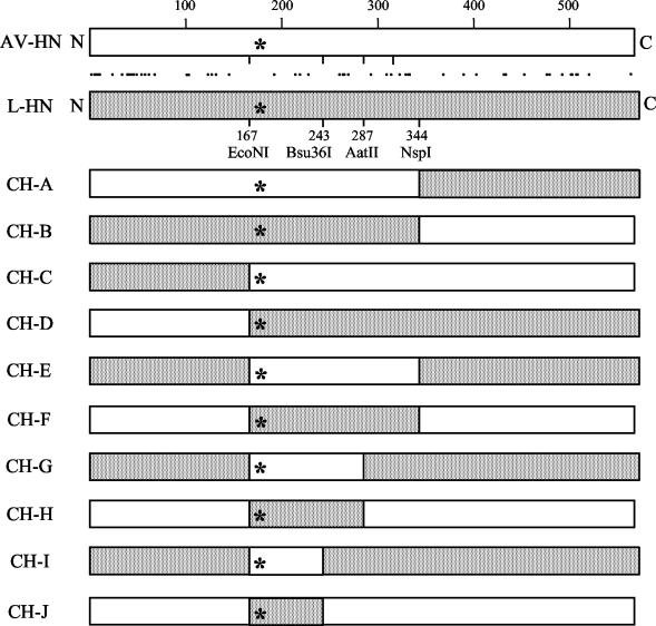
Using unique, homologous restriction enzyme sites in the AV-HN and L-HN genes, an initial pair of chimeric HN genes (CH-A and CH-B) was constructed (Fig. 1). Taking advantage of NspI sites in the HN gene and in the vector, C-terminal fragments of the two HN genes were isolated. The fragments were ligated into the heterologous HN gene previously digested with the same enzyme. This resulted in chimeras CH-A and CH-B, in which the N-terminal 344 residues are derived from AV and L HN, respectively, and the rest of the protein is derived from the heterologous HN protein.
FIG. 1.
Construction of chimeras of HN glycoproteins of the AV and L strains of NDV. Chimeras were constructed between the HN of the AV strain (white bars) and that of the L strain (stippled bars). This was made possible by the use of unique restriction enzyme sites in the two genes corresponding to amino acid residues 167, 243, 287, and 344. Residue 175 in each protein is indicated by an asterisk. Residues that are not conserved between the two proteins are indicated by dots between the sequences of the two parent proteins. Also, L-HN has six additional residues at its C terminus relative to AV-HN.
To exchange the N-terminal 167 amino acids of AV-HN and L-HN, we took advantage of a unique EcoNI site in L-HN. The same site was introduced into AV-HN by using the primer 5′-GAATTTTATACCAGCACCTACTACAGGATCAG-3′. This did not result in any amino acid changes in the AV-HN protein. The mutated HN genes were cut with HindIII and EcoNI, and the fragments were ligated into the heterologous HN gene that had been digested with the same enzymes to produce chimeras CH-C and CH-D.
To exchange the amino acids between residues 168 and 344 in AV-HN and L-HN, chimeras CH-C and CH-D were cut with NspI and the fragments were ligated into the heterologous chimeric HN gene previously digested with the same enzyme. This resulted in chimeras CH-E and CH-F.
To exchange the amino acids between residues 168 and 288 in AV-HN and L-HN, chimeras CH-C and CH-D were cut with AatII and SacI and the fragments were ligated into the heterologous, chimeric HN genes that had been digested with the same enzymes. This resulted in chimeras CH-G and CH-H.
To exchange the amino acids between residues 168 and 244 in AV-HN and L-HN, we took advantage of unique, homologous Bsu36I sites in the two genes. Chimeras CH-C and CH-D were cut with this enzyme and SacI, and the fragments were ligated into the heterologous HN genes that had been digested with the same enzymes, resulting in chimeras CH-I and CH-J.
Transient expression systems.
wt and mutant HN proteins were expressed in BHK-21 cells (American Type Culture Collection, Manassas, Va.) by using the vaccinia-T7 RNA polymerase expression system (6). All experiments, except NA assays, were performed in 35-mm-diameter plates seeded a day earlier at 4 × 10⁵ cells/well. Maintenance of cells, infection with the vaccinia recombinant, and transfection were performed as described previously (2), with 1 μg of each plasmid for transfection.
HN and F proteins in the pCAGGS vector were expressed in HeLa cells that were seeded in six-well plates a day earlier at 4 × 10⁵ cells/well. DNA (1.5 μg/well) was transfected into the cells with PolyFect transfection agent (Qiagen Inc., Valencia, Calif.) according to protocols provided by the company. Assays were performed 48 h posttransfection.
Cell surface expression.
The amount of each HN mutant at the cell surface was determined by flow cytometry with a mixture of monoclonal antibodies (MAbs), including at least three that recognize epitopes conserved in the globular domains of each of the HN proteins used in this study (9, 10, 14-16).
HAd assay.
The HAd activity of HN proteins was assayed by the ability of expressed proteins to adsorb guinea pig erythrocytes (Crane Laboratories, Syracuse, N.Y.). HN-expressing monolayers were incubated for 30 min at 4°C with a 2% suspension of erythrocytes in phosphate-buffered saline (PBS) supplemented with 1% CaCl2 and MgCl2. After extensive washing, adsorbed erythrocytes were lysed in 50 mM NH4Cl and the lysate was clarified by centrifugation. HAd activity was quantitated by measuring the absorbance at 540 nm minus the background obtained with vector-expressing cells.
NA assay.
NA assays were performed on 12-well (22.6-mm diameter) plates seeded a day earlier at 1.6 × 10⁵ cells/well and transfected with 0.5 μg of DNA. Monolayers were incubated at 37°C for 20 min with 0.5 ml of 625-μg/ml neuraminlactose (Sigma Chemical Co., St. Louis, Mo.) in 0.1 M sodium acetate (pH 6), and NA activity was determined as described previously (20). Data were corrected for differences in cell surface expression.
Content-mixing assay for fusion.
The ability of the mutated HN proteins to complement the F protein in the promotion of fusion was quantitated by a content-mixing assay which measures β-galactosidase activity in target cells following fusion induced by HN-F-expressing effector cells (2).
Rescue of fusion-promoting activity of HN mutants by coexpression with another HN protein.
Using a modification of an assay described previously (11), we determined whether the fusion-promoting activity of functionally deficient mutants could be rescued through the contribution of NA activity by another coexpressed HN protein. The HN protein chosen for these experiments was a human parainfluenza virus type 3 (hPIV3)-NDV chimera, called CH1(+11) (5). This chimera has 152 N-terminal amino acids derived from hPIV3 HN and does not oligomerize with NDV HN. The rest of the protein, including the entire globular domain, is derived from NDV-AV HN. It retains approximately 10% of wt NDV-AV HN NA activity.
To make it possible to selectively inhibit the attachment activity of the chimera, an E347G substitution was introduced into each of the functionally deficient mutants as well as the control wt protein. This substitution renders the protein unrecognizable by MAbs to antigenic site 14, which inhibits the attachment and fusion activities of the rescuing chimera (9, 14). The substitution itself has no effect on HN function (9, 16).
To determine the fusion-promoting activity of mutants coexpressed with CH1(+11), the content-mixing assay was used. The HN-F-expressing cells (105 cells in 100 μl of medium) were treated with 200 μg of MAb 14f for 30 min at 37°C prior to mixing with the target cells containing the β-galactosidase gene. This antibody completely blocks the attachment and fusion activities of the chimera but has no effect on the mutants carrying the E347G substitution. The HAd and fusion-promoting activities of the monolayers were then determined. The data are expressed as percentages of the activities obtained with monolayers expressing wt HN carrying the E347G substitution after subtraction of the background from vector-expressing cells.
Coimmunoprecipitation assay.
The ability of wt and mutated HN proteins to interact with F at the surface of transfected BHK cells was assayed at 16 h posttransfection, by a modification of a coimmunoprecipitation assay described previously (4). Because the amount of the F protein immunoprecipitated from the cell surface is greatly affected by the extent of fusion, we used a cleavage site mutant form of F for which cleavage activation and fusion are dependent on trypsin treatment. Our laboratory has previously shown that this mutated form of F interacts efficiently with the HN protein in the coimmunoprecipitation assay (4).
Equal numbers of cells were starved and labeled as described previously (4) and washed three times with cold PBS supplemented with 0.1 mM CaCl2 and 1 mM MgCl2. Cell surface proteins were biotinylated with sulfo-NHS-SS-biotin (Pierce, Rockford, Ill.) for 30 min in the cold with gentle agitation, followed by removal of the excess reagent by washing twice with PBS supplemented with 0.1 mM CaCl2 and 1 mM MgCl2. Cells were lysed in DH buffer (50 mM HEPES [pH 7.2], 10 mM dodecyl-β-d maltoside [United States Biochemical], 150 mM NaCl) containing 1 mM phenylmethylsulfonyl fluoride for 45 min in the cold. Lysates were split into two aliquots and immunoprecipitated for 90 min in the cold with either a MAb specific for the F protein only or this MAb plus a mixture of anti-HN MAbs. After centrifugation, the supernatant was added to protein A-agarose beads and incubated for 1 h in the cold, after which immunobeads were washed four times with DH buffer. Bound proteins were released by boiling for 5 min in 10 μl of 10% SDS. Samples were brought up to 0.5 ml with DH buffer, and the beads were pelleted. Proteins were reprecipitated from the supernatant with streptavidin beads, and the immunoprecipitates were displayed by SDS-polyacrylamide gel electrophoresis. The percentage of total cell surface HN that coimmunoprecipitated with F was quantified with a Bio-Rad Fluor-S multi-imager (Hercules, Calif.). Cells expressing vector, HN, or cleavage site mutant F alone served as controls to ensure that the coimmunoprecipitation of HN by the F antibody occurred only through its interaction with the F protein.
RESULTS
Attachment-deficient mutants lack detectable fusion-promoting activity.
It was previously demonstrated that several substitutions in the NA active site completely abolish the HAd and NA activities of NDV AV-HN, despite efficient expression of the protein at the cell surface. These mutations include I175E, D198R, K236R, Y526L, and E547Q (4, 11). D198R-mutated HN is also unable to complement the homologous F protein in the promotion of fusion (4). We have now examined the fusion-promoting capability of HN carrying each of the other active site mutations. Similar to D198R-HN, none of these mutants, including I175E-AV-HN, exhibits significant fusion-promoting activity. All promote fusion less than 3% of that of wt HN. Results for each substitution mutant are as follows (results are percentages of wt AV-HN): I175E, 1.9 ± 0.7; D198R, 1.1 ± 0.1; K236R, 1.2 ± 0.6; E547Q, 2.7 ± 0.2. No fusion was detected for Y526L.
I175E-mutated AV-HN interacts with F, despite its lack of attachment and fusion-promoting activities.
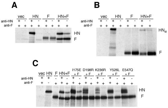
To assay for the presence of an interaction between the HN and F proteins at the cell surface, we use a coimmunoprecipitation assay that determines the amount of HN brought down through its interaction with F by an antibody to the latter. Figure 2A shows the coimmunoprecipitation results for wt AV-HN and F. To determine the total amount of each protein present at the cell surface, each set of monolayers was immunoprecipitated with a mixture of anti-HN MAbs and an anti-F MAb (the first lane in each set). The percentage of cell surface HN that interacts with F is quantitated by determining the amount of HN that coimmunoprecipitates with F, with only the anti-F MAb (the second lane in each set). The last lane in this gel shows that a protein comigrating with HN is coimmunoprecipitated by the F antibody from the surface of cells coexpressing the HN and F proteins.
FIG. 2.
Coimmunoprecipitation of AV-HN and fusion-deficient mutants derived from it with the homologous F protein. Equal numbers of cells were transfected as indicated. The F protein used in these experiments is a cleavage site mutant of NDV-AV F that is not cleaved and does not promote fusion (4). After 16 h, the cells were starved and labeled as described previously (4). The cell surface proteins were biotinylated, and the cells were lysed by treatment with dodecyl maltoside. The lysate was split into two aliquots and immunoprecipitated with either a combination of an anti-F antibody and a mixture of anti-HN antibodies (the first lane in each pair) or an anti-F antibody alone (the second lane in each pair). The gels in panels A and C were run under reducing conditions. The gel in panel B was run under nonreducing conditions. +, present; −, absent; vec, vector; HNdi, HN dimer.
To verify that the coimmunoprecipitated protein is authentic HN, a duplicate experiment of that in Fig. 2A was performed and the gel was run under nonreducing conditions (Fig. 2B). Under these conditions, the protein coimmunoprecipitated with F comigrates near the top of the gel with the disulfide-linked dimer of HN precipitated by the anti-HN antibody. This verifies that the protein coimmunoprecipitating with F is HN.
The mean of five experiments identical to that shown in Fig. 2A reveals that the percentage of total cell surface HN that coimmunoprecipitates with F in this assay is 32.6% ± 7.6%. Critical controls for this experiment show that HN is not brought down by the F antibody in the absence of the F protein and is not present in precipitates from cells expressing only the F protein (Fig. 2A). These controls ensure that the coimmunoprecipitation of HN occurs only through its interaction with F.
Figure 2C shows coimmunoprecipitation results for the functionally deficient I175E, D198R, K236R, Y526L, and E547Q mutated HN proteins. Consistent with previous findings (4), D198R-HN does not interact with F in this assay. Similarly, no interaction can be detected between the F protein and HN carrying either the K236R or E547Q substitution. A band comigrating with HN is barely detectable with Y526L mutated HN, but it is less than 1% of the total amount of Y526L mutated HN at the cell surface and is probably not significant. However, I175E-HN does coimmunoprecipitate with F. The mean of three determinations shows that 14.3% ± 4.7% of the total amount of the mutant at the cell surface coimmunoprecipitates with F. Again, this mutant HN-F interaction takes place despite negligible HAd, NA, and fusion.
The NA deficiency of each of the HN mutants is evident from the slower migration rate of the F protein coexpressed with any of the mutants, even I175E, compared to that of F coexpressed with wt HN. F coexpressed with the mutants comigrates with F expressed in the absence of HN. The comparatively faster migration rate of F coexpressed with HN results from the removal of sialic acid from its glycosyl groups mediated by the wt HN NA activity, which is lacking in the mutated proteins.
I175E-AV-HN is not dominant negative for fusion; its fusion-promoting activity can be rescued by NA activity contributed by a coexpressed protein.
Since the interaction between I175E-HN and F results in a complex that is nonfusogenic, the possibility exists that I175E mutated HN may be dominant negative for fusion when coexpressed with the wt HN protein. The mutated protein could tie up F protein spikes in a nonfusogenic complex, precluding their association with wt F and decreasing the extent of fusion. To test this possibility, we cotransfected the mutant along with wt HN and F and stained the monolayers for fusion. The presence of the mutant, even up to a threefold excess over wt HN, has no effect on the extent of fusion (data not shown).
The HAd activity of proteins carrying any of the above substitutions in the NA active site, including I175E, can be rescued by the contribution of NA either exogenously or by coexpression with another protein (11). Thus, we have explored the possibility that the ability of these mutants to complement the F protein in the promotion of fusion can also be rescued in a similar fashion. To address this question, we have used a modification of a chimera rescue assay described previously (11). In this assay, the attachment-deficient mutant is coexpressed with an hPIV3-NDV HN chimera, whose attachment and fusion-promoting activities are blocked by an NDV-HN-specific MAb. The mutant carries an innocuous substitution that renders it unrecognizable by the inhibiting antibody. In the content-mixing assay for fusion, the cells coexpressing the attachment-deficient HN mutant, the rescuing chimera, and NDV F are treated with the anti-HN inhibiting antibody prior to mixing with the target cells to initiate fusion. This ensures that any attachment and fusion in the system is mediated through the rescue of the attachment and fusion activities of the mutant.
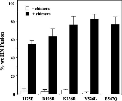
The results of this experiment for the NDV-AV HN mutants are shown in Fig. 3. As expected, none of the mutants mediates a significant level of fusion in this assay. However, when each mutant is coexpressed with the chimera, extensive fusion can be detected. In the presence of the rescuing chimera, I175E-HN promotes 54.8% of the positive control, whereas it promotes only 3.1% of wt fusion in the absence of the chimera. Similarly, by coexpression with the chimera, the other mutants, D198R, K236R, Y526L and E547Q, gain the ability to promote 63.1, 75.9, 81.9, and 76.2% of wt HN fusion-promoting activity, respectively, though they all promote less than 5% of wt fusion in the absence of the chimera. Thus, each of the normally fusion-deficient mutants gains the ability to promote highly significant amounts of fusion via the contribution of NA from a coexpressed HN protein.
FIG. 3.
Rescue of fusion-promoting activity of attachment and fusion-deficient mutants by coexpression with an hPIV3-NDV HN chimera. The figure shows the fusion-promoting activity exhibited by NDV-AV mutants I175E, D198R, K236R, Y526L, and E547Q. The mutated HN proteins carry an innocuous mutation (E347G) that renders them unrecognizable by an antibody to antigenic site 14. Cells coexpressing the mutated HN protein, the rescuing chimera, and the NDV F protein are pretreated for 30 min at room temperature with 200 μg of site 14 antibody in 100 μl of medium just before mixing with the target cells in the reporter gene content-mixing assay. Data are expressed as percentages of wt HN carrying the site 14 mutation and treated in the same way. Each bar represents the average of the results from at least four determinations. +, plus; −, without.
Phenotypic differences between I175E-mutated AV-HN and L-HN are not related to the use of different cells or expression systems.
The phenotype of I175E-mutated AV-HN is quite different from that of Kansas-HN carrying the same substitution (1). The latter retains 40% of the HAd activity and 13% of the NA activity exhibited by the wt protein. Most importantly, it promotes fusion approximately 50% more effectively than the wt parent protein. To try to gain insight into the changes in the structure of I175E-mutated AV-HN responsible for its unique phenotype, we have compared it to the corresponding mutated form of Kansas HN.
Possibilities that could account for the phenotypic difference between AV-HN and Kansas-HN, both carrying I175E mutations, are the use of different cells and/or expression systems. The AV-HN study was performed with BHK cells with protein expression driven by the vaccinia-T7 system (11). On the other hand, the Kansas-HN data was generated with 293T (HAd and NA) and HeLa (fusion) cells with the pCAGGS expression plasmid, which uses the β-chicken actin promoter (1).
The L strain of NDV is closely related to the Kansas isolate used in the Connaris et al. (1) study, and its I175E mutated form exhibits a phenotype similar to that of the Kansas HN (Table 1). To determine whether the phenotypic differences between the I175E mutated forms of AV-HN and L-HN are related to the use of different expression systems and/or cells, we have compared the activities of each mutant expressed in BHK cells with the vaccinia-T7 system and in HeLa cells with the pCAGGS expression system. Table 1 shows the cell surface expression and HAd, NA, and fusion-promoting (with AV-F) activities of each of the proteins expressed in both cell types. I175E mutated AV-HN is functionally deficient whether it is expressed in the vaccinia-T7-BHK system or in the pCAGGS-HeLa system, promoting negligible levels of HAd, NA, and fusion, despite efficient expression at the cell surface.
TABLE 1.
Phenotypic difference between I175E mutated forms of L-HN and AV-HN is not due to use of different cells or expression systems
| HN function | Result for mutant and cell type
|
|||
|---|---|---|---|---|
| I175E L-HNa
|
175E AV-HNb
|
|||
| BHKc | HeLad | BHK | HeLa | |
| Expression | 91.3 ± 8.1 | 93.7 ± 7.8 | 84.1 ± 12.9 | 89.5 ± 10.6 |
| HAd | 54.0 ± 2.8 | 51.7 ± 6.1 | 0.7 ± 0.9 | 0.8 ± 0.3 |
| NA | 5.6 ± 0.4 | 8.8 ± 0.9 | NDe | ND |
| Fusion | 138.8 ± 12.6 | 146.0 ± 6.4 | 1.9 ± 0.7 | 1.5 ± 0.4 |
All data are expressed as percentages of wt L-HN.
All data are expressed as percentages of wt AV-HN.
Expression in BHK cells driven by the vaccinia-T7 system.
Expression in HeLa cells with the pCAGGS expression system.
ND, none detected.
Similarly, expression of I175E mutated L-Kansas HN either in BHK cells with the vaccinia-T7 system or in HeLa cells with the pCAGGS system gave similar results (Table 1). The mutant hemadsorbs approximately 50% of wt and exhibits 5 to 10% of wt NA activity in either system. Also, it promotes fusion nearly 50% more efficiently than the wt protein in both systems. These levels of activity are very similar to those previously reported with the pCAGGS-HeLa expression system (1).
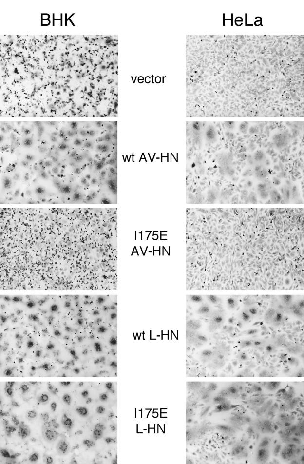
Figure 4 compares the extent of syncytium formation in monolayers in which the wt AV-HN and L-HN proteins, as well as the I175E mutated form of each, are coexpressed with NDV F in the pBSK-BHK and pCAGGS-HeLa expression systems. The two wt proteins promote significant amounts of syncytium formation in either BHK or HeLa cells. Similarly, I175E-L-HN promotes more fusion than its parent wt protein in either system and monolayers expressing I175E-AV-HN are indistinguishable from vector-expressing cells in either system. Thus, the phenotypic differences between AV-HN and L-Kansas HN, both carrying an I175E mutation, are not related to the use of different expression systems or cells.
FIG. 4.
Syncytium formation induced by wt AV and L HN and I175E mutated forms of the two proteins in BHK and HeLa cells with different transient expression systems. Panels on the left show BHK cell monolayers, and panels on the right show HeLa cells. Except for the top two monolayers, in which only vector is expressed, cells coexpress wt NDV-AV F with the indicated HN protein. At 22 h posttransfection, the monolayers were fixed with methanol and stained with Giemsa stain.
Functional activities of AV-HN/L-HN chimeras carrying the I175E mutation.
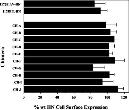
The data in Table 1 suggest that the effect of the I175E mutation on HN function is strain specific, i.e., dependent on the HN background in which the mutation is introduced. There are 52 amino acid differences between the AV and L HN proteins (89.3% homology), scattered over the entire length of the protein (Fig. 1). In addition, the L-HN protein also has 6 more amino acids at its C terminus. To identify those amino acid differences between the two proteins that account for their phenotypic properties, we have constructed a series of chimeric HN proteins, all of which carry the I175E mutation (Fig. 1). All chimeras in the series are expressed at the cell surface in amounts ranging between 82.5 and 112.1% of the wt protein (Fig. 5).
FIG. 5.
Cell surface expression and antigenic integrity of I175E mutated HN chimeras. The cell surface expression of the chimeras was quantitated by flow cytometry, with a panel of at least 3 MAbs specific for different antigenic sites on the globular domain of the protein. The results shown represent the mean of the results from at least four determinations.
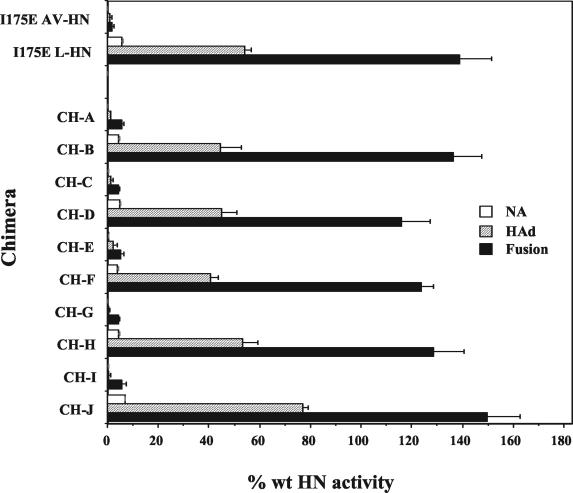
The HAd and NA activities of the chimeras are shown in Fig. 6. CH-A has 318 residues at its N terminus derived from I175E-AV-HN, and the rest of the protein is derived from L-HN (Fig. 1). It has undetectable NA activity, similar to that of I175E-AV HN, whereas the NA of CH-B is slightly higher, 4.7% of L-HN, similar to that of I175E-L-HN. Similar results are found with respect to HAd and fusion. Chimera CH-A and I175E-AV-HN exhibit similar low HAd activities, approximately 1% of wt AV-HN, although CH-A does promote marginally higher levels of fusion, 5.5% compared to 1.9% (Fig. 6).
FIG. 6.
HAd, NA, and fusion-promoting activities of HN chimeras. The NA activity of each chimera was assayed at 22 h posttransfection by determining the ability of each to cleave sialic acid from neuraminlactose. The NA data are corrected for differences in cell surface expression (Fig. 5) and are expressed as a percentage of the NA of the respective wt HN protein. They represent the means of the results from a minimum of three determinations. The HAd activity was determined by the ability of the expressed proteins to hemadsorb guinea pig erythrocytes. For HAd activity, the data represent the means of the results from at least four determinations. The ability to complement the wt NDV-AV F protein in the promotion of fusion was determined by the content-mixing assay. Each value is the mean of the results from a minimum of three determinations.
CH-B is the reciprocal chimera with 318 N-terminal residues from I175E mutated L-HN and the rest of the protein from AV-HN. It has similar NA (4.2 versus 5.6%), HAd (44.4 versus 54.0%), and fusion activities (136.1 versus 138.8%) to wt L-HN (Fig. 6). Thus, in both cases, the phenotype of the parent I175E mutated protein segregates with the N-terminal segment. This indicates that amino acids that lie C-terminal to residue 318 do not play a role in the phenotypic differences between the I175E mutated forms of AV-HN and L-HN.
Chimeras CH-C and CH-D address the possibility that the N-terminal 167 residues of HN contribute to the phenotypic differences between the I175E mutants. Chimera CH-C, with 167 residues at its N terminus derived from L-HN and the rest of the protein from AV-HN, behaves like I175E mutated AV-HN. It has barely detectable HAd and NA activities (Fig. 6) and promotes fusion at only 4.4% of the wt level (Fig. 6). Similarly, the reciprocal chimera CH-D exhibits a phenotype very similar to that of I175E mutated L-HN (Fig. 6). Thus, data from the analyses of chimeras A to D point to one or more residues between 168 and 318 as being accountable for the phenotypic differences between AV-HN and L-HN carrying the I175E mutation.
To confirm this, we have constructed chimeras CH-E and CH-F, in which only this central domain is derived from the heterologous HN protein. As expected from the results obtained with the first four chimeras in the series, CH-E, with residues 168 to 318 derived from AV-HN, has a phenotype that corresponds to that of I175E-AV-HN: 0.2% NA activity, 2.0% HAd, and 5.3% fusion relative to wt AV HN (Fig. 6). Similarly, CH-F, with the central 168 to 318 domain from L-HN and the rest of the protein from AV-HN, exhibits a phenotype similar to that of I175E-L-HN: approximately 4% NA, 40% HAd, and approximately 125% fusion relative to the control (Fig. 6). These findings confirm that the strain-specific phenotypic differences in I175E mutated HN map to residues 168 to 318.
Two additional sets of chimeras with shorter central heterologous domains were constructed. CH-G and CH-H have only residues 168 to 287 derived from the heterologous HN protein (Fig. 1). These chimeras behave very similarly to the chimeras from which they were derived, CH-E and CH-F, respectively (Fig. 6). CH-G has barely detectable HAd and NA and fuses only 4.4% of the wt. Thus, it corresponds phenotypically to both CH-E and I175E-AV-HN. CH-H, with only residues 168 to 287 derived from L-HN, has 4.5% NA, 53.2% HAd, and 128.5% fusion relative to wt L-HN (Fig. 6). Thus, this set of chimeras further maps the I175E-HN phenotype to these 120 residues.
The final set of chimeras in the series includes CH-I and CH-J (Fig. 1). These two chimeras have only residues 168 to 243 derived from the heterologous HN protein. As before, the central heterologously derived segment determines the phenotype of these chimeras. CH-I, with an AV-HN heterologous segment, exhibits an I175E-AV-HN-like phenotype, with HAd and NA less than 1% and fusion only 5.8% of the wt. In contrast, CH-J, with a heterologous domain from the L-HN protein, exhibits a phenotype like I175E-L-HN. It has 4.2% of wt NA activity but mediates both HAd and fusion extremely well, 76.8 and 149.8% of the wt, respectively (Fig. 6). These data map the residues responsible for the phenotypic difference between the I175E mutated HN proteins to residues 168 to 243, a domain consisting of 76 residues.
Phenotypic differences between I175E mutated AV and L-HN are due to amino acid differences at four residues.
Comparison of the sequences of AV-HN and L-HN between residues 168 and 243 reveals only four amino acid differences. Residue 193 is a phenylalanine in AV-HN and a leucine in L-HN. Residue 214 is a serine in AV-HN and a threonine in L-HN. Residue 219 is a valine in AV-HN and an isoleucine in L-HN. Lastly, residue 228 is an asparagine in AV-HN and a serine in L-HN.
To evaluate the contribution of each of these amino acid differences between the two strains to their phenotypic differences, we have mutated the residues at each position in I175E-AV-HN to the residues found in L-HN and determined the effect on HN function. Initially, mutations F193L and N228S were introduced individually and S214T and V219I were introduced in tandem. The properties of these mutants are listed in Table 2. All three mutated proteins are efficiently expressed at the cell surface. The N228S mutated protein is very similar to the parent protein (I175E-AV-HN), with negligible HAd, NA, and fusion activities. The F193L change and the S214T and V219I changes do have some effect on the HAd and fusion of I175E-AV-HN, each resulting in a 10-fold increase in HAd and a 5-fold increase in fusion. However, these activities are still much less than the HAd and fusion activities of I175E-L-HN.
TABLE 2.
Mutation of I175E AV-HN at residues 193, 214, 219, and 228 converts its phenotype to that of I175E L-HN
| Strain | Substitution(s) | Result fora:
|
|||
|---|---|---|---|---|---|
| Expression | HAd | NA | Fusion | ||
| AV | I175E | 84.1 ± 12.9 | 0.7 ± 0.9 | NDb | 1.9 ± 0.7 |
| AV | I175E, F193L | 80.5 ± 8.9 | 6.6 ± 1.8 | 0.4 ± 0.3 | 8.9 ± 0.8 |
| AV | I175E, S214T, V219I | 95.6 ± 6.7 | 5.7 ± 1.3 | 0.1 ± 0.1 | 9.1 ± 6.1 |
| AV | I175E, N228S | 77.6 ± 3.7 | 0.6 ± 0.5 | ND | 3.4 ± 0.5 |
| AV | I175E, F193L, S214T, V219I | 79.7 ± 4.2 | 12.8 ± 3.2 | 1.1 ± 0.9 | 28.5 ± 2.1 |
| AV | I175E, F193L, S214T, V219I, N228S | 103.4 ± 2.6 | 53.8 ± 3.8 | 6.0 ± 2.2 | 141.6 ± 11.4 |
| L | I175E | 91.3 ± 8.1 | 54.0 ± 2.8 | 5.6 ± 0.4 | 138.8 ± 12.6 |
Data are expressed as percentages of the corresponding wt HN protein. The data for AV and L HN carrying only the I175E substitution are repeated from Table 1 for convenience.
ND, none detected.
Next, the F193L, S214T, and V219I mutations were introduced together into I175E-AV-HN. This triple mutant shows a >10-fold increase in HAd (12.8 versus 0.7%), NA (1.1 versus 0.1%), and fusion (28.5 versus 1.9%) relative to the parent protein. However, only when all four mutations relative to L-HN (F193L, S214T, V219I, and N228S) are introduced in I175E-AV-HN does the protein acquire the phenotype of I175E-L-HN. I175E-AV-HN carrying all four substitutions has HAd (53.8 versus 54.0%), NA (6.0 versus 5.6%), and fusion (141.6 versus 138.8%) activities that are remarkably similar to those of I175E mutated L-HN. Thus, all four amino acid differences between residues 168 and 243 in AV-HN and L-HN together are responsible for the phenotypic differences between the two proteins. It is likely that the increased HAd and fusion activities exhibited by the mutant are secondary effects of the increased NA activity. In other words, HAd and fusion are dependent on NA activity.
Strain comparisons confirm the importance of residues T214, I219, and N228 to the high-fusion phenotype of I175E mutated HN.
Figure 7 compares the sequences of the HN proteins of NDV strains AV, L, BC, and B1 between residues 168 and 243. With the lone exception of the tyrosine at position 203 in B1-HN, there is complete homology at all positions except 193, 214, 219, and 228. In fact, B1-HN is completely homologous to AV-HN at the latter three positions, whereas BC-HN is completely homologous to L-HN at these three residues. AV-HN is, thus far, unique among NDV strains in having a phenylalanine at position 193 (26).
FIG. 7.
Sequence alignment of amino acids 175 to 230 in the HN glycoproteins of NDV strains AV, L, BC, and B1. Dashes indicate amino acid identities. The four residues responsible for the phenotypic difference between the I175E AV-HN and I175E L-HN proteins are indicated in boldface type. Sequence alignment was performed by using the Megalign program in Lasergene (DNASTAR).
To confirm our findings with respect to the importance of residues 214, 219, and 228 to the phenotype of I175E mutated HN, we have created NDV-BC HN carrying an I175E substitution and compared its phenotype to that of I175E-L-HN (Table 3). As expected from its homology to L-HN at positions 214, 219, and 228, I175E mutated BC-HN has a similar phenotype, promoting fusion more than 25% more efficiently than wt BC-HN.
TABLE 3.
Effect of I175E substitutions in other NDV HN proteins
| NDV strain | Result fora:
|
|||
|---|---|---|---|---|
| Expression | HAd | NA | Fusion | |
| BC | 108.1 ± 7.6 | 44.2 ± 2.8 | 8.0 ± 0.9 | 125.2 ± 10.8 |
| B1 | 103.2 ± 12.4 | 24.4 ± 5.1 | 1.9 ± 0.8 | 6.2 ± 1.8 |
All data are expressed as percentages of wt AV-HN.
Because of the leucine at position 193 in B1-HN, the phenotype of I175E mutated B1-HN is most accurately compared to that of I175E mutated AV-HN carrying an F193L substitution (Table 2). Indeed, these two proteins promote fusion to similar extents (6.2 and 8.9% of wt) (Tables 2 and 3). Thus, the relationship between the phenotype of I175E-HN and residues 214, 219, and 228 is confirmed by analysis of naturally occurring strains of the virus.
DISCUSSION
The promotion of membrane fusion by most paramyxoviruses, including NDV, is dependent on the recognition of receptors by HN (18). Indeed, the strength of the HN-receptor interaction can determine the extent of fusion (12, 23). However, lectins cannot replace HN in fusion (22). Also, MAbs to antigenic sites 3 and 4 on NDV HN inhibit fusion but not attachment (13, 14). Thus, HN′s role in fusion clearly involves something more than attachment. A second role for HN in fusion is mediated by a virus-specific interaction between it and the homologous F protein (8).
However, the relationship between HN′s recognition of receptors and its interaction with F is not clear. At present, there are two hypotheses. One is that the two proteins interact intracellularly. In this model, HN is thought to maintain F in a prefusion state until receptor recognition at the cell surface induces a conformational change in F (29). An intracellular interaction between the two proteins is supported by data indicating that coexpression of HN with F alters the conformation of the latter independent of receptor binding (19). On the other hand, the demonstration that paramyxovirus HN or F proteins tagged for retention in the endoplasmic reticulum do not affect transport of the other protein argues against a strong interaction between the two proteins, at least in this cellular compartment (24). The alternative hypothesis proposes that the HN-F interaction takes place only after the two proteins have arrived at the cell surface, triggered by HN′s recognition of receptors (17). In this model, HN and F would not interact either intracellularly or in the absence of receptor binding. Of course, the two hypotheses are not mutually exclusive, and the possibility exists that HN and F interact intracellularly and the intracellular interaction is not fusion related.
HN proteins deficient in receptor recognition activity provide an opportunity to begin to discriminate between these two mechanisms. If the two proteins do arrive at the cell surface already associated with each other, the interaction would presumably take place independent of receptor recognition and might even be stabilized in its absence. In this case, one would expect attachment-deficient HN and F to be coimmunoprecipitated from the cell surface. On the other hand, if the HN-F interaction is triggered at the cell surface by HN′s recognition of receptors, this would result in a lack of HN-F complex formation with an HN protein that fails to bind receptors.
By the introduction of single-amino-acid substitutions in the NA active site, we have produced several NDV HN proteins with single-amino-acid substitutions which lack detectable receptor recognition and fusogenic activity. Four of these mutants, carrying substitutions of D198R, K236R, Y526L, and E547Q, fail to interact with the NDV F protein in a coimmunoprecipitation assay. This strongly suggests that HN and F do not arrive at the cell surface as a complex, at least one stable under the conditions used for the coimmunoprecipitation. The simplest explanation for these findings is that the HN-F interaction takes place at the cell surface, triggered by HN′s recognition of receptors.
Among HN active site mutations, I175E appears to be unique with respect to its relationship to F. Similar to the others, this NA active site mutation virtually eliminates attachment, NA, and fusion. However, unlike the others, the I175E substitution converts HN to a form of the protein that interacts with the F protein. This mutation separates the HN-F interaction from receptor recognition. It apparently converts HN to a fusion-ready conformation, though it is still unable to promote fusion due to the lack of receptor recognition activity. The reduced amount of I175E-HN coimmunoprecipitating with F relative to wt HN indicates that the mutant complex is either formed less efficiently or is less stable.
An interaction between I175E-AV-HN and F is consistent with previous findings obtained with I175E-Kansas HN (1). The latter mutant promotes fusion 50% more effectively than the parent wt protein, despite exhibiting a more than 50% reduction in HAd activity. It was postulated that the I175E mutation in this protein acts by promoting a conformational change in HN that is propagated to the dimer interface through residue R174. This change in the dimer association would then trigger the HN-F interaction necessary for fusion.
The mechanism by which this change in the structure of the dimer converts HN to its F-interactive form is still not clear. An understanding of this process is dependent on the precise location of the site in HN that mediates the interaction with F. It has been postulated that residues in the dimer interface, exposed by the change in dimer association, may directly interact with the F protein (1). Subsequently, mutations in the HN dimer interface were identified that severely reduced fusion (31). However, a mutational analysis of a domain at the membrane-proximal end of the dimer interface has revealed that loss of fusion-promoting activity correlates with a destabilization of the interaction between HN and its receptor(s) (2). This suggests that the fusion deficiency resulting from at least some mutations in the dimer interface may be due to a defect in receptor binding, rather than to a direct effect on fusion.
Moreover, the possibility that dimer interface residues in the globular domain could directly mediate the interaction with F is inconsistent with data obtained by the analysis of chimeras composed of segments from heterologous HN proteins. These studies have shown that specificity for the homologous F protein segregates with the stalk region of HN (5, 32, 33). HN chimeras with heterologously derived globular domains fuse only with the F protein homologous to the HN stalk segment. It is difficult to reconcile how dimer interface residues could mediate specificity in these chimeras. They are neither conserved nor located in the domain that determines F specificity. To this end, it will be informative to determine the F-interactive capacity of fusion-deficient HN carrying mutations in the dimer interface. It is quite possible that changes at the dimer interface could be part of the conformational change that takes place in HN in its conversion from a form that does not interact with F to one that does.
A more recent peptide-based study suggests that the NDV HN-F interaction is mediated by amino acids 124 to 152 in the globular domain of HN and the membrane-proximal heptad repeat in F (7). This finding may be consistent with the demonstration that an NDV-hPIV3 HN chimera with 141 NDV-derived N-terminal residues complements NDV F in fusion and that one with only 125 NDV-derived N-terminal residues does not (5). However, it is inconsistent with the failure of several other chimeras with residues 124 to 152 intact to fuse with NDV F. In fact, chimera CH1(−6), which has NDV-derived residues 120 to 571 intact, not only does not fuse with NDV F but fuses quite efficiently with hPIV3 F (5). While it is possible that the F-interactive domains in the NDV and hPIV3 HN proteins could be totally different, it seems unlikely that the F-interactive residues reside in the stalk in one HN protein and in the globular domain in the other. Clearly, to understand the changes in HN that take place upon its conversion to the F-interactive form, we must first more definitively identify the region of the protein that mediates that interaction.
One question that can be answered is why I175E-mutated HN fails to promote fusion, despite its efficient interaction with the F protein. This appears to be the result of its lack of receptor recognition activity, which is, in turn, a result of its lack of NA activity. When the protein is supplied with NA activity, it gains both significant attachment (11) and fusion-promoting (Fig. 3) activities.
Thus, perhaps a potentially more informative question to pose is why I175E-NDV HN lacks attachment activity in the first place. NDV-Kansas, carrying the I175E mutation, retains almost 50% of wt HAd activity and promotes fusion 50% more efficiently than wt HN (1). We have shown that the amino acid differences between the HN proteins from the two strains of the virus that are responsible for their phenotypic differences with respect to HAd and fusion are at positions 193, 214, 219, and 228. When I175E-HN is further mutated at these positions to those residues present in NDV-Kansas HN (F193L, S214T, V219I, and N228S), the phenotype of the protein becomes virtually indistinguishable from that of I175E-Kansas HN, including enhanced fusogenic activity. The importance of these residues to HN function was also supported by the analysis of other strains of the virus.
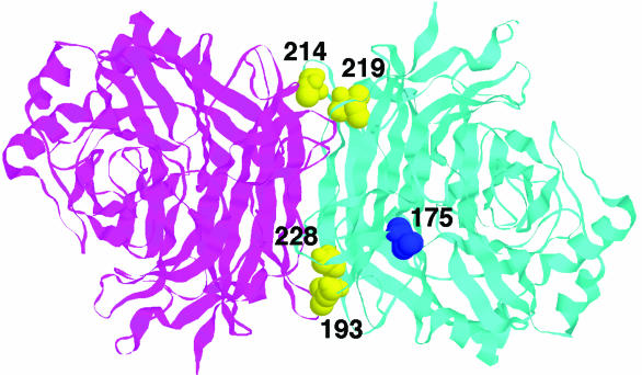
Figure 8 shows the position in one monomer of each of these residues relative to both residue I175 and the dimer interface. All four residues reside at the interface, with residues 193 and 228 at one end and residues 214 and 219 at the other. With their close proximity to the dimer interface, our data argue for the importance of the integrity of the dimer interface to the receptor recognition activity of NDV HN. This is consistent with earlier conclusions based on a mutational analysis of residues 218 to 226 (2). Substitutions at positions 220, 222, and 224 destabilize the interaction between HN and its receptor(s). In addition, a V219A substitution results in a 60% enhancement of HAd activity at 37°C relative to wt HN (2).
FIG. 8.
Locations of HN residues 193, 214, 219, and 228 relative to the dimer interface and residue 175. The figure shows a top view of the Kansas HN dimer with the pH 6.5 structure of Crennell at al. (3). The two monomers in the dimer are shown in ribbon mode in magenta and cyan. Important residues are shown in space-filling mode in one of the monomers. Residue 175 is shown in dark blue. Residues 193, 214, 219, and 228 are shown in yellow. The figure was generated with RasMol.
A role for residue 193 in the receptor recognition activity of HN is also consistent with our previous findings. AV-HN is unique in having a phenylalanine at residue 193, with leucine being much more common at this position in NDV HN from different strains (26). Substitutions at this position can have profound effects on the strength of the HN-receptor interaction. MAb-selected variants and temperature-sensitive mutants of NDV-AV carrying an F193L or I substitution in HN (often in combination with a mutation at I175) can be shown to exhibit increased avidity for receptors and acquisition of the ability to promote fusion from without, an activity normally lacking in the parent wt NDV-AV virus (12, 15). This supports the idea that residues 175 and 193 are functionally related.
In conclusion, we have shown that four different mutations that abolish the receptor recognition activity of NDV HN also abolish its ability to interact with the homologous F protein at the cell surface. This is consistent with the idea that HN interacts with F only after binding to its receptor. Also, we have identified a point mutation (I175E) in the NA active site that converts HN to a form that is capable of interacting with the homologous F protein at the cell surface. The ability of the latter to complement F in fusion is still dependent on its ability to bind receptors. Thus, the I175E-HN-F complex could represent a fusion intermediate in which HN and F are associated and primed for the promotion of fusion.
Acknowledgments
We gratefully acknowledge Judith Alamares, Elizabeth Corey, Paul Mahon, Vanessa Melanson, and Nikhat Parveen for critical reading of the manuscript. We also thank Robert Lamb for the NDV-AV F gene, Trudy Morrison for the NDV-AV HN gene, Bernard Moss for the recombinant vaccinia virus, Walter Demkowicz for wt vaccinia virus, Richard A. Morgan for the pGINT7β-gal plasmid, Peter Emmerson for the BC HN gene, and Kemal Karaca for B1 HN gene.
This work was made possible by grant AI-49268 from the National Institutes of Health.
REFERENCES
- 1.Connaris, H., T. Takimoto, R. Russell, S. Crennell, I. Moustafa, A. Portner, and G. Taylor. 2002. Probing the sialic acid binding site of the hemagglutinin-neuraminidase of Newcastle disease virus: Identification of key amino acids involved in cell binding, catalysis, and fusion. J. Virol. 76:1816-1824. [DOI] [PMC free article] [PubMed] [Google Scholar]
- 2.Corey, E. A., A. M. Mirza, E. Levandowsky, and R. M. Iorio. 2003. Fusion deficiency induced by mutations at the dimer interface in the Newcastle disease virus hemagglutinin-neuraminidase is due to a temperature-dependent defect in receptor binding. J. Virol. 77:6913-6922. [DOI] [PMC free article] [PubMed] [Google Scholar]
- 3.Crennell, S., T. Takimoto, A. Portner, and G. Taylor. 2000. Crystal structure of the multifunctional paramyxovirus hemagglutinin-neuraminidase. Nat. Struct. Biol. 7:1068-1074. [DOI] [PubMed] [Google Scholar]
- 4.Deng, R., Z. Wang, P. J. Mahon, M. Marinello, A. M. Mirza, and R. M. Iorio. 1999. Mutations in the NDV HN protein that interfere with its ability to interact with the homologous F protein in the promotion of fusion. Virology 253:43-54. [DOI] [PubMed] [Google Scholar]
- 5.Deng, R., Z. Wang, A. M. Mirza, and R. M. Iorio. 1995. Localization of a domain on the paramyxovirus attachment protein required for the promotion of cellular fusion by its homologous fusion protein spike. Virology 209:457-469. [DOI] [PubMed] [Google Scholar]
- 6.Fuerst, T. R., E. G. Niles, F. W. Studier, and B. Moss. 1986. Eucaryotic transient expression system based on recombinant vaccinia virus that synthesizes bacteriophage T7 RNA polymerase. Proc. Natl. Acad. Sci. USA 83:8122-8126. [DOI] [PMC free article] [PubMed] [Google Scholar]
- 7.Gravel, K., and T. G. Morrison. 2003. Interacting domains of the HN and F proteins of Newcastle disease virus. J. Virol. 77:11040-11049. [DOI] [PMC free article] [PubMed] [Google Scholar]
- 8.Hu, X., R. Ray, and R. W. Compans. 1992. Functional interactions between the fusion protein and hemagglutinin-neuraminidase of human parainfluenza viruses. J. Virol. 66:1528-1534. [DOI] [PMC free article] [PubMed] [Google Scholar]
- 9.Iorio, R. M., J. B. Borgman, R. L. Glickman, and M. A. Bratt. 1986. Genetic variation within a neutralizing domain on the haemagglutinin-neuraminidase glycoprotein of Newcastle disease virus. J. Gen. Virol. 67:1393-1403. [DOI] [PubMed] [Google Scholar]
- 10.Iorio, R. M., and M. A. Bratt. 1983. Monoclonal antibodies to Newcastle disease virus: delineation of four epitopes on the HN glycoprotein. J. Virol. 48:440-450. [DOI] [PMC free article] [PubMed] [Google Scholar]
- 11.Iorio, R. M., G. M. Field, J. M. Sauvron, A. M. Mirza, R. Deng, P. J. Mahon, and J. Langedijk. 2001. Structural and functional relationship between the receptor recognition and neuraminidase activities of the Newcastle disease virus hemagglutinin-neuraminidase protein: receptor recognition is dependent on neuraminidase activity. J. Virol. 75:1918-1927. [DOI] [PMC free article] [PubMed] [Google Scholar]
- 12.Iorio, R. M., and R. L. Glickman. 1992. Fusion mutants of Newcastle disease virus selected with monoclonal antibodies to the hemagglutinin-neuraminidase. J. Virol. 66:6626-6633. [DOI] [PMC free article] [PubMed] [Google Scholar]
- 13.Iorio, R. M., R. L. Glickman, and J. P. Sheehan. 1992. Inhibition of fusion by neutralizing monoclonal antibodies to the haemagglutinin-neuraminidase glycoprotein of Newcastle disease virus. J. Gen. Virol. 73:1167-1176. [DOI] [PubMed] [Google Scholar]
- 14.Iorio, R. M., R. L. Glickman, A. M. Riel, J. P. Sheehan, and M. A. Bratt. 1989. Functional and neutralization profile of seven overlapping antigenic sites on the HN glycoprotein of Newcastle disease virus: monoclonal antibodies to some sites prevent viral attachment. Virus Res. 13:245-262. [DOI] [PubMed] [Google Scholar]
- 15.Iorio, R. M., R. J. Syddall, R. L. Glickman, A. M. Riel, J. P. Sheehan, and M. A. Bratt. 1989. Identification of amino acid residues important to the neuraminidase activity of the HN glycoprotein of Newcastle disease virus. Virology 173:196-204. [DOI] [PubMed] [Google Scholar]
- 16.Iorio, R. M., R. J. Syddall, J. P. Sheehan, M. A. Bratt, R. L. Glickman, and A. M. Riel. 1991. Neutralization map of the HN glycoprotein of Newcastle disease virus: domains recognized by monoclonal antibodies that prevent receptor recognition. J. Virol. 65:4999-5006. [DOI] [PMC free article] [PubMed] [Google Scholar]
- 17.Lamb, R. A. 1993. Paramyxovirus fusion: a hypothesis for changes. Virology 197:1-11. [DOI] [PubMed] [Google Scholar]
- 18.Lamb, R. A., and D. Kolakofsky. 1996. Paramyxoviridae: the viruses and their replication, p. 1305-1340. In B. N. Fields, D. M. Knipe, and P. M. Howley (ed.), Fields virology, 4th ed. Lippincott-Raven, Philadelphia, Pa.
- 19.McGinnes, L. W., K. Gravel, and T. G. Morrison. 2002. Newcastle disease virus HN protein alters the conformation of the F protein at cell surfaces. J. Virol. 76:12622-12633. [DOI] [PMC free article] [PubMed] [Google Scholar]
- 20.Mirza, A. M., R. Deng, and R. M. Iorio. 1994. Site-directed mutagenesis of a conserved hexapeptide in the paramyxovirus hemagglutinin-neuraminidase glycoprotein: effects on antigenic structure and function. J. Virol. 68:5093-5099. [DOI] [PMC free article] [PubMed] [Google Scholar]
- 21.Mirza, A. M., J. P. Sheehan, L. W. Hardy, R. L. Glickman, and R. M. Iorio. 1993. Structure and function of a membrane anchor-less form of the hemagglutinin-neuraminidase glycoprotein of Newcastle disease virus. J. Biol. Chem. 268:21425-21431. [PubMed] [Google Scholar]
- 22.Moscona, A., and R. W. Peluso. 1991. Fusion properties of cells persistently infected with human parainfluenza virus type 3: participation of hemagglutinin-neuraminidase in membrane fusion. J. Virol. 65:2772-2777. [DOI] [PMC free article] [PubMed] [Google Scholar]
- 23.Moscona, A., and R. W. Peluso. 1993. Relative affinity of the human parainfluenza virus type 3 hemagglutinin-neuraminidase for sialic acid correlates with virus-induced fusion activity. J. Virol. 67:6463-6468. [DOI] [PMC free article] [PubMed] [Google Scholar]
- 24.Paterson, R. G., M. L. Johnson, and R. A. Lamb. 1997. Paramyxovirus fusion (F) protein and hemagglutinin-neuraminidase (HN) protein interactions: intracellular retention of F and HN does not affect transport of the homotypic HN or F protein. Virology 237:1-9. [DOI] [PubMed] [Google Scholar]
- 25.Porotto, M., M Murrell, O. Greengard, and A. Moscona. 2003. Triggering of human parainfluenza virus 3 fusion protein (F) by the hemagglutinin-neuraminidase (HN) protein: an HN mutation diminishes the rate of F activation and fusion. J. Virol. 77:3647-3654. [DOI] [PMC free article] [PubMed] [Google Scholar]
- 26.Sakaguchi, T., T. Toyoda, B. Gotoh, N. M. Inocencio, K. Kuma, T. Miyada, and Y. Nagai. 1989. Newcastle disease virus evolution. I. Multiple lineages defined by sequence variability of the hemagglutinin-neuraminidase gene. Virology 169:260-272. [DOI] [PubMed] [Google Scholar]
- 27.Sergel, T., L. W. McGinnes, and T. G. Morrison. 1993. The attachment function of the Newcastle disease virus hemagglutinin-neuraminidase protein can be separated from fusion-promotion by mutation. Virology 193:717-726. [DOI] [PubMed] [Google Scholar]
- 28.Sheehan, J. P., R. M. Iorio, R. J. Syddall, R. L. Glickman, and M. A. Bratt. 1987. Reducing agent-sensitive dimerization of the hemagglutinin-neuraminidase glycoprotein of Newcastle disease virus correlates with the presence of cysteine at residue 123. Virology 161:603-606. [DOI] [PubMed] [Google Scholar]
- 29.Stone-Hulslander, J., and T. G. Morrison. 1997. Detection of an interaction between the HN and F proteins in Newcastle disease virus-infected cells. J. Virol. 71:6287-6295. [DOI] [PMC free article] [PubMed] [Google Scholar]
- 30.Stone-Hulslander, J., and T. G. Morrison. 1999. Mutational analysis of heptad repeats in the membrane-proximal region of Newcastle disease virus HN protein. J. Virol. 73:3630-3637. [DOI] [PMC free article] [PubMed] [Google Scholar]
- 31.Takimoto, T., G. L. Taylor, H. C. Connaris, S. J. Crennell, and A. Portner. 2002. Role of the hemagglutinin-neuraminidase protein in the mechanism of paramyxovirus-cell membrane fusion. J. Virol. 76:13028-13033. [DOI] [PMC free article] [PubMed] [Google Scholar]
- 32.Tanabayashi, K., and R. W. Compans. 1996. Functional interaction of paramyxovirus glycoproteins: identification of a domain in Sendai virus HN which promotes cell fusion. J. Virol. 70:6112-6118. [DOI] [PMC free article] [PubMed] [Google Scholar]
- 33.Tsurodome, M., M. Kawano, T. Yuasa, N. Tabata, M. Nishio, H. Komada, and Y. Ito. 1995. Identification of regions on the hemagglutinin-neuraminidase protein of human parainfluenza virus type 2 important for promoting cell fusion. Virology 213:190-203. [DOI] [PubMed] [Google Scholar]


