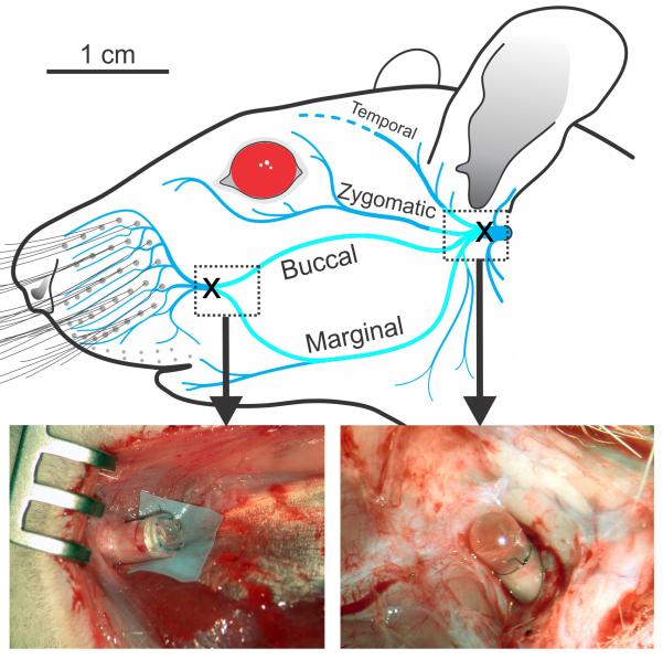
Figure 2.
Diagram of the rat facial nerve showing where the nerve was unilaterally transected (X), and sutured into dead-end silicone tubes at the main trunk (right photo), as well as at the distal convergence of the buccal and marginal mandibular branches (left photo) for the VII Resected group (N=10). The region of nerve extirpated between the transection locations is shown as a lighter shade of blue.

