Abstract
Mucosa-associated Invariant T (MAIT) cells are an evolutionarily conserved innate-like T cell subset that recognizes antigens presented by MR1 molecules. These antigens include vitamin B derivatives shared by many potentially pathogenic microbes, including Mycobacterium tuberculosis and Candida albicans. It was recently discovered that MAIT cells decay numerically and functionally in HIV-1 infection, and that they fail to recover despite several years of effective suppression of viral replication by antiretroviral therapy (ART). Here, we briefly discuss the roles of MAIT cells and their loss in HIV immunopathogenesis. We furthermore propose that the persistence of MAIT cell loss on ART needs to be taken into account when assessing the immunological response to treatment, and when treatment should commence. The importance of this T cell subset in HIV-1 infection needs further study, and interventions to restore the MAIT cell compartment should be considered.
Keywords: HIV-1, antiretroviral therapy, MAIT cells, MR1, IL-17
Introduction
Mucosa-associated Invariant T (MAIT) cells are a relatively recently described subset of T cells, representing some 2–10% of peripheral blood T cells in healthy donors [1]. They are mostly CD8+, although CD4/8 double negative or CD4+ populations also exist [2, 3], and can comprise up to 10–25% of T cells in the liver and at intestinal sites [4]. MAIT cells are CD161hi, characterized by their Vα7.2-containing semi-invariant T cell receptor, and respond to antigens presented by the evolutionarily conserved MHC class Ib-related protein 1 (MR1) [5–7]. They respond rapidly in an innate-like fashion and with a predominant Th1/Th17 profile [4]. MAIT cells display tropism for mucosal sites with expression of chemokine receptors such as CCR6, CCR9, and CXCR6, and in mucosa they express the tissue-protective cytokine IL-22 [3, 4]. The type of antigens presented by MR1 molecules was long elusive, but recently described to include derivatives of the microbial vitamin B biosynthetic pathway [8]. As components of these pathways are shared by many microbes but not mammals, they represent good targets of a conserved immune mechanism at mucosal sites where such microbes are encountered. In line with this, MAIT cells are emerging as an important component of the healthy mucosal immune system.
Irreversible loss of MAITs in HIV-1 infection: How, where and when?
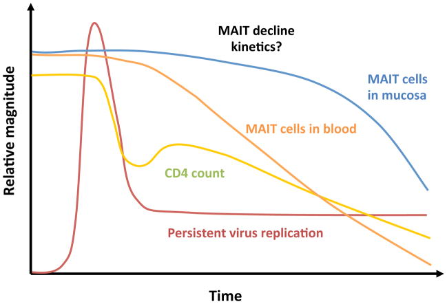
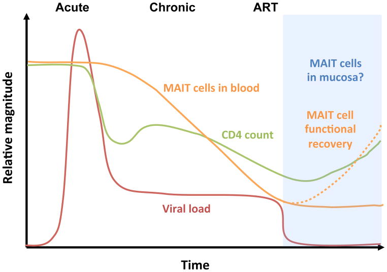
Given the characteristics of MAIT cells it is reasonable to hypothesize a role for these cells in patients that suffer from acquired immunodeficiency and impaired control of microbial infections. Two recent studies investigated the impact of HIV-1 infection on the MAIT cell compartment [9, 10]. These studies together indicate that MAIT cells decline numerically in peripheral blood of HIV-1 infected subjects, whereas the loss of these cells appears to be slower in gut associated lymphoid tissue (Fig. 1). The function of residual MAIT cells is reduced with lower cytokine production in response to E. coli, where in particular IL-17 production is essentially completely lost. Importantly, peripheral blood MAIT cells do not recover significantly in numbers as patients initiate combination antiretroviral therapy (ART), even when viral load is suppressed to undetectable levels for several years and the CD4 compartment recovers (Fig. 2). Suppression of viremia does however benefit the status of residual MAIT cells, as evidenced by a partial recovery of cytokine production.
Fig. 1. MAIT cell loss in HIV-1 infection.
MAIT cells are activated, exhausted and persistently depleted in chronic HIV-1 infection. The exact kinetics of MAIT cell loss in blood and mucosa are unknown.
Fig. 2. The loss of MAIT cells is not reversed by successful ART.
The function of MAIT cells recovers partly when patients go on ART. The effect of ART on mucosal MAIT cells is so far unknown.
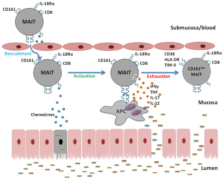
Loss of MAIT cells in HIV infection occurs despite that they are predominantly CD8-positive cells and appear to be relatively resistant to direct infection by the virus [10]. The data support a model wherein the MAIT cell compartment is engaged in immune responses and declines as a consequence of persistent activation, exhaustion and apoptosis [9, 10]. Notably, however, MAIT cells probably do not respond to viral antigens [11], leaving the open question what the driving force of activation and exhaustion may be. Given what we know about the nature of MR1-presented antigens, it is reasonable to speculate that persistent exposure to bacterial and fungal antigens might drive these changes. Loss of immune control of bacteria and fungi at mucosal surfaces may lead to an enhanced burden on MAIT cells at these sites (Fig. 3). Furthermore, microbial translocation products may also engage MAIT cells not only in the mucosal epithelium and lamina propria, but also in the liver. In this context the paucity of unequivocal markers of microbial translocation is problematic [12]. In fact, the same vitamin B metabolites recognized by MAIT cells may potentially serve as novel microbial translocation markers in plasma.
Fig. 3.
Hypothetical model of MAIT cell recruitment, activation, exhaustion and loss at the mucosal frontlines.
According to the exhaustion model of MAIT cell loss, the loss would occur as they are exhausted and consumed at the “frontlines” of exposure to microbes (Fig. 3). Data from matched blood and rectal biopsy material support the possibility that the decline of peripheral blood MAIT cells may in part be driven by recruitment to the intestinal mucosa. Significant loss of MAIT cells in the mucosa may then become more apparent as their levels in peripheral blood and other non-mucosal sites become depleted. It will be important to investigate mucosal sites other than the rectal mucosa to fully understand these events. Yet another possibility to consider is that HIV may actively perturb MR1-mediated antigen presentation. This is the case for both classical HLA class I molecules and MHC class I-like molecules such as CD1d [13–15]. If similar mechanisms exist to interfere with MR1 pathways, then this may contribute to MAIT cell dysfunction in HIV-1 infection.
The exact kinetics of MAIT cell loss in HIV-1 infected patients remain unclear. The study by Cosgrove et al. observed that MAIT cell loss was pronounced already early in infection, whereas a negative correlation between MAIT cell percentage and estimated time since infection was found in our study. This apparent discrepancy may be explained by differences between the cohorts. If our model is correct and MAIT cell exhaustion and loss occurs at mucosal sites, the rate of loss from blood can probably be influenced by the total infectious disease burden and environmental factors.
MAIT cells overlap at least partly with the CD8 T cell population described as Tc17 cells. The Tc17 population express CD161 and have the capacity to produce IL-17. Our data suggest that the majority of these cells are MAIT cells as defined by the expression of the Vα7.2 T cell receptor (unpublished observations). Interestingly, it was recently reported that Tc17 cells are lost in HIV infection [16, 17], in concordance with the observations on MAIT cells [9, 10].
Consequences and implications: An argument for early ART
With the data suggesting that MAIT cell numbers do not recover on ART, we have reason to believe that the damage that has been done to this immune cell compartment will persist over time. On the positive side, however, the data suggest that suppression of viremia with effective ART prevents further decay of the MAIT cells. These patterns may have important implications for immune defence against several microbes relevant in HIV immunopathogenesis. For example, both M. tuberculosis and C. albicans are targets of MAIT cell responses [11], and MAIT cells probably make up a substantial fraction of the CD8 T cell response against these pathogens. Importantly, effective ART allows the residual MAIT cells to regain a relative level of functionality, although not to levels fully comparable to uninfected healthy controls. In this context the ability of tissue MAIT cells to produce IL-22 may be important, given the recent data suggesting that this cytokine has a protective role in maintenance of mucosal epithelium [18].
The relatively early decay of the MAIT cell compartment thus adds an argument in favor of treating HIV-1 infection as early as possible. When MAIT cells are preserved they may contribute to a better mucosal immune status and barrier function. Poor recovery of CD4 counts on ART can be associated with remaining elevated immune activation and microbial translocation markers [19–21]. Early treatment and the resulting maintenance of relatively healthy levels of MAIT cells may positively influence the clinical recovery.
Can the MAIT cell compartment be restored?
The persisting defect in MAIT cells in patients on ART suggests that many of the patients treated for HIV-1 infection today have a persistent weakness in the ability to handle bacterial and fungal infections, including opportunistic infections. With this understanding we should consider ways to restore MAIT cells in vivo by immunotherapeutic intervention. Cytokine treatment has overall had limited success in the context of HIV infection. However, another innate-like T cell subset, the invariant natural killer T (iNKT) cells, was previously shown to expand in vivo in response to IL-2 treatment in combination with effective ART [22]. It may thus be interesting to evaluate the effect of IL-2 therapy on MAIT cells in HIV-1 infected patients. MAIT cells express receptors for both IL-7 and IL-18, suggesting that the prospect of these cytokines in treatment should also be evaluated.
A more direct approach is to test the potential of probiotic bacteria in stimulating MAIT cell recovery. Species of Lactobacillus, which are included in many probiotic products, are stimulatory to these cells and may have a positive effect [11]. Other bacteria that are part of the normal commensal gut flora would also be of interest. Stimulation of MAIT cell recovery by such “friendly” microbes would be consistent with the expansion seen during the first year of life when the normal flora is established. Finally, MAIT cells may need both suitable antigens and cytokines to expand, suggesting that a mixed cytokine plus probiotics approach may be a viable way forward.
Concluding remarks
MAIT cells are part of the CD8 T cell compartment and their numerical and functional decline in HIV-1 infection was at the first instance unexpected. Their loss is likely to put a significant dent in the human host defence against microbes at mucosal surfaces. The persistence of MAIT cell loss on ART needs to be taken into account when assessing the immunological response to treatment, and when treatment should commence. The importance of this innate-like T cell subset in HIV-1 infection needs further study, and interventions to restore MAIT cell numbers should be considered.
Acknowledgments
This work was supported by grants from the Swedish Research Council, the Swedish Cancer Foundation, the Stockholm County Council, and NIH/NIAID R01-AI057020. EL is a Swedish Institute Postdoctoral Fellow. JD is supported by a PhD fellowship from the Fundação para a Ciência e a Tecnologia (FCT). JKS is a Senior Research Fellow of the Swedish Research Council.
Footnotes
All authors contributed to the writing of this article.
The authors report no conflict of interest.
NOTE: This is a non-final version of an article published in final form in:
AIDS. 2013 Oct 23;27(16):2501-4. doi: 10.1097/QAD.0b013e3283620726.
The final version of this article appears at: http://journals.lww.com/aidsonline/Citation/2013/10230/Will_loss_of_your_mucosa_associated_invariant_T.1.aspx
References
- 1.Le Bourhis L, Guerri L, Dusseaux M, Martin E, Soudais C, Lantz O. Mucosal-associated invariant T cells: unconventional development and function. Trends Immunol. 2011;32:212–218. doi: 10.1016/j.it.2011.02.005. [DOI] [PubMed] [Google Scholar]
- 2.Martin E, Treiner E, Duban L, Guerri L, Laude H, Toly C, et al. Stepwise development of MAIT cells in mouse and human. PLoS Biol. 2009;7:e54. doi: 10.1371/journal.pbio.1000054. [DOI] [PMC free article] [PubMed] [Google Scholar]
- 3.Walker LJ, Kang YH, Smith MO, Tharmalingham H, Ramamurthy N, Fleming VM, et al. Human MAIT and CD8alphaalpha cells develop from a pool of type-17 precommitted CD8+ T cells. Blood. 2012;119:422–433. doi: 10.1182/blood-2011-05-353789. [DOI] [PMC free article] [PubMed] [Google Scholar]
- 4.Dusseaux M, Martin E, Serriari N, Peguillet I, Premel V, Louis D, et al. Human MAIT cells are xenobiotic-resistant, tissue-targeted, CD161hi IL-17-secreting T cells. Blood. 2011;117:1250–1259. doi: 10.1182/blood-2010-08-303339. [DOI] [PubMed] [Google Scholar]
- 5.Treiner E, Duban L, Bahram S, Radosavljevic M, Wanner V, Tilloy F, et al. Selection of evolutionarily conserved mucosal-associated invariant T cells by MR1. Nature. 2003;422:164–169. doi: 10.1038/nature01433. [DOI] [PubMed] [Google Scholar]
- 6.Huang S, Martin E, Kim S, Yu L, Soudais C, Fremont DH, et al. MR1 antigen presentation to mucosal-associated invariant T cells was highly conserved in evolution. Proc Natl Acad Sci U S A. 2009;106:8290–8295. doi: 10.1073/pnas.0903196106. [DOI] [PMC free article] [PubMed] [Google Scholar]
- 7.Tsukamoto K, Deakin JE, Graves JA, Hashimoto K. Exceptionally high conservation of the MHC class I-related gene, MR1, among mammals. Immunogenetics. 2013;65:115–124. doi: 10.1007/s00251-012-0666-5. [DOI] [PubMed] [Google Scholar]
- 8.Kjer-Nielsen L, Patel O, Corbett AJ, Le Nours J, Meehan B, Liu L, et al. MR1 presents microbial vitamin B metabolites to MAIT cells. Nature. 2012;491:717–723. doi: 10.1038/nature11605. [DOI] [PubMed] [Google Scholar]
- 9.Leeansyah E, Ganesh A, Quigley MF, Sonnerborg A, Andersson J, Hunt PW, et al. Activation, exhaustion, and persistent decline of the antimicrobial MR1-restricted MAIT-cell population in chronic HIV-1 infection. Blood. 2013;121:1124–1135. doi: 10.1182/blood-2012-07-445429. [DOI] [PMC free article] [PubMed] [Google Scholar]
- 10.Cosgrove C, Ussher JE, Rauch A, Gartner K, Kurioka A, Huhn MH, et al. Early and nonreversible decrease of CD161++/MAIT cells in HIV infection. Blood. 2013;121:951–961. doi: 10.1182/blood-2012-06-436436. [DOI] [PMC free article] [PubMed] [Google Scholar]
- 11.Le Bourhis L, Martin E, Peguillet I, Guihot A, Froux N, Core M, et al. Antimicrobial activity of mucosal-associated invariant T cells. Nat Immunol. 2010;11:701–708. doi: 10.1038/ni.1890. [DOI] [PubMed] [Google Scholar]
- 12.Leeansyah E, Malone DF, Anthony DD, Sandberg JK. Soluble biomarkers of HIV transmission, disease progression and comorbidities. Curr Opin HIV AIDS. 2013;8:117–124. doi: 10.1097/COH.0b013e32835c7134. [DOI] [PubMed] [Google Scholar]
- 13.Schwartz O, Marechal V, Le Gall S, Lemonnier F, Heard JM. Endocytosis of major histocompatibility complex class I molecules is induced by the HIV-1 Nef protein. Nat Med. 1996;2:338–342. doi: 10.1038/nm0396-338. [DOI] [PubMed] [Google Scholar]
- 14.Chen N, McCarthy C, Drakesmith H, Li D, Cerundolo V, McMichael AJ, et al. HIV-1 down-regulates the expression of CD1d via Nef. Eur J Immunol. 2006;36:278–286. doi: 10.1002/eji.200535487. [DOI] [PubMed] [Google Scholar]
- 15.Moll M, Andersson SK, Smed-Sorensen A, Sandberg JK. Inhibition of lipid antigen presentation in dendritic cells by HIV-1 Vpu interference with CD1d recycling from endosomal compartments. Blood. 2010;116:1876–1884. doi: 10.1182/blood-2009-09-243667. [DOI] [PMC free article] [PubMed] [Google Scholar]
- 16.Guillot-Delost M, Le Gouvello S, Mesel-Lemoine M, Cherai M, Baillou C, Simon A, et al. Human CD90 identifies Th17/Tc17 T cell subsets that are depleted in HIV-infected patients. J Immunol. 2012;188:981–991. doi: 10.4049/jimmunol.1101592. [DOI] [PubMed] [Google Scholar]
- 17.Gaardbo JC, Hartling HJ, Thorsteinsson K, Ullum H, Nielsen SD. CD3+CD8+CD161high Tc17 cells are depleted in HIV-infection. AIDS. 2013;27:659–662. doi: 10.1097/QAD.0b013e32835b8cb3. [DOI] [PubMed] [Google Scholar]
- 18.Kim CJ, Nazli A, Rojas OL, Chege D, Alidina Z, Huibner S, et al. A role for mucosal IL-22 production and Th22 cells in HIV-associated mucosal immunopathogenesis. Mucosal Immunol. 2012;5:670–680. doi: 10.1038/mi.2012.72. [DOI] [PubMed] [Google Scholar]
- 19.Hunt PW, Martin JN, Sinclair E, Bredt B, Hagos E, Lampiris H, Deeks SG. T cell activation is associated with lower CD4+ T cell gains in human immunodeficiency virus-infected patients with sustained viral suppression during antiretroviral therapy. J Infect Dis. 2003;187:1534–1543. doi: 10.1086/374786. [DOI] [PubMed] [Google Scholar]
- 20.Jiang W, Lederman MM, Hunt P, Sieg SF, Haley K, Rodriguez B, et al. Plasma levels of bacterial DNA correlate with immune activation and the magnitude of immune restoration in persons with antiretroviral-treated HIV infection. J Infect Dis. 2009;199:1177–1185. doi: 10.1086/597476. [DOI] [PMC free article] [PubMed] [Google Scholar]
- 21.Fernandez S, Tanaskovic S, Helbig K, Rajasuriar R, Kramski M, Murray JM, et al. CD4+ T-cell deficiency in HIV patients responding to antiretroviral therapy is associated with increased expression of interferon-stimulated genes in CD4+ T cells. J Infect Dis. 2011;204:1927–1935. doi: 10.1093/infdis/jir659. [DOI] [PubMed] [Google Scholar]
- 22.Moll M, Snyder-Cappione J, Spotts G, Hecht FM, Sandberg JK, Nixon DF. Expansion of CD1d-restricted NKT cells in patients with primary HIV-1 infection treated with interleukin-2. Blood. 2006;107:3081–3083. doi: 10.1182/blood-2005-09-3636. [DOI] [PMC free article] [PubMed] [Google Scholar]


