
Fig. 4.
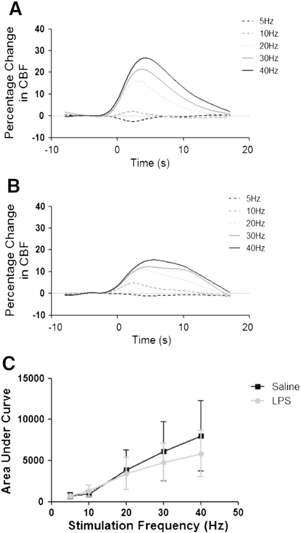
Effect of LPS treatment on neurovascular coupling and functional hyperaemia. Time course of cerebral blood flow (CBF) changes recorded in primary somatosensory MCx in response to electrical stimulation of the contralateral somatosensory MCx in (A) saline treated and (B) LPS treated animals (n = 3 per group). Stimuli were delivered using carbon fibre electrodes positioned overlying the somatosensory MCx and the stimulus evoked CBF changes were recorded using laser Doppler flowmetry probe positioned over the corresponding contralateral MCx. Stimuli consisted of a 2-s train of 0.3 ms 1.5 mA pulses at one of 5 frequencies (5, 10, 20, 30 and 40 Hz). (C) CBF responses over the range of stimulation frequencies were quantified by determining area under the curve for the mean response to each condition for each animal.