Abstract
In Europe, human dirofilariasis refers to a group of autochtonous parasitic infections caused by tissue nematodes of the genus Dirofilaria, responsible for two distinct clinical presentations: Dirofilaria immitis usually presenting as pulmonary lesions and Dirofilaria repens as subcutaneous nodules. Rare in humans, genital involvement manifests itself as pseudotumor nodules affecting the scrotum, epididymis, or spermatic cord. We report on two cases of Dirofilaria repens infections, involving the spermatic cord and epididymis.
Keywords: Human dirofilariasis, Dirofilaria repens, Genital involvement
Introduction
Human dirofilariasis is an infection caused by parasitic nematodes that occurs in a wide geographic distribution. Dirofilaria repens is an endemic nematode in the Old World with a predominant localization along the Mediterranean coast while Dirofilaria tenuis, Dirofilaria ursi, and Dirofilaria lutrae are endemic in the New World and Dirofilaria immitis has been reported on all continents.1,2 Their natural hosts include dogs, cats and foxes. The mosquito species Aedes, Culex, and Anopheles have been identified as vectors of these nematodes.2 Infection by D. repens presents itself clinically as subcutaneous and conjunctival nodules. Lesions involving the male sexual organs may mimic malignant tumors and the diagnosis is based on histological examination. Genital localization is rare however, with only 20 cases ever reported in the male sexual organs (scrotum, epididymis, spermatic cord).2–6
Case Report
Case 1
A 29-year-old man without travel history who lived in Saint Tropez consulted an urologist on January 2007 due to a nodule in the left epididymis. It had appeared in April of 2006 and progressively increased in size. Upon clinical examination there was erythema involving the two testicles, extending into the inguinal region. Routine laboratory analysis revealed hyperleucocytosis without eosinophilia. Ultrasound analysis showed normal testicles and a solid 13 mm nodule, with central hypoechogenic images facing the head of the epidydimis. Nodule resection was performed followed by a histological diagnosis of dirofilariosis.
Case 2
A 66-year-old man, living in the French Var region (Fréjus) who never left the South of France in his lifetime, complained in February 2010 of a nodule on the left spermatic cord. Clinical examination revealed an inflammatory, firm and movable nodule attached to the spermatic cord. Laboratory analyses were normal. Ultrasound examination aimed at excluding a malignant tumor showed a 15×10 mm hypoechogenic lesion. As above, nodule resection was performed followed by a histological diagnosis of dirofilariasis.
Histological Description
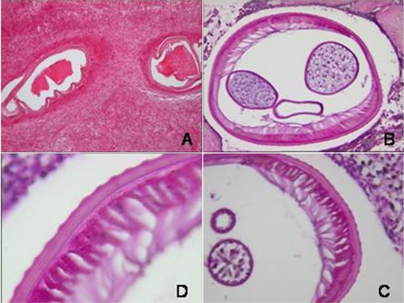
In both cases, histological examination revealed several transversal sections of the parasite species D. repens (Fig. 1A). Its maximal section was estimated at 470 μm. The parasite’s cuticle was approximately 10 μm thick with spikes on the entire outer surface with crests measuring 3 μm in height. The internal tubular organs were located in a pseudocoelomic cavity comprising an intestine and a double uterus containing oocytes (Fig. 1B). The polymyarian type muscle structure appeared to be well-developed (Fig. 1C and D).
Figure 1.
(A) (HES×10): Granuloma with two longitudinal sections of the worm. (B) (HES×20): Transversal section of the nematode showing two uterine sections and one central intestine section in the pseudocoele. (C) (HES×40) and (D) (HES×100): Partial view of a transversal section showing the scalloped cuticle and muscular layer.
Discussion
Dirofilariasis of the male genitalia remains a rare event with only 20 cases reported in the literature involving the scrotum, epididymis, or spermatic cord.2–10 The Mediterranean basin, particularly in Italy and the south of France, represents an endemic area of this infection caused by D. repens. Since the resulting infectious nodule in the male genitalia is frequently indistinguishable clinically from a malignant neoplasm, it is usually removed surgically. Cases of D. repens can also present themselves as pulmonary, pancreatic, tubal, and peritoneal nodules.2,6,8 The localisation in genital organs likely occurs after migration of the adult worm in subcutaneous tissue.
Cases of D. repens dissemination are rare as reproduction requires the simultaneous presence of both sexes. Dissemination may be suspected with the presence of a gravid adult uterus filled with microfilariae.4 In general, the clinical features of the parasitic infection are not specific, consisting of minor cutaneous signs such as edema and sometimes pain. More rarely, patient anamnesis reveals the presence of a migration route in the form of a mobile erythematous cord, preceding encystment of the worm. Laboratory analyses provide few specific clues to diagnosis, in the absence of hyperleucocytosis and eosinophilia. Filariasis serologies are not very specific, and mostly negative.1 Diagnosis is primarily based on histopathological characteristics of the D. repens worm. With a diameter ranging from 220 to 660 μm, D. repens exhibits a thick cuticle (5 to 20 μm), which is spiked with hundreds of longitudinal, regularly dispersed crests arranged in a typical annular pattern. These crests measure 2 to 6 μm in height. The distance separating them is 12 μm on average providing the hallmark of the diagnosis.11,12 In the region of the lateral cords, the worm’s cuticle exhibits an internal lateral bulge, which is triangular in size. In most cases, a female worm is found, presenting a double uterus, usually without microfilariae.2,12 The male worm is characterized by a single sex organ.
The host inflammatory response can occasionally destroy parasite morphology, making identification of the parasite difficult. Precise identification of Dirofilaria species may be achieved at the molecular level with DNA analysis using the polymerase chain reaction. For the histological differential diagnosis, other species of Dirofilariae must be ruled out, as well as conventional African filariae. D. immitis exhibits a smooth cuticle, which, in the male worm, is spiked at its posterior extremity by longitudinal crests and is not to be confused with those of D. repens. To avoid this pitfall, it is mandatory that several histological slides be examined.4,12 If a subcutaneous localization is found in a North American patient, a D. lutrae infection (derived from an infestation of the river otter population) must be discussed as its cuticle displays similar features.2,10 Another differential diagnosis to consider is the raccoon parasite D. tenuis; however, its longitudinal crests are closer together (10 μm) than those of D. repens.2,3 The bear parasite D. ursi, and D. striata found in wild felines, are difficult to morphologically distinguish from D. repens. Their preferential localization in North America and Canada combined with their typical crest features which are less numerous and more spaced out than those of D. repens facilitate the diagnosis.2,11,12 Loa loa, transmitted to humans by horsefly bites, displays a cuticle with irregularly dispersed dents.12 Wuchereria bancrofti, a tropical filaria present on all continents, exhibits a fine (2 μm) laminated cuticle, devoid of any ornaments, which may at times be delicately annulated.12 Onchocerca volvulus which is transmitted via midges and causes onchocercosis, presents similar features but its cuticle is annulated.6,12
References
- 1.Pampiglione S, Rivasi F, Gustinelli A. Dirofilarial human cases in the Old World, attributed to Dirofilaria immitis: a critical analysis. Histopathology. 2009;54:192–204. doi: 10.1111/j.1365-2559.2008.03197.x. [DOI] [PubMed] [Google Scholar]
- 2.Raccurt CP. La dirofilariose, zone émergente et méconnue en France. Med Trop. 1999;59:389–400. [PubMed] [Google Scholar]
- 3.Fleck R, Kurz W, Quade B, Geginat G, Hof H. Human dirofilariasis due to Dirofilaria repens mimicking a srotal tumor. Urology. 2009;73:209e1–3. doi: 10.1016/j.urology.2008.02.015. [DOI] [PubMed] [Google Scholar]
- 4.Pampiglione S, Peraldi R, Burelli JP. Dirofilaria repens en Corse: un nouveau cas humain localisé à la verge. Bull Soc Pathol Exot, Addendum. 1999;92:308. [PubMed] [Google Scholar]
- 5.Marty P. Human dirofilariasis due to Dirofilaria repens in France. A review of reported cases. Parassitologia. 1997;39:383–6. [PubMed] [Google Scholar]
- 6.Pampiglione S, Rivasi F. Angeli G, Boldorini R, Incensati RM, Pastormerlo M, et al. Dirofilariasis due to Dirofilaria repens in Italy, an emergent zoonosis: report of 60 new cases. Histopathology. 2001;38:344–54. doi: 10.1046/j.1365-2559.2001.01099.x. [DOI] [PubMed] [Google Scholar]
- 7.Munichor M, Gold D, Lengy J, Linn R, Merzbach D. An unusual case of Dirofilaria conjunctivae infection suspected to be malignancy of the spermatic cord. IMAJ. 2001;3:860–1. [PubMed] [Google Scholar]
- 8.Elek G, Minik K, Pajor L, Parlagi G, Varga I, Vetesi F, et al. New human dirofilarioses in Hungary. Pathol Oncol Res. 2000;6:141–5. doi: 10.1007/BF03032365. [DOI] [PubMed] [Google Scholar]
- 9.Pampiglione S, Montevecchi R, Lorenzini P, Pucetti M. Dirofilaria (Nochtiella) repens in the spermatic cord: a new case in Italy. Bull Soc Pathol Exot. 1997;90:22–4. [PubMed] [Google Scholar]
- 10.Singh R, Shwetha JV, Samantaray JC, Bando G. Dirofilariasis: a rare case report. Indian J Med Microbiol. 2010;28:75–7. doi: 10.4103/0255-0857.58739. [DOI] [PubMed] [Google Scholar]
- 11.Cordonnier C, Chatelain D, Nevze G, Sevestre H, Gontier MF, Raccurt CP. Problèmes soulevés par le diagnostic de la dirofilariose humaine à distance d’une région enzootique connue. Rev Med Int. 2002;23:71–6. doi: 10.1016/s0248-8663(01)00513-6. [DOI] [PubMed] [Google Scholar]
- 12.Guttierrez Y. Diagnostic pathology of parasitic infections with clinical correlations. 2nd ed. London: Oxford University Press; 2000. pp. 480–507. p. [Google Scholar]


