Abstract
Background
Coexistence of thyroid-stimulating hormone (TSH)-secreting pituitary adenoma (TSHoma) with Graves' disease has been rarely reported. We describe a female patient displaying TSHoma with Graves' disease and who presented initially with inappropriate TSH values.
Case Report
A 36-year-old woman presented with signs of thyrotoxicosis, small and vascular goiter and mild bilateral exophthalmos. Thyroid function tests showed hyperthyroxinemia and normal TSH values despite the use of different assays. Heterophile antibody testing result was negative. The patient underwent total right lobectomy with partial left lobectomy after 18 months of carbimazole treatment. Histology confirmed Graves' disease. Symptoms of thyrotoxicosis recurred 2 months later. Thyroid function tests showed hyperthyroxinemia and elevated TSH values. Investigations were consistent with a 10-mm TSHoma. The patient underwent a trans-sphenoidal tumor resection following preoperative lanreotide preparation. Histological examination and immunocytochemistry concluded to a pure TSH-producing tumor. There was no evidence of tumor recurrence after 18 years of follow-up.
Conclusion
Association of TSHoma with Graves' disease should be carefully taken into account, especially when TSH values are not compatible with either the clinical history or other thyroid functions tests.
Key Words : Thyrotropin adenoma, Graves’ hyperthyroidism, Inappropriate secretion of thyroid-stimulating hormone, Somatostatin analogs, Trans-sphenoidal surgery
What Is Known about This Topic
• Coexistence of TSHoma with Graves' disease is uncommon with only a few cases being reported. In most of these cases, TSHoma diagnosis preceded the diagnosis of Graves' disease.
What This Case Report Adds
• We report a case of Graves' disease and inappropriately normal TSH values. Co-existent TSHoma was detected after thyroid surgery, while recurrent hyperthyroidism was not caused by Graves' disease.
Introduction
Thyroid-stimulating hormone (TSH)-secreting pituitary adenoma (TSHoma) is a rare tumor and represents less than 2% of all pituitary tumors [1,2,3]. The coexistence of autoimmune thyroid disease and TSHoma is rarely reported. Very few cases of coexistence of TSHoma with hyperthyroidism due to Graves' disease have been reported [4,5,6,7,8,9]. Here, we describe a female patient displaying TSHoma with Graves' disease who presented initially with inappropriate TSH values.
Case Report
The patient was a 36-year-old woman who had consulted at a non-university department for tachycardia, tremor, thermophobia, polyuria, and polydipsia. She had an unremarkable past history. She had no previous history of vaccination or blood transfusion. She reached menarche at 12 years of age, and she had regular menstrual periods. There was no family history of thyroid or autoimmune diseases. On physical examination, she was found to be clinically hyperthyroid. Her blood pressure was 130/70 mm Hg, and her pulse was regular at 88 bpm. Her height was 150 cm, body weight 46 kg, with a BMI of 20.4. She had a small, homogeneous and vascular goiter. Examination of her eyes showed mild bilateral exophthalmos. Her serum-free triiodothyronine (FT3) was 9.9 pmol/l (range 3.3-6.1 pmol/l) and free thyroxine (FT4) was 37.6 pmol/l (range 9.0-24.5 pmol/l). TSH levels, measured from different laboratories, were consistently normal (between 1.2 and 1.8 µU/ml; radioimmunometric and immunoenzymatic methods). Assay interference from anti-TSH antibodies was suspected; however, not proven.
TSH measurements were repeated after sample incubation in heterophile-blocking tubes (Scantibodies Laboratory). The results did not differ significantly from those obtained in the untreated samples.
Sex hormone-binding globulin was elevated (228 nmol/l, normal range 30-60 nmol/l). TSH receptor antibodies were positive (14 IU/ml, normal range <2 IU/ml). Antithyroid peroxidase antibodies were raised at 576 IU/ml (reference interval 0-100 IU/ml). Antithyroglobulin antibodies were negative. Thyroid ultrasonography showed heterogeneous, hypervascular, and hypoechoic parenchyma. Radionuclide scan showed diffusely increased uptake. Graves' disease was considered, and the patient was commenced on 45 mg/day of carbimazole and 80 mg/day of propranolol. At subsequent follow-up examinations, the patient showed good compliance with carbimazole and was clinically asymptomatic. TSH levels fluctuated between 4.4 and 18.8 µU/ml; FT3 between 6.6 and 8.6 pmol/l, and FT4 between 11 and 35.5 pmol/l.
Wishing a quick and speedy recovery, the patient desired surgical intervention. She underwent total right lobectomy with partial left lobectomy after 18 months of medical treatment. Histological examination of the surgical specimen showed glandular hyperplasia and lymphocytic infiltration of the thyroid tissue consistent with Graves' disease.
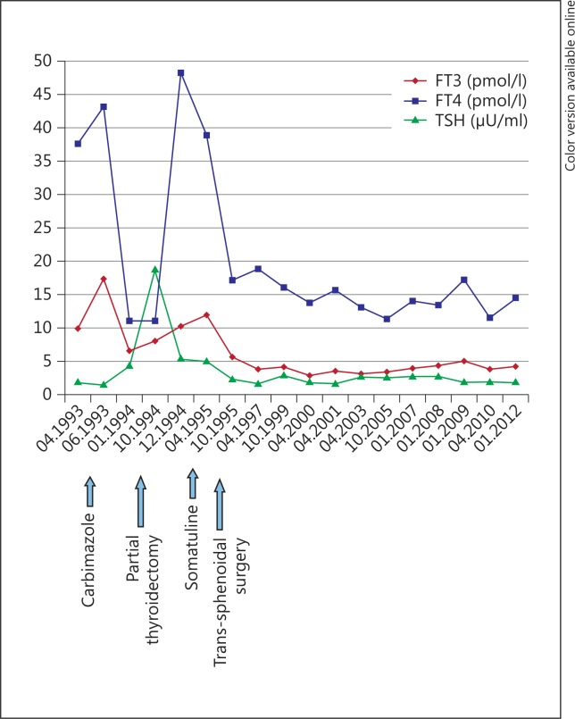
After a transient amelioration, symptoms of thyrotoxicosis recurred 2 months later, and the patient was referred to our university department. Thyroid function tests after immuno-precipitation were as follow: FT3 10.3 pmol/l; FT4 48.3 pmol/l, and TSH 5.4 µU/ml. Serum concentration of the α-TSH was elevated at 1.3 IU/l (normal range 0-0.9 IU/l), and the α-TSH/TSH molar ratio was also elevated at 2.4 (normal range <1). TSH levels were not effectively increased after TRH injection (250 µg, intravenous injection) [baseline 5.4 µIU/ml; 15 min (maximal TSH response) 6.1 µIU/ml]. The diagnosis of inappropriate secretion of TSH due to TSHoma was suggested. After administration of octreotide (octreotide acetate 50 µg s.c.), TSH concentrations decreased significantly [baseline 5.1 µIU/ml, 4 h (nadir) 2.4 µIU/ml]. After 24-hour subcutaneous injection of octreotide (200 µg), FT4 decreased from 35.8 to 26.6 pmol/l, FT3 from 12 to 5.1 pmol/l and TSH from 3.9 to 1.56 µU/ml.
Levels of basal growth hormone, insulin-like growth factor 1, and prolactin were normal (0.4 ng/ml, 0.87 IU/l and 7 ng/ml, respectively). Basal plasma ACTH level was in the normal range (44 pg/ml; normal range 10-55 pg/ml), with normal plasma cortisol (19 µg/100 ml; normal range 9-22 µg/100 ml). Gonadotropin and estradiol levels were also normal.
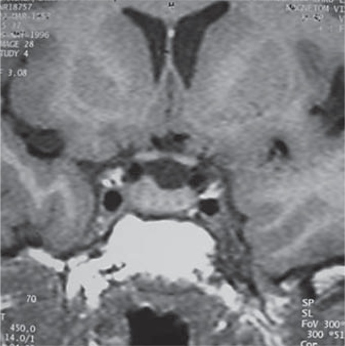
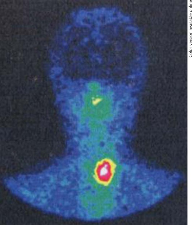
Thyroid ultrasound revealed an enlarged, hypoechoic and homogeneous left lobe (60 × 25 × 21 mm). Magnetic resonance imaging (MRI) revealed a pituitary lesion with a diameter of 10 mm and located on the right side of the sella. This lesion was associated with an elevation of the diaphragma sellae. No clear sign of invasion of the cavernous sinuses was evident (fig. 1). The mass showed isosignal on T1-weighted images; slight hypersignal on T2-weighted images and homogeneous enhancement after gadolinium administration. The patient had no symptoms of headache or visual disturbances, and her visual field was normal. Pituitary indium-111 (DTPA-octreotide) scintigraphy showed focal area of uptake within the sella turcica corresponding to the pituitary mass (fig. 2). Based on these findings, the patient was diagnosed as a case of pituitary TSHoma. Because of the somatostatin receptor positivity of the tumour, a 6-month preoperative treatment with lanreotide (Somatuline® LP, IPSEN, France) was planned, with 30 mg/10 days i.m. This treatment resulted in significant clinical improvement and normalization of thyroid function tests (FT3 5.7 pmol/l; FT4 17 pmol/l, and TSH 2.4 µU/ml). Thyroid ultrasound showed reduction in the size of the left lobe (45 × 20 × 19 mm). There was no change on 6-month follow-up pituitary MRI. Total resection of the pituitary tumor by trans-sphenoidal neurosurgery was performed. Postoperative recovery was uneventful. Histological examination revealed a pituitary adenoma. Immunocytochemistry showed diffuse positive staining for TSH, whereas it was negative for LH, FSH, PRL and ACTH. The final diagnosis was pure TSH-secreting pituitary adenoma. During 18 years of follow-up, she remained clinically well with normal thyroid function tests (fig. 3). Recent thyroid ultrasonography showed a slightly hypoechoic vascular left lobe measuring 44 × 16 × 16 mm. Repeated pituitary MRI showed no sign of tumor recurrence.
Fig. 1.

T1-weighted nonenhanced coronal MRI scan showing right-sided isointense pituitary mass.
Fig. 2.
Post-partial thyroidectomy indium-111 (DTPA-octreotide) scintigraphy showing an uptake in the left thyroid lobe along with focus of increased activity within the sella turcica.
Fig. 3.
Evolution of thyroid function tests during follow-up.
Discussion
We report the case of a female patient who suffered from a precocious recurrent hyperthyroidism following subtotal thyroidectomy for Graves' disease.
Initially, our patient presented with high levels of TSH receptor antibodies and antithyroid peroxidase antibodies associated with hyperthyroxinemia and unexpectedly normal TSH values. This led us to suspect assay interference. However, the concordance of TSH values, being persistently within the reference range despite use of different TSH assays, made this diagnosis less likely. Antibody interference in thyroid hormone immunoassays may result either from autoantibodies or heterophile antibodies [10,11]. The hypothesis of heterophilic antibody interference might be excluded in our patient because TSH values were not modified by serum treatment with heterophilic blocking tubes.
Autoantibodies have been reported mainly after injections of bovine TSH [12], and their existence is also less likely in our patient as she had unremarkable past history.
Later, our patient underwent extensive investigations that concluded to a TSHoma. TSHomas are rare tumors and account for less than 2% of all pituitary tumors [1,2,3]. They are part of the syndrome of inappropriate secretion of TSH. Normal or elevated TSH levels in hyperthyroid patients are characteristic of TSH-secreting pituitary adenoma. The lack of response of TSH to TRH is a hallmark of TSHoma. Other diagnostic findings are high sex hormone-binding globulin levels, high alpha-subunit levels, and a high alpha-subunit/TSH molar ratio [1,2,3]. Our patient fulfilled all these diagnostic criteria.
Most patients present mild signs of thyrotoxicosis and possible neurological features due to the pituitary mass. Goiter is present in almost all patients [1,2]. Our patient had recurrence of thyrotoxicosis signs and thyroid enlargement of the left lobe following total right lobectomy with partial left lobectomy. One explanation for this is that thyroid residue may regrow as a consequence of the continuous thyrotropin hyperstimulation [1]. A large body of evidence suggests that TSH is the main factor involved in the control of proliferation of thyrocytes [13].
In our case, the adenoma was immunohistologically confirmed as a pure TSH-producing tumor. About 50-70% of TSHomas are reportedly pure TSH-producing tumors. In one third of cases, TSHoma produces other hormones, especially growth hormone and prolactin [1].
The inhibitory effect of octreotide on TSH levels seen in our patient is a typical character in TSHoma. The octreotide test is useful before surgery to predict whether the drug could be used as therapy if surgery alone was not curative. The different response is due to the high concentration of somatostatin receptors on TSH-secreting tumors [14].
Association of TSHoma with Graves' disease is extremely rare. Previously, only six cases of such association and with histological confirmation have been reported [4,5,6,7,8,9] (table 1). Five cases were reported in female patients. Also, in 5 patients, pituitary adenoma precedes the occurrence of the Graves' diseases. Trans-sphenoidal tumor resection, administration of octreotide and pituitary irradiation were identified as possible etiological factors for this association [4,5,6,7,8].
Table 1.
Summary of case reports of association of thyrotropin adenoma with Graves’ disease (we included cases with histological confirmation of a thyrotropin adenoma)
| Authors | Sex, age, years | Clinical presentation | Initial diagnosis | Possible etiological factor(s) | Pituitary tumor size, mm | Hormonal cosecretion |
|---|---|---|---|---|---|---|
| O'Donnell et al. [4], 1973 | male, 25 | thyrotoxicosis signs visual field defect | TSHoma | tumor resection | macroadenoma | ? |
| Sandler [5], 1976 | female, 56 | thyrotoxicosis signs acromegalic features | TSHoma | pituitary irradiation | ? | GH |
| Kamoi et al. [7], 1985 | female, 46 | thyrotoxicosis signs galactorrhea | TSHoma | tumor resection | ? | PRL |
| Koriyama et al. [8], 2004 | female, 31 | thyrotoxicosis signs | TSHoma | tumor resection octreotide administration | 15 | no |
| Ogawa and Tominaga [9], 2013 | female, 32 | thyrotoxicosis signs exophthalmos | Graves’ disease | ? | 5 | plurihormonal expression |
| Present case | female, 36 | thyrotoxicosis signs exophthalmos | Graves’ disease | ? | 10 | No |
One case was omitted from this table because no data could be collected from the reference [6].
TSH is known as an important hormone that plays the major role not only in the maintenance of normal physiology but also in the regulation of immunomodulatory gene expression in thyrocytes. Fas antigen is functionally expressed on the surface of thyrocytes, and TSH inhibits Fas antigen-mediated apoptosis of thyrocytes resulting in the promotion of growth of the thyroid gland [15]. Intercellular adhesion molecule-1 and class II MHC have been implicated as contributing factors for numerous diseases, including autoimmune thyroid diseases, and TSH could downregulate their interferon-γ-mediated expression [16,17,18,19]. Therefore, a rapid reduction in the TSH levels after trans-sphenoidal tumor resection, pituitary irradiation, or octreotide administration may induce apoptosis and activate autoimmune responses against the thyroid gland as a result of increased expression of various cell surface markers (Fas, intercellular adhesion molecule-1, MHC class II) on thyrocytes [8]. In line with these hypotheses, Kageyama et al. [19] reported the case of a 21-year-old woman with TSHoma who had increases in both anti-TSH receptors antibody and thyroid-stimulating antibody after tumor resection.
Paradoxically, a patient with Graves' disease who subsequently developed a TSH-secreting pituitary adenoma was recently reported [9]. Although this could be incidental occurrence, anti-thyroid medication administered under a misdiagnosis of Graves' disease may carry the risk of promotion of TSH-secreting pituitary adenoma due to the positive feedback system.
In conclusion, we report an exceptional case of Graves' disease associated with thyrotropin adenoma in a female patient who presented initially inappropriate TSH values. Association of TSHoma with Graves' disease should be carefully taken into account, especially when TSH values are not compatible with either the clinical history or other thyroid functions tests.
Disclosure Statement
The authors have nothing to declare.
References
- 1.Losa M, Fortunato M, Molteni L, Peretti E, Mortini P. Thyrotropin-secreting pituitary adenomas: biological and molecular features, diagnosis and therapy. Minerva Endocrinol. 2008;3:329–340. [PubMed] [Google Scholar]
- 2.Beck-Peccoz P, Persani L, Mannavola D, Campi I. Pituitary tumours: TSH-secreting adenomas. Best Pract Res Clin Endocrinol Metab. 2009;23:597–606. doi: 10.1016/j.beem.2009.05.006. [DOI] [PubMed] [Google Scholar]
- 3.Beck-Peccoz P, Lania A, Beckers A, Chatterjee K, Wemeau JL. 2013 European Thyroid Association guidelines for the diagnosis and treatment of thyrotropin-secreting pituitary tumors. Eur Thyroid J 2013, DOI: 10.1159/000351007. [DOI] [PMC free article] [PubMed]
- 4.O'Donnell J, Hadden DR, Weaver JA, Montgomery DA. Thyrotoxicosis recurring after surgical removal of a thyrotrophin-secreting pituitary tumour. Proc R Soc Med. 1973;66:441–442. [PMC free article] [PubMed] [Google Scholar]
- 5.Sandler R. Recurrent hyperthyroidism in an acromegalic patient previously treated with proton beam irradiation: Graves' disease as probable etiology based on follow-up observations. J Clin Endocrinol Metab. 1976;42:163–168. doi: 10.1210/jcem-42-1-163. [DOI] [PubMed] [Google Scholar]
- 6.Azukizawa M, Morimoto S, Miyai K, Miki T, Yabu Y, Amino N, et al. TSH-producing pituitary adenoma associated with Graves' disease; in Stockigt JR, Nagataki S (eds): Thyroid Research. Canberra, Australian Academy of Science, 1980, vol VII, pp 645-648.
- 7.Kamoi K, Mitsuma T, Sato H, Yokoyama M, Washiyama K, Tanaka R, et al. Hyperthyroidism caused by a pituitary thyrotrophin-secreting tumour with excessive secretion of thyrotrophin-releasing hormone and subsequently followed by Graves' disease in a middle-aged woman. Acta Endocrinol (Copenh) 1985;110:373–382. doi: 10.1530/acta.0.1100373. [DOI] [PubMed] [Google Scholar]
- 8.Koriyama N, Nakazaki M, Hashiguchi H, Aso K, Ikeda Y, Kimura T, et al. Thyrotropin-producing pituitary adenoma associated with Graves' disease. Eur J Endocrinol. 2004;151:587–594. doi: 10.1530/eje.0.1510587. [DOI] [PubMed] [Google Scholar]
- 9.Ogawa Y, Tominaga T. Thyroid-stimulating hormone-secreting pituitary adenoma presenting with recurrent hyperthyroidism in post-treated Graves' disease: a case report. J Med Case Rep. 2013;7:27. doi: 10.1186/1752-1947-7-27. [DOI] [PMC free article] [PubMed] [Google Scholar]
- 10.Sakai H, Fukuda G, Suzuki N, Watanabe C, Odawara M. Falsely elevated thyroid-stimulating hormone (TSH) level due to macro-TSH. Endocr J. 2009;56:435–440. doi: 10.1507/endocrj.k08e-361. [DOI] [PubMed] [Google Scholar]
- 11.Tate J, Ward G. Interferences in immunoassay. Clin Biochem Rev. 2004;25:105–120. [PMC free article] [PubMed] [Google Scholar]
- 12.Sapin R, d'Herbomez M, Schlienger JL, Wemeau JL. Anti-thyrotropin antibody interference in thyrotropin assays. Clin Chem. 1998;44:2557–2559. [PubMed] [Google Scholar]
- 13.Fiore E, Vitti P. Serum TSH and risk of papillary thyroid cancer in nodular thyroid disease. J Clin Endocrinol Metab. 2012;97:1134–1145. doi: 10.1210/jc.2011-2735. [DOI] [PubMed] [Google Scholar]
- 14.Caron P, Arlot S, Bauters C, Chanson P, Kuhn JM, Pugeat M, et al. Efficacy of the long-acting octreotide formulation (octreotide-LAR) in patients with thyrotropin-secreting pituitary adenomas. J Clin Endocrinol Metab. 2001;86:2849–2853. doi: 10.1210/jcem.86.6.7593. [DOI] [PubMed] [Google Scholar]
- 15.Kawakami A, Eguchi K, Matsuoka N, Tsuboi M, Kawabe Y, Ishikawa N, et al. Thyroid-stimulating hormone inhibits Fas antigen-mediated apoptosis of human thyrocytes in vitro. Endocrinology. 1996;137:3163–3169. doi: 10.1210/endo.137.8.8754734. [DOI] [PubMed] [Google Scholar]
- 16.Chung J, Park ES, Kim D, Suh JM, Chung HK, Kim J, et al. Thyrotropin modulates interferon-gamma-mediated intercellular adhesion molecule-1 gene expression by inhibiting Janus kinase-1 and signal transducer and activator of transcription-1 activation in thyroid cells. Endocrinology. 2000;141:2090–2097. doi: 10.1210/endo.141.6.7507. [DOI] [PubMed] [Google Scholar]
- 17.Park ES, You SH, Kim H, Kwon OY, Ro HK, Cho BY, et al. Hormone-dependent regulation of intercellular adhesion molecule-1 gene expression: cloning and analysis of 5′-regulatory region of rat intercellular adhesion molecule-1 gene in FRTL-5 rat thyroid cells. Thyroid. 1999;9:601–612. doi: 10.1089/thy.1999.9.601. [DOI] [PubMed] [Google Scholar]
- 18.Kim H, Suh JM, Hwang ES, Kim DW, Chung HK, Song JH, et al. Thyrotropin-mediated repression of class II trans-activator expression in thyroid cells: involvement of STAT3 and suppressor of cytokine signaling. J Immunol. 2003;171:616–627. doi: 10.4049/jimmunol.171.2.616. [DOI] [PubMed] [Google Scholar]
- 19.Kageyama K, Ikeda H, Sakihara S, Nigawara T, Terui K, Tsutaya S, et al. A case of thyrotropin-producing pituitary adenoma, accompanied by an increase in anti-thyrotropin receptor antibody after tumor resection. J Endocrinol Invest. 2007;30:957–961. doi: 10.1007/BF03349244. [DOI] [PubMed] [Google Scholar]


