

Abstract
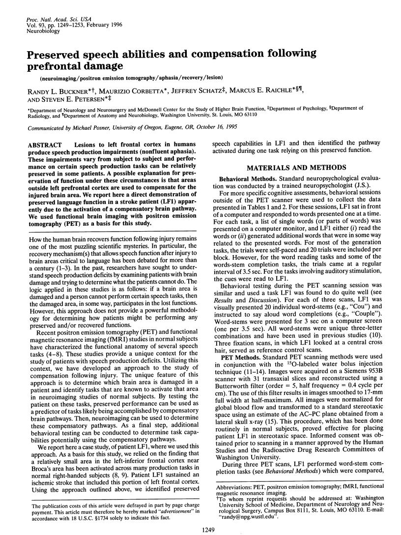
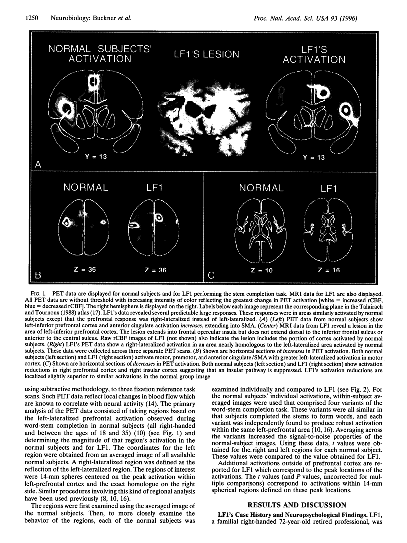
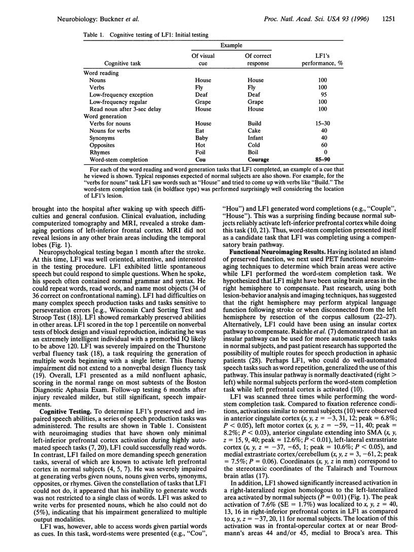
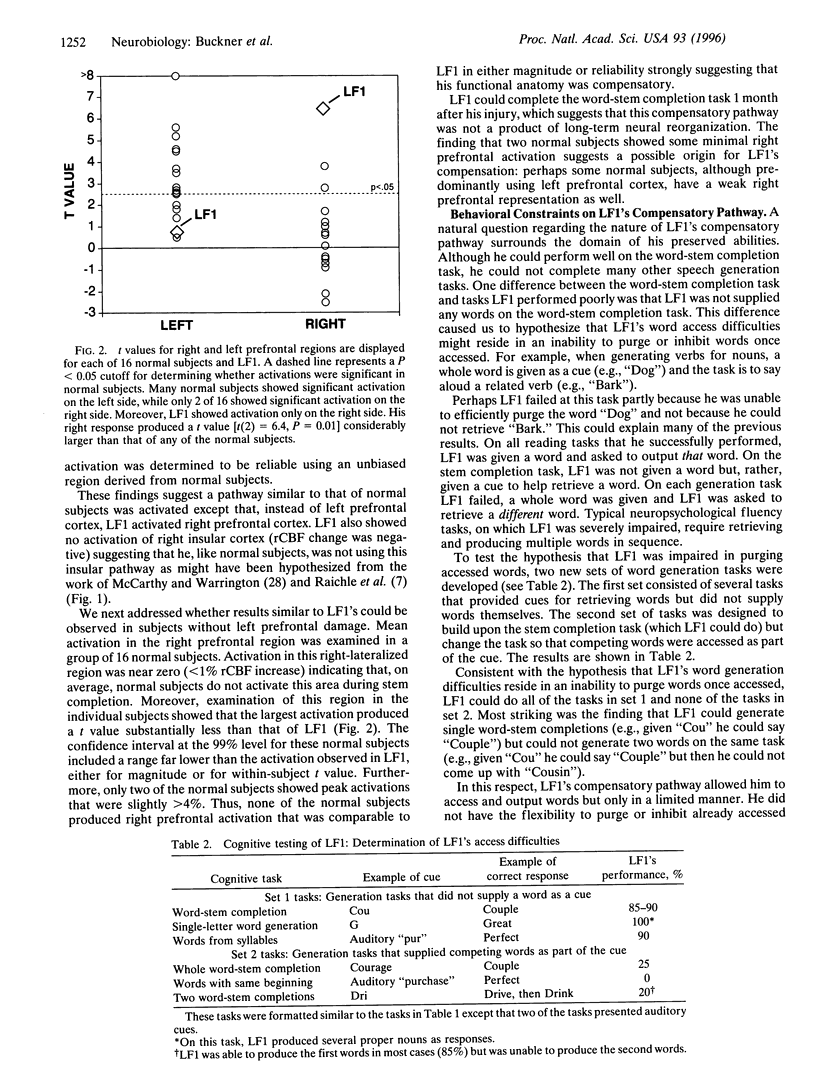
Lesions to left frontal cortex in humans produce speech production impairments (nonfluent aphasia). These impairments vary from subject to subject and performance on certain speech production tasks can be relatively preserved in some patients. A possible explanation for preservation of function under these circumstances is that areas outside left prefrontal cortex are used to compensate for the injured brain area. We report here a direct demonstration of preserved language function in a stroke patient (LF1) apparently due to the activation of a compensatory brain pathway. We used functional brain imaging with positron emission tomography (PET) as a basis for this study.
Full text
PDF
Images in this article
Selected References
These references are in PubMed. This may not be the complete list of references from this article.
- Buckner R. L., Petersen S. E., Ojemann J. G., Miezin F. M., Squire L. R., Raichle M. E. Functional anatomical studies of explicit and implicit memory retrieval tasks. J Neurosci. 1995 Jan;15(1 Pt 1):12–29. doi: 10.1523/JNEUROSCI.15-01-00012.1995. [DOI] [PMC free article] [PubMed] [Google Scholar]
- Buckner R. L., Raichle M. E., Petersen S. E. Dissociation of human prefrontal cortical areas across different speech production tasks and gender groups. J Neurophysiol. 1995 Nov;74(5):2163–2173. doi: 10.1152/jn.1995.74.5.2163. [DOI] [PubMed] [Google Scholar]
- Fox P. T., Mintun M. A. Noninvasive functional brain mapping by change-distribution analysis of averaged PET images of H215O tissue activity. J Nucl Med. 1989 Feb;30(2):141–149. [PubMed] [Google Scholar]
- Fox P. T., Perlmutter J. S., Raichle M. E. A stereotactic method of anatomical localization for positron emission tomography. J Comput Assist Tomogr. 1985 Jan-Feb;9(1):141–153. doi: 10.1097/00004728-198501000-00025. [DOI] [PubMed] [Google Scholar]
- Gazzaniga M. S. Right hemisphere language following brain bisection. A 20-year perspective. Am Psychol. 1983 May;38(5):525–537. doi: 10.1037//0003-066x.38.5.525. [DOI] [PubMed] [Google Scholar]
- Gazzaniga M. S., Volpe B. T., Smylie C. S., Wilson D. H., LeDoux J. E. Plasticity in speech organization following commissurotomy. Brain. 1979 Dec;102(4):805–815. doi: 10.1093/brain/102.4.805. [DOI] [PubMed] [Google Scholar]
- Herscovitch P., Markham J., Raichle M. E. Brain blood flow measured with intravenous H2(15)O. I. Theory and error analysis. J Nucl Med. 1983 Sep;24(9):782–789. [PubMed] [Google Scholar]
- Howard D., Patterson K., Wise R., Brown W. D., Friston K., Weiller C., Frackowiak R. The cortical localization of the lexicons. Positron emission tomography evidence. Brain. 1992 Dec;115(Pt 6):1769–1782. doi: 10.1093/brain/115.6.1769. [DOI] [PubMed] [Google Scholar]
- Jones-Gotman M., Milner B. Design fluency: the invention of nonsense drawings after focal cortical lesions. Neuropsychologia. 1977;15(4-5):653–674. doi: 10.1016/0028-3932(77)90070-7. [DOI] [PubMed] [Google Scholar]
- Kinsbourne M. The minor cerebral hemisphere as a source of aphasic speech. Arch Neurol. 1971 Oct;25(4):302–306. doi: 10.1001/archneur.1971.00490040028003. [DOI] [PubMed] [Google Scholar]
- McCarthy G., Blamire A. M., Rothman D. L., Gruetter R., Shulman R. G. Echo-planar magnetic resonance imaging studies of frontal cortex activation during word generation in humans. Proc Natl Acad Sci U S A. 1993 Jun 1;90(11):4952–4956. doi: 10.1073/pnas.90.11.4952. [DOI] [PMC free article] [PubMed] [Google Scholar]
- McCarthy R., Warrington E. K. A two-route model of speech production. Evidence from aphasia. Brain. 1984 Jun;107(Pt 2):463–485. doi: 10.1093/brain/107.2.463. [DOI] [PubMed] [Google Scholar]
- Mintun M. A., Fox P. T., Raichle M. E. A highly accurate method of localizing regions of neuronal activation in the human brain with positron emission tomography. J Cereb Blood Flow Metab. 1989 Feb;9(1):96–103. doi: 10.1038/jcbfm.1989.13. [DOI] [PubMed] [Google Scholar]
- Petersen S. E., Fox P. T., Posner M. I., Mintun M., Raichle M. E. Positron emission tomographic studies of the cortical anatomy of single-word processing. Nature. 1988 Feb 18;331(6157):585–589. doi: 10.1038/331585a0. [DOI] [PubMed] [Google Scholar]
- Raichle M. E., Fiez J. A., Videen T. O., MacLeod A. M., Pardo J. V., Fox P. T., Petersen S. E. Practice-related changes in human brain functional anatomy during nonmotor learning. Cereb Cortex. 1994 Jan-Feb;4(1):8–26. doi: 10.1093/cercor/4.1.8. [DOI] [PubMed] [Google Scholar]
- Raichle M. E., Martin W. R., Herscovitch P., Mintun M. A., Markham J. Brain blood flow measured with intravenous H2(15)O. II. Implementation and validation. J Nucl Med. 1983 Sep;24(9):790–798. [PubMed] [Google Scholar]
- Squire L. R., Ojemann J. G., Miezin F. M., Petersen S. E., Videen T. O., Raichle M. E. Activation of the hippocampus in normal humans: a functional anatomical study of memory. Proc Natl Acad Sci U S A. 1992 Mar 1;89(5):1837–1841. doi: 10.1073/pnas.89.5.1837. [DOI] [PMC free article] [PubMed] [Google Scholar]
- Weiller C., Isensee C., Rijntjes M., Huber W., Müller S., Bier D., Dutschka K., Woods R. P., Noth J., Diener H. C. Recovery from Wernicke's aphasia: a positron emission tomographic study. Ann Neurol. 1995 Jun;37(6):723–732. doi: 10.1002/ana.410370605. [DOI] [PubMed] [Google Scholar]
- Wise R., Chollet F., Hadar U., Friston K., Hoffner E., Frackowiak R. Distribution of cortical neural networks involved in word comprehension and word retrieval. Brain. 1991 Aug;114(Pt 4):1803–1817. doi: 10.1093/brain/114.4.1803. [DOI] [PubMed] [Google Scholar]