Abstract
Aim:
YCP, a novel (1,4)-α-D-glucan, was isolated from the mycelium of the marine filamentous fungus Phoma herbarum YS4108. In this work, we investigated a YCP-binding cellular receptor expressed by macrophages and the intracellular signal transduction pathways involved in YCP-induced macrophage activation.
Methods:
Fluorescence-labeled YCP (fl-YCP) was prepared using the CDAP-activation method. Fluorescence confocal laser microscopy and fluorescence-activated cell sorting (FACS) were used to analyze the effect of fl-YCP on macrophages. To characterize the properties of the YCP receptor, carbohydrates and antibodies were used to inhibit the binding of fl-YCP to macrophages. Moreover, we investigated the role of membrane receptors Toll-like receptor 2 (TLR2), Toll-like receptor 4 (TLR4), Toll-like receptor 6 (TLR6) and complement receptor 3 (CR3). We also examined the role of the p38 kinase pathway in mediating nitric oxide (NO) production.
Results:
YCP had an in vitro stimulatory effect on the release of NO in macrophage, and fl-YCP can bind directly to receptors on the surface of macrophages in a time- and dose-dependent manner. Competition studies show that LPS, laminarin, anti-TLR4 antibody and anti-CD11b (CR3) antibody could inhibit fl-YCP binding to macrophages. Conversely, mannose, anti-TLR2 and anti-TLR6 antibody could not. Treatment of RAW264.7 cells with YCP resulted in significant activation of p38 in a time-dependent manner. The specific p38 inhibitor SB203580 abrogated YCP-induced NO generation. Treatment of RAW264.7 cells with anti-TLR4 antibody and anti-CR3 antibody significantly reduced YCP-induced NO production and p38 activation.
Conclusion:
We have demonstrated that YCP-induced NO production occurs through the TLR4 and CR3 membrane receptors in a p38 kinase-dependent manner in macrophages.
Keywords: YCP, Phoma herbarum, macrophages, nitric oxide, receptor, p38 kinase
Introduction
YCP is a polysaccharide with a mean molecular weight of 2.4×10³ kDa purified from the mycelium of Phoma herbarum YS4108, a marine filamentous fungus that inhabits the sediment at a depth of 50 m in the Yellow Sea near Sheyang Port, Yancheng, China. (YCP is the acronym of Yancheng and polysaccharide.) In an earlier study, YCP was found to be able to increase phagocytic activity of mice in vitro and in vivo, indicating that it may activate macrophages1. YCP could significantly increase the production of NO and interleukin-1 (IL-1) by macrophages. It also significantly increased phagocytic activity of macrophages2. However, the YCP-binding cellular receptor expressed by macrophages and the intracellular signal transduction pathways involved in YCP-induced macrophage activation are poorly understood.
β-glucans are potent stimulators of the innate immune system in invertebrates, while in mammals they are potent activators of the complement system. Soluble and particulate β-glucans interact with cognate receptors on macrophages to stimulate the synthesis of cytokines, chemokines and reactive oxygen intermediates. Even though several papers describing the immunostimulatory properties of β-glucans are available in the literature3, α-glucans have been scarcely investigated to date. We have reported previously on the isolation and characterization of a novel (1,4)-α-D-glucan from the mycelium of the marine filamentous fungus Phoma herbarum YS41081. Immunostimulatory properties of β-glucans have been ascribed to their β-glycosidic linkages, degree of branching and solution conformation3. YCP is water soluble, and its backbone likely possesses a linear α-(1,4)-linked glucose main chain with an α-(1,6)-linked side chain1.
Macrophages are one of the most important immune cell types, and they play a pivotal role in host defense and immunomodulation. Macrophages are major sources of inflammatory cytokines and proinflammatory radicals such as nitric oxide (NO) and superoxide. These inflammatory mediators can be both produced and utilized by macrophages in an autocrine fashion. When activated, macrophages can release cytokines, such as IL-1 and NO, to defend against microbial infection and lyse tumor cells4.
Although the membrane receptor for YCP is not known, some membrane proteins are assumed to act as receptors in macrophages. Possible examples are Toll-like receptors and CR3. Toll-like receptors are a family of membrane receptors that has a relatively broad affinity for polysaccharides isolated from a variety of sources5. Complement receptor CR3 (also called CD11b) is a β-glucan-specific receptor for opsonized polysaccharides in macrophages. Receptor engagement leads to macrophage activation and an inflammatory response3. The mitogen-activated protein kinase (MAPK) family member p38 kinase is activated in response to lipopolysaccharide (LPS) and plays a central role in NO production in macrophages6. To study the molecular basis of macrophage activation by YCP, we investigated the effect of YCP on activation of p38 kinase as well as the involvement of the Toll-like receptors and CR3 in YCP-mediated macrophage activation.
Materials and methods
Materials
YCP was isolated and characterized as described previously7. Briefly, a hot water (80 °C) extract was prepared from the harvested mycelium of P herbarum YS4108. The active fraction, YCP, was then isolated by a combination of fractionation procedures, including ethanol precipitation and chromatography on DEAE-32 anion exchange and Sephacryl S-400 columns. YCP appeared to be endotoxin-free. Necessary precautions were taken to avoid endotoxin contamination throughout the investigation by using endotoxin-free buffers, reagents and sterile water. SB203580 was purchased from Calbiochem (San Diego, CA, USA). Reagents used for cell culture were purchased from Gibco (Grand Island, NY, USA). Anti-mouse/human TLR2, anti-human TLR4, anti-human TLR6 and anti-mouse CD11b antibodies (Abs) were purchased from eBioscience (San Diego, CA, USA).
Cell culture
An established stable murine macrophage line, RAW264.7, was used (kindly donated by Professor Ping Li). Cells were grown in Dulbecco's modified Eagle's medium supplemented with 10% fetal bovine serum, 4-(2-hydroxyethyl)-1-piperazineethanesulfonic acid (15 mmol/L), glucose (25 mmol/L), 100 U/mL penicillin and 100 μg/mL streptomycin at 37 °C in an atmosphere containing 5 % CO2+95 % air. Cultures were passaged twice weekly at 1:4 split with the medium mentioned above.
Preparation of fluorescence-labeled YCP
A solution containing 1-cyano-4-dimethylaminopyridine tetrafluoroborate (CDAP) (Sigma, USA)8, 9 and hydroxyl group activator (10 mg/0.1 mL acetonitrile) was added to an aqueous solution of YCP (30 mg/2 mL) while stirring. After 1 min, pH was raised to 9.0 by adding 0.25 mol/L NaOH. The activation reaction proceeded for 2.5 min. Then, the CDAP-activated YCP was mixed with 2 mg fluoresceinamine [2 mg/2 mL phosphate-buffered saline (PBS)], and pH was adjusted to 8.0 using 0.25 mol/L NaOH. The reaction was carried out in the dark for 18 h at room temperature. The reaction mixture was centrifuged at 4000 r/min for 8 min, and the supernatant was collected. Next, fluorescence-labeled polysaccharide was separated from free fluoresceinamine by gel filtration on a Sephacryl S-400 column, and elution fractions (15 min/tube) were collected. The amount of fluorescence-labeled polysaccharide was determined by phenol-sulfuric acid assay, and the content of fluoresceinamine was determined by measuring absorbance at 440 nm. A standard curve was prepared using serially diluted glucose. The standard curve was constructed using serially diluted fluoresceinamine in PBS.
Confocal laser microscopy
Living macrophages growing on glass cover slips were incubated with fluorescence-labeled YCP (fl-YCP) for 1 h at 4 °C. After a brief wash with PBS, cells were observed through fluorescence confocal laser microscopy (Leica) with 450 nm excitation and 520 nm emission wavelengths.
Flow cytometry
Cell suspensions were centrifuged, and the cell pellets (1×10⁶/tube) were resuspended with 50 μL of fl-YCP (100 μg/mL) and incubated for 1 h at 4 °C. The cells were washed twice in PBS and resuspended in 500 μL of PBS for flow cytometric analysis on a fluorescence-activated cell sorter (FACS) Calibur (Becton–Dickinson).
Carbohydrate and antibody inhibition experiments
All carbohydrates were obtained from Sigma (USA). To perform inhibition assays, the cells (1×10⁶/tube) were treated with laminarin, mannose or LPS at 1 mg/mL for 1 h and then fl-YCP (100 μg/mL) for another hour at 4 °C. After two washes with PBS, the cells were resuspended in 500 μL of PBS for flow cytometric analysis.
The antibody inhibition experiments were performed similarly, except that the cells were incubated with the monoclonal antibodies in PBS for 1 h and then washed extensively before the determination of fl-YCP binding10.
Nitrite quantitation
NO2− accumulation was used as an indicator of NO production as previously described11. Cells were plated at 4×10⁶ cells/mL in 96-well culture plates and stimulated with YCP for 24 h. The isolated supernatants were mixed with an equal volume of Griess reagent (1% sulfanilamide, 0.1% naphthylethylenediamine dihydrochloride and 2% phosphoric acid) and incubated at room temperature for 10 min. Using NaNO2 to generate a standard curve, nitrite production was estimated by measuring optical density at 550 nm.
Western immunoblot analysis
Whole cell lysates (20 μg, for phospho-p38 and p38) were separated by 10% sodium dodecyl sulfate-polyacrylamide gel electrophoresis (SDS-PAGE) and then electro-transferred to nitrocellulose membranes (Amersham International, Buckinghamshire, UK). The membranes were preincubated for 1 h at room temperature in Tris-buffered saline (TBS) containing 0.05% Tween 20 and 3% bovine serum albumin at pH 7.6. The nitrocellulose membranes were incubated with p38- and phosphorylated p38-specific antibodies (Beyotime Institute of Biotechnology, China). Immunoreactive bands were then detected by incubation with conjugates of anti-mouse antibody with horseradish peroxidase and enhanced chemiluminescence reagents (Amersham)12.
Statistical analysis
The mean±SD was determined for each treatment group in a given experiment. When significant differences occurred, treatment groups were compared with the vehicle control using a Dunnett's two-tailed t-test.
Results
Fluorescence labeling of YCP
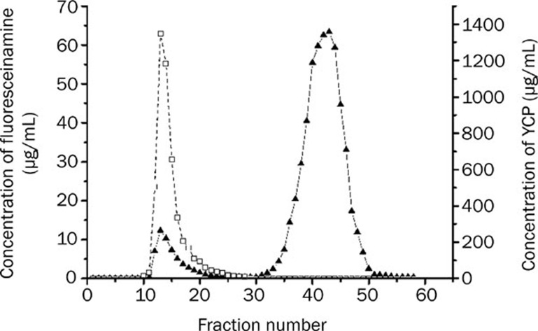
In order to be able to identify cells expressing specific receptor(s) for YCP, YCP was conjugated with fluoresceinamine using the CDAP-activation method as described in Materials and methods. The resultant fluorescence-labeled YCP (fl-YCP) was separated from free fluoresceinamine in the labeling mixture by filtration on a Sephacryl S-400 column. All fractions were monitored for carbohydrate and fluoresceinamine concentration. As shown in Figure 1, a small amount of fluoresceinamine co-eluted with polysaccharides, while free fluoresceinamine eluted much more slowly. Fluorescence-labeled polysaccharide fractions were mixed, adjusted to a concentration of 1 mg/mL and stored at -20 °C for future use.
Figure 1.
Polysaccharide labeling with fluoresceinamine. CDAP-activated YCP reacted with fluoresceinamine for 18 h at room temperature. The mixtures were fractionated on a Sephacryl S-400 column, concentration (μg/mL) of fluoresceinamine (▴) and YCP (□) in each fraction were determined. n=3.
Time and concentration dependence of fl-YCP binding to cells
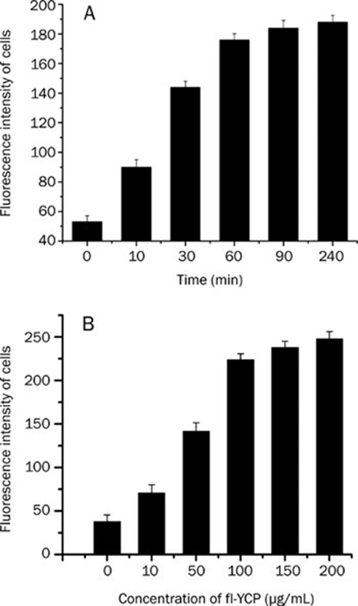
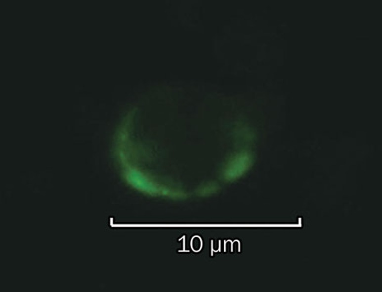
Initial experiments tested the time (Figure 2A) and concentration (Figure 2B) dependence of fl-YCP binding to macrophages. Saturable fl-YCP binding to macrophages occurred within 60 min at 100 μg/mL. Saturable binding to macrophages at 4 °C was demonstrated by Scatchard analysis and indicated a class of specific receptors with a Bmax (fluorescence intensity of 1×10⁶ cells) of 263 and a Kd of 1.1×10⁻⁸ mol/L. Specific binding of fl-YCP to macrophages was also observed using a confocal laser-scanning microscope (Figure 3).
Figure 2.
(A) Time dependence of fl-YCP binding in macrophages. RAW264.7 cells were stained with fl-YCP at 100 μg/mL for 0, 10, 30, 60, 90, or 240 min at 4 °C for flow cytometric analysis. n=3. (B) Concentration dependence of fl-YCP binding in macrophages. RAW264.7 cells were stained with fl-YCP at 0, 10, 50, 100, 150, or 200 μg/mL for 60 min at 4°C for flow cytometric analysis. n=3.
Figure 3.
Live macrophage observed through confocal laser microscopy after incubation with fl-YCP for 1 h. Excitation and emission wavelengths were 450 nm and 520 nm, respectively. n=2.
Competitive inhibition of fl-YCP binding to cells by laminarin, mannose or LPS
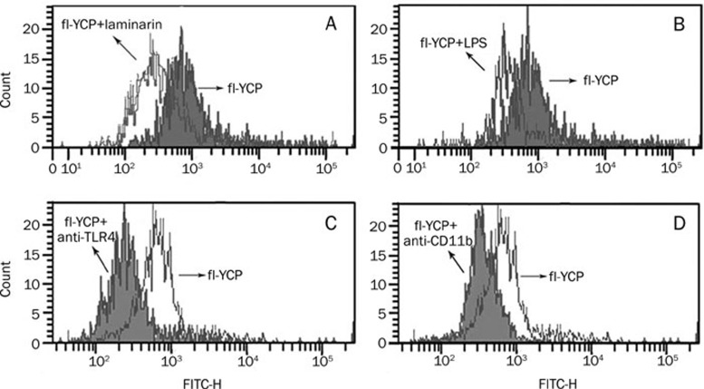
To address whether laminarin, mannose or LPS binds the same cellular receptor(s) as YCP, the ability of laminarin, mannose or LPS to inhibit the binding of fl-YCP to target cells was measured by flow cytometric analysis. Figure 4A and Figure 4B show that laminarin and LPS effectively, although not completely, inhibited fl-YCP binding to RAW264.7 cells. In contrast, mannose did not exhibit any significant inhibitory effect in similar experiments (data not shown).
Figure 4.
Competitive inhibition of fl-YCP binding to cells by laminarin (A) or LPS (B). RAW264.7 cells were stained with fl-YCP in the presence or absence of laminarin or LPS at 1 mg/mL for 1 h at 4 °C for flow cytometric analysis. n=3. Inhibition of fl-YCP binding to macrophages by anti-TLR4 (C) or anti-CD11b (D) Ab. RAW264.7 cells were treated with anti-mouse/human TLR4 or anti-mouse CD11b for 1 h at 4 °C followed by fl-YCP staining. n=3.
Role of TLR2, TLR4, TLR6, and CD11b in YCP-mediated activation of macrophages
In order to determine if TLR2, TLR4, TLR6, or CD11b plays a role in YCP-mediated activation of macrophages, RAW264.7 cells were incubated with anti-TLR2, anti-TLR4, anti-TLR6, and anti-CD11b Abs for 1 h and then with fl-YCP (100 μg/mL) for another hour at 4 °C. As shown in Figure 4C and Figure 4D, anti-TLR4 and anti-CD11b Abs significantly inhibited the ability of fl-YCP to bind to macrophages, suggesting a direct interaction between YCP and TLR4 or CD11b on the cell surface. By contrast, anti-TLR2 and TLR6 did not exhibit any significant inhibitory effect in similar experiments (data not shown).
Activation of p38 kinase in response to YCP in RAW264.7 cells
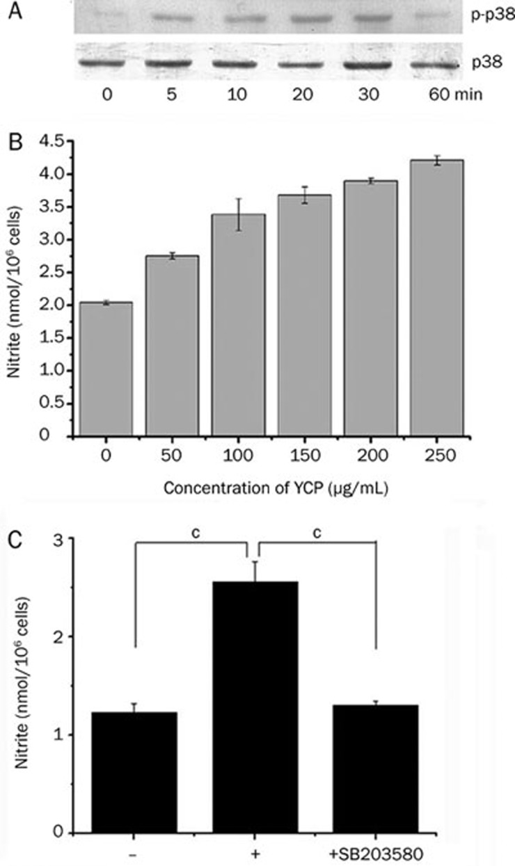
To further investigate the molecular mechanism of YCP-mediated activation of macrophages, we focused on signal transduction pathways. Because p38 kinase activity has been shown to be required for iNOS induction by LPS in RAW264.7 macrophages, we assessed the effect of YCP on p38 kinase activation. Activation of p38 kinase requires phosphorylation at threonine and tyrosine residues. Immunoblot analysis with phospho-p38-specific (Thr180/Tyr182) antibody was performed. Time course experiments showed the peak activation of p38 occurred 20 min after treatment, and activity declined to basal levels within 60 min of treatment (Figure 5A).
Figure 5.
(A) Activation of p38 by YCP in RAW264.7 cells. RAW264.7 cells were stimulated with YCP (100 μg/mL) for the indicated times. Cell extracts were then prepared and subjected to Western immunoblotting using antibodies specific for p38 or phosphorylated p38. n=3. (B) Effect of YCP on NO production in RAW264.7 cells. RAW264.7 cells were incubated with YCP (50, 100, 150, 200, or 250 μg/mL) for 24 h. Levels of nitrite in supernatants were determined using Griess reagent. n=3. (C) Effect of SB203580 on nitrite production in YCP-stimulated RAW264.7 cells. RAW264.7 cells were pretreated with SB203580 (30 μmol/L) for 30 min before incubation with YCP (100 μg/mL) for 24 h. Nitrite generation was determined from the culture supernatant. n=2. cP<0.01.
Effect of p38 inhibition on nitrite production in macrophages
YCP has been reported to activate mouse peritoneal macrophages1. In the present study, we confirmed the effect of YCP on macrophage activation using RAW264.7 cells. Upon stimulation with YCP (50, 100, 150, 200, or 250 μg/mL), nitrite generation by RAW264.7 cells was increased in a dose-dependent manner (Figure 5B).
Since YCP activated p38, we further investigated whether the p38 kinase pathway was involved in YCP-induced nitrite generation. We specifically blocked the p38 kinase pathway and monitored nitrite production when RAW264.7 cells were challenged with YCP. SB203580, a bicyclic imidazole compound, is a specific inhibitor of p3813. SB203580 treatment almost completely blocked YCP-induced nitrite generation (Figure 5C). This result suggests that p38 kinase activity is important in the regulation of nitrite production by YCP.
Effects of anti-TLR2, anti-TLR4, anti-TLR6, and anti-CD11b Abs treatments on YCP-induced nitrite production and p38 activation
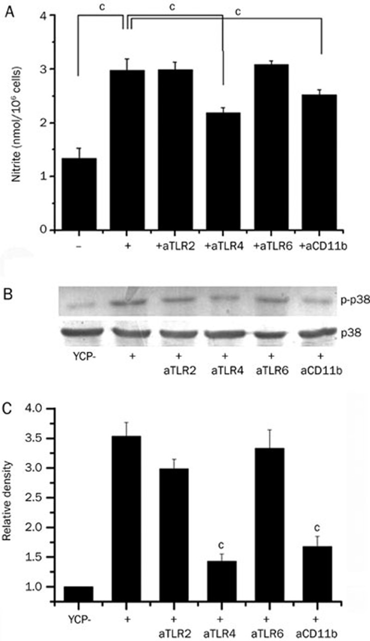
To further characterize the membrane receptor of YCP, we assessed the effects of anti-TLR2, anti-TLR4, anti-TLR6, and anti-CD11b Abs treatments on YCP-induced production of nitrite. Treatment of RAW264.7 cells with anti-TLR4 or anti-CD11b Abs reduced YCP-induced nitrite production by 48% and 27%, respectively (Figure 6A). Treatment with anti-TLR4 and anti-CD11b antibodies reduced nitrite production by 84%±6% (data not shown). These results suggest that YCP induces nitrite production through the activation of TLR4 and CD11b.
Figure 6.
(A) Effects of anti-TLR2, anti-TLR4, anti-TLR6 and anti-CD11b Ab treatments on YCP-induced nitrite production. RAW264.7 cells were treated with anti-mouse/human TLR2 (5 μg/mL), anti-human TLR4 (5 μg/mL), anti-human TLR6 (5 μg/mL) or anti-mouse CD11b (5 μg/mL) for 1 h before stimulation with YCP (100 μg/mL). Nitrite generation was determined after 24 h of culture. n=3. cP<0.01. (B) Effects of anti-TLR2, anti-TLR4, anti-TLR6 and anti-CR3 Ab treatments on YCP-induced p38 activation. RAW264.7 cells were treated with anti-mouse/human TLR2 (5 μg/mL), anti-human TLR4 (5 μg/mL), anti-human TLR6 (5 μg/mL) or anti-mouse CR3 (5 μg/mL) for 1 h before stimulation with YCP (100 μg/mL) for 20 min. Cell extracts were then prepared and subjected to Western immunoblotting using antibodies specific for p38 or phosphorylated p38. n=3. cP<0.01 vs YCR group.
We further analyzed the relationship between membrane receptor and p38 kinase pathways. Treatment of RAW264.7 cells with anti-TLR4 Ab and anti-CR3 Ab blocked YCP-induced p38 phosphorylation (Figure 6B). These results suggest that YCP-mediated activation of TLR4 and CR3 induces p38 kinase activity.
Discussion
We have previously demonstrated that YCP could bind to receptors on monocytes and macrophages14 and stimulate macrophages to produce cytokines such as NO and IL-12. However, the intracellular signal transduction pathways involved in YCP-induced cytokine synthesis by macrophages are poorly understood. In the present study, we demonstrate that YCP is capable of inducing nitrite release from RAW264.7 cells in a TLR4- and CR3-dependent manner. Additionally, we show that p38 kinase activation is required for NO production in YCP-stimulated RAW264.7 cells.
In this paper, we show that the organic cyanylating reagent 1-cyano-4-dimethylaminopyridine tetrafluoroborate (CDAP) can be used to synthesize fluorescence-labeled polysaccharides. CDAP9, an organic cyanylating reagent, is useful for activating soluble polysaccharides in a manner similar to the better known reagent cyanogen bromide (CNBr). In comparison to CNBr, CDAP is easier to use, can be employed at a lower pH and has fewer side effects. In order to be able to identify cells expressing specific receptor(s) for YCP, YCP was conjugated with fluoresceinamine using the CDAP-activation method.
Toll-like receptors (TLRs) are pattern recognition receptors that primarily recognize characteristic molecular patterns present in microbes. TLRs are expressed by various cells of the immune system. Monocytes, mast cells, dendritic cells, macrophages, neutrophils, NK cells, B cells and T cells are some of the prominent cell types showing an immune response to TLR agonists15. The TLR family now consists of 10 different members (TLR1-TLR10)16. TLR4, a mammalian homologue of the Drosophila Toll receptor, has been identified as an important membrane receptor of macrophages17. It mediates macrophage activation by transmitting a variety of extracellular signals12. Complement receptor CR3 (also called Mac-1, CD11b/CD18, and αMβ2-integrin) has been identified as the leukocyte membrane receptor for β-glucans18. They are expressed on the surface of neutrophils, monocytes, macrophages, and NK cells and are involved in numerous cell-cell and cell-substrate interactions. Multiple proteins have been implicated in the initial steps of LPS signaling. These include TLRs, heat shock proteins, chemokine receptors and the caspase-activator Nod 119. Mannose signaling occurs through the mannose receptor/DC-SIGN/MBL/SP-A/SP-D pathway20. Signaling induced by laminarin, a low molecular weight neutral β-glucan polymer, occurs through CR318. To determine which receptor is specific for YCP, we assessed the effect of LPS (ligand of TLR4), mannose (ligand of mannose receptors), and laminarin (ligand of CR3) on fl-YCP binding. The ability of LPS or laminarin to competitively inhibit the binding of YCP to macrophages suggests that the receptors of LPS and laminarin are also involved in fl-YCP binding to target cells. The ability of anti-TLR4 and anti-CR3 monoclonal antibodies to inhibit fl-YCP binding also supports this notion. TLR4 signaling pathways may play a role in immune cell activation by mediating polysaccharide-based biological response modifiers21. Local or systemic infections are likely to activate complement and TLRs at the same time, suggesting that complement receptor pathways may intersect with TLR pathways22. We propose that, although TLR4 is likely the primary receptor, YCP might interact with other receptors, such as CR3.
Nitric oxide (NO) is one of the inflammatory mediators produced by various cell types including macrophages in response to infection. NO is a short-lived free radical synthesized from L-arginine by nitric oxide synthase (NOS)23. NOSs can be grouped into two constitutive types (cNOS) in neuronal and epithelial cells and one inducible type (iNOS) produced by immune cells during inflammation. It is well documented that iNOS is overproduced in several cell types after stimulation by bacterial components during sepsis, which subsequently leads to the overproduction of NO24. NO contributes to the killing of microorganisms and tumor cells by activated macrophages and mediates a variety of biological functions as an intracellular messenger molecule. LPS is a well-known inducer of NO production in inflammatory cells. In the present study, we have shown that YCP induces NO production from macrophages through the membrane receptors TLR4 and CR3.
LPS signals through TLR4, which activates downstream molecules such as myeloid differentiation factor 88 (MyD88) to induce production of inflammatory cytokines25. Signals transmitted through TLR4 trigger activation of several members of the MAPK family26. A major role of MAPKs is the transmission of extracellular signals to the nucleus, where the transcription of specific genes is induced by the phosphorylation and activation of transcription factors. The MAPK family is a group of serine/threonine-specific, proline-directed protein kinases. LPS induces tyrosine phosphorylation and activation of MAPK family members such as ERK1/2, p38, and SAPK/JNK in macrophages. The p38 kinase is an important mediator of stress-induced gene expression6. Signaling by p38 is required to transduce signals leading to NF-κB/Rel activation, NOS expression and NO production in macrophages. The activation of NF-κB is regulated by cellular kinases, including p38. NF-κB is located in the cytoplasm of unstimulated cells and linked to the inhibitory protein IκB. NF-κB activation results from pro-inflammatory stimuli, which coincide with IκB phosphorylation and degradation via the IKK signalosome complex. This process results in the release of free NF-κB dimers (p50 and p65) that translocate to the nucleus where they are involved in the transcription of target genes such as iNOS, TNF-α, and IL-1β4. The p38 MAPK signaling pathway plays an important role in promoting inflammatory disease27. Activation of p38 induces the production of key inflammatory mediators. In particular, p38 is activated by LPS stimulation and has been postulated to play an important role in the control of iNOS expression4. We have shown that the p38 kinase pathway is an important mediator of NO production by YCP in two ways. First, YCP causes maximal activation of p38 kinase in RAW264.7 macrophages after 20 min. Second, specific inhibitors of p38 kinase (SB203580) completely blocked YCP-induced NO release.
In summary, these experiments demonstrate that YCP is capable of inducing NO production from macrophages through TLR4 and CR3 pathways. Based on our findings, the most likely mechanism that can account for this effect involves the activation of p38 kinase pathways. However, further studies should be undertaken to fully understand the intracellular processes involved in YCP-mediated macrophage activation. It is possible that YCP may be used clinically as a biological response modifier to activate macrophages.
Author contribution
Song CHEN, Deng-ke YIN, Wen-bing YAO, and Xiang-dong GAO designed the research; Song CHEN, Yi-dan WANG, and Yi-ran ZHANG performed the research; Wen-bing YAO and Xiang-dong GAO contributed new reagents and analytical tools; Song CHEN and Deng-ke YIN analyzed data; Song CHEN and Deng-ke YIN wrote the paper.
Acknowledgments
This work was co-financed by the Ministry of Science and Technology (National '863' project 2006AA090302), the National Natural Science Foundation (No 30672479, 30873201), the Specialized Research Fund for the Doctoral Program of Higher Education (No 20060316001) and the National Found for Fostering Talents of Basic Science (No J0630858).
References
- Yang XB, Gao XD, Han F, Xu BS, Song YC, Tan RX. Purification, characterization and enzymatic degradation of YCP, a polysaccharide from marine filamentous fungus Phoma herbarum YS4108. Biochimie. 2005;87:747–54. doi: 10.1016/j.biochi.2005.03.004. [DOI] [PubMed] [Google Scholar]
- Yao JF, Wu L, Wu XZ, Han F, Gao XD.Immuno-enhancing effect of YCP obtained from a marine filamentous fungus Keissleriella sp. YS 4108 on mice peritoneal macrophage J China Pharm Univ 200435573–5.Chinese. [Google Scholar]
- Nair PKR, Melnick SJ, Ramachandran R, Escalon E, Ramachandran C. Mechanism of macrophage activation by (1,4)-α-D-glucan isolated from Tinospora cordifolia. Int Immunopharmacol. 2006;6:1815–24. doi: 10.1016/j.intimp.2006.07.028. [DOI] [PubMed] [Google Scholar]
- Yun KJ, Kim JY, Kim JB, Lee KW, Jeong SY, Park HJ, et al. Inhibition of LPS-induced NO and PGE2 production by asiatic acid via NF-κB inactivation in RAW 264.7 macrophages: possible involvement of the IKK and MAPK pathways. Int Immunopharmacol. 2008;8:431–41. doi: 10.1016/j.intimp.2007.11.003. [DOI] [PubMed] [Google Scholar]
- Yoon YD, Han SB, Kang JS, Lee CW, Park SK, Lee HS, et al. Toll-like receptor 4-dependent activation of macrophages by polysaccharide isolated from the radix of Platycodon grandiflorum. Int Immunopharmacol. 2003;3:1873–82. doi: 10.1016/j.intimp.2003.09.005. [DOI] [PubMed] [Google Scholar]
- Jeon YJ, Han SB, Lee SH, Kim HC, Ahn KS, Kim HM. Activation of mitogen-activated protein kinase pathways by angelan in murine macrophages. Int Immunopharmacol. 2001;1:237–45. doi: 10.1016/s1567-5769(00)00003-5. [DOI] [PubMed] [Google Scholar]
- Yang XB, Gao XD, Han F, Tan RX. Sulfation of a polysaccharide produced by a marine filamentous fungus Phoma herbarum YS4108 alters its antioxidant properties in vitro. Biochim Biophys Acta. 2005. 1725. pp. 120–7. [DOI] [PubMed]
- Shafer DE, Toll B, Schuman RF, Nelson BL, Mond JJ, Lees A. Activation of soluble polysaccharides with 1-cyano-4-dimethylaminopyridinium tetrafluoroborate (CDAP) for use in protein-polysaccharide conjugate vaccines and immunological reagents. II. Selective crosslinking of proteins to CDAP-activated polysaccharides. Vaccine. 2000;18:1273–81. doi: 10.1016/s0264-410x(99)00370-9. [DOI] [PubMed] [Google Scholar]
- Bystrický S, Paulovicova E, Machova E. Candida albicans mannan-protein conjugate as vaccine candidate. Immunol Lett. 2003;85:251–5. doi: 10.1016/s0165-2478(02)00241-9. [DOI] [PubMed] [Google Scholar]
- Brown GD, Taylor PR, Reid DM, Willment JA, Williams DL, Pomares LM, et al. Dectin-1 is a major β-glucan receptor on macrophages. J Exp Med. 2002;196:407–12. doi: 10.1084/jem.20020470. [DOI] [PMC free article] [PubMed] [Google Scholar]
- Lee KY, You HJ, Jeong HG, Kang JS, Kim HM, Rhee SD, et al. Polysaccharide isolated from Poria cocos sclerotium induces NF-κB/Rel activation and iNOS expression through the activation of p38 kinase in murine macrophages. Int Immunopharmacol. 2004;4:1029–38. doi: 10.1016/j.intimp.2004.03.014. [DOI] [PubMed] [Google Scholar]
- Yoon YD, Kang JS, Han SB, Park SK, Lee HS, Kang JS, et al. Activation of mitogen-activated protein kinases and AP-1 by polysaccharide isolated from the radix of Platycodon grandiflorum in RAW 264.7 cells. Int Immunopharmacol. 2004;4:1477–87. doi: 10.1016/j.intimp.2004.06.012. [DOI] [PubMed] [Google Scholar]
- Jeon YJ, Han SB, Lee SH, Kim HC, Ahn KS, Kim HM. Activation of mitogen-activated protein kinase pathways by angelan in murine macrophages. Int Immunopharmacol. 2001;1:237–45. doi: 10.1016/s1567-5769(00)00003-5. [DOI] [PubMed] [Google Scholar]
- Wang YD, Yin DK, Zhu J, Zhang H, Zhao H, Gao XD.Screening of the types of immunocytes bound to YCP obtained from marine filamentous Phoma herbarum YS4108 in vitro J China Pharm Univ 200839463–7.Chinese. [Google Scholar]
- Sodhi A, Tarang S, Kesherwani V. Concanavalin A induced expression of Toll-like receptors in murine peritoneal macrophages in vitro. Int Immunopharmacol. 2007;7:454–63. doi: 10.1016/j.intimp.2006.11.014. [DOI] [PubMed] [Google Scholar]
- Xu WY, Wang L, Wang HM, Wang YQ, Liang YF, Zhao TT, et al. TLR2 and TLR4 agonists synergistically up-regulate SR-A in RAW264.7 through p38. Mol Immunol. 2007;44:2315–23. doi: 10.1016/j.molimm.2006.11.013. [DOI] [PubMed] [Google Scholar]
- Han SB, Yoon YD, Ahn HJ, Lee HS, Lee CW, Yoon WK, et al. Toll-like receptor-mediated activation of B cells and macrophages by polysaccharide isolated from cell culture of Acanthopanax senticosus. Int Immunopharmacol. 2003;3:1301–12. doi: 10.1016/S1567-5769(03)00118-8. [DOI] [PubMed] [Google Scholar]
- Feng J, Zhao LH, Yu QQ. Receptor-mediated stimulatory effect of oligochitosan in macrophages. Biochem Biophys Res Commun. 2004;317:414–20. doi: 10.1016/j.bbrc.2004.03.048. [DOI] [PubMed] [Google Scholar]
- Dobrovolskaia MA, Vogel SN. Toll receptors, CD14, and macrophage activation and deactivation by LPS. Microbes Infect. 2002;4:903–14. doi: 10.1016/s1286-4579(02)01613-1. [DOI] [PubMed] [Google Scholar]
- Kerrigan AM, Brown GD.C-type lectins and phagocytosis Immunobiology 2009. doi: 10.1016/j.imbio.2008.11.003 [DOI] [PMC free article] [PubMed]
- Kim JY, Yoon YD, Ahn JM, Kang JS, Park SK, Lee K, et al. Angelan isolated from Angelica gigas Nakai induces dendritic cell maturation through toll-like receptor 4. Int Immunopharmacol. 2007;7:78–87. doi: 10.1016/j.intimp.2006.08.017. [DOI] [PubMed] [Google Scholar]
- Hawlisch H, Kohl J. Complement and Toll-like receptors: Key regulators of adaptive immune responses. Mol Immunol. 2006;43:13–21. doi: 10.1016/j.molimm.2005.06.028. [DOI] [PubMed] [Google Scholar]
- Lee MH, Lee JM, Jun SH, Lee SH, Kim NW, Lee JH, et al. The anti-inflammatory effects of Pyrolae herba extract through the inhibition of the expression of inducible nitric oxide synthase (iNOS) and NO production. J Ethnopharmacol. 2007;112:49–54. doi: 10.1016/j.jep.2007.01.036. [DOI] [PubMed] [Google Scholar]
- Zhu XY, Liu YJ, Liu SJ, Diao F, Xu RB, Ni X. Lipopolysaccharide primes macrophages to increase nitric oxide production in response to Staphylococcus Aureus. Immunol Lett. 2007;112:75–81. doi: 10.1016/j.imlet.2007.06.007. [DOI] [PubMed] [Google Scholar]
- Hosoi T, Wada S, Suzuki S, Okuma Y, Akira S, Matsuda T, et al. Bacterial endotoxin induces IL-20 expression in the glial cells. Mol Brain Res. 2004;130:23–9. doi: 10.1016/j.molbrainres.2004.07.005. [DOI] [PubMed] [Google Scholar]
- Sato F, Imaizumi T, Sashinami H, Yoshida H, Kusumi T, Mori F, et al. Upregulation of vascular endothelial growth factor by heat-killed Listeria monocytogenes in macrophages. Biochem Biophys Res Commun. 2007;354:608–12. doi: 10.1016/j.bbrc.2007.01.027. [DOI] [PubMed] [Google Scholar]
- Kawahara KI, Biswas KK, Unoshimab M, Ito T, Kikuchi K, Morimoto Y, et al. CRP induces high-mobility group box-1 protein release through activation of p38 MAPK in macrophage RAW264.7 cells. Cardiovasc Pathol. 2008;17:129–38. doi: 10.1016/j.carpath.2007.08.006. [DOI] [PubMed] [Google Scholar]


