

Abstract
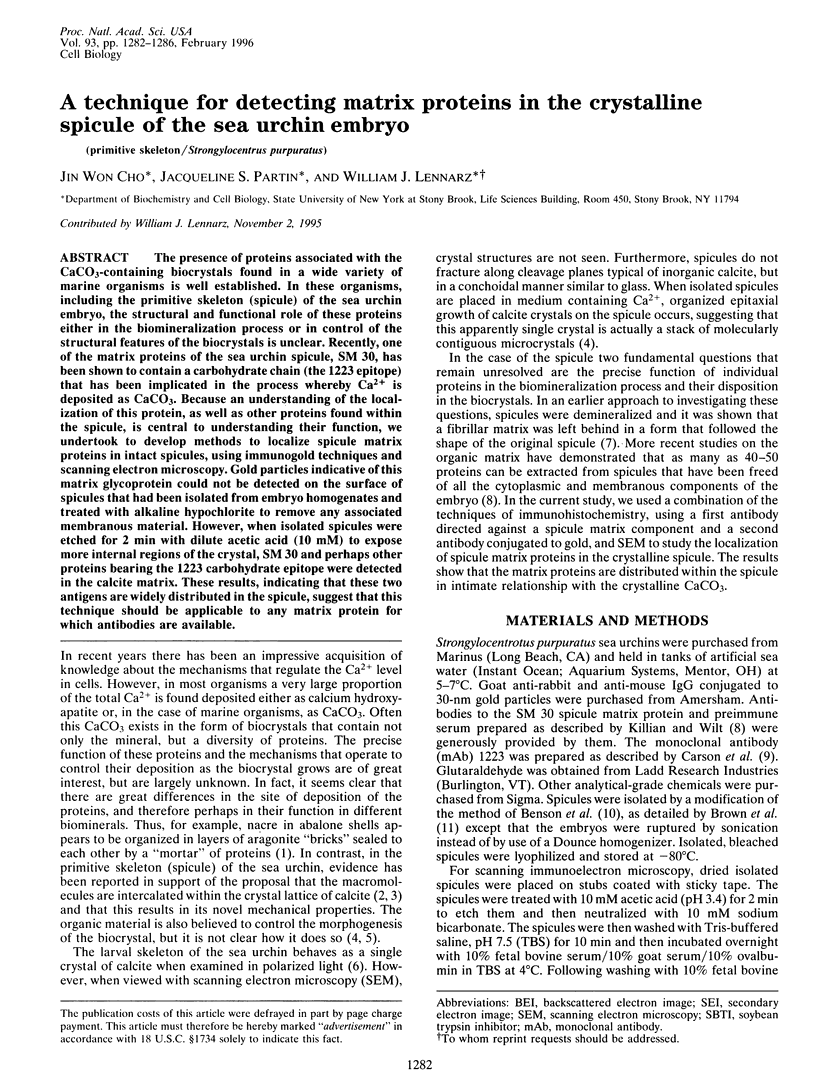
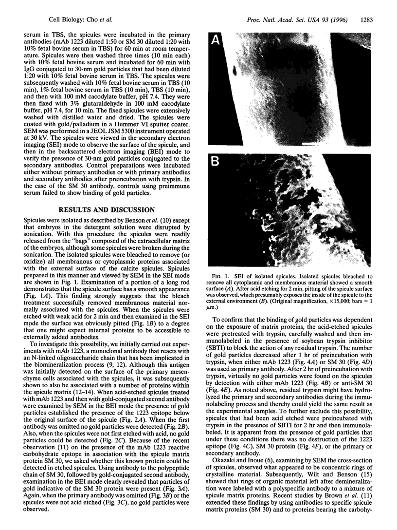
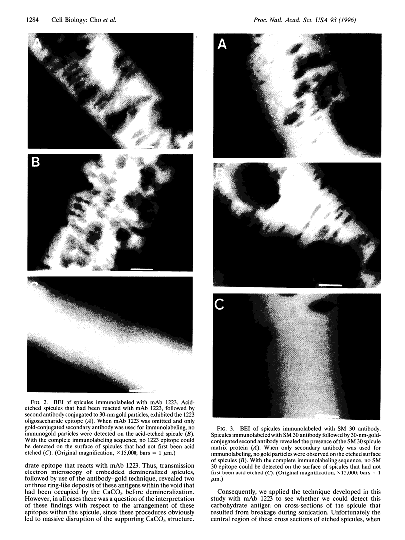
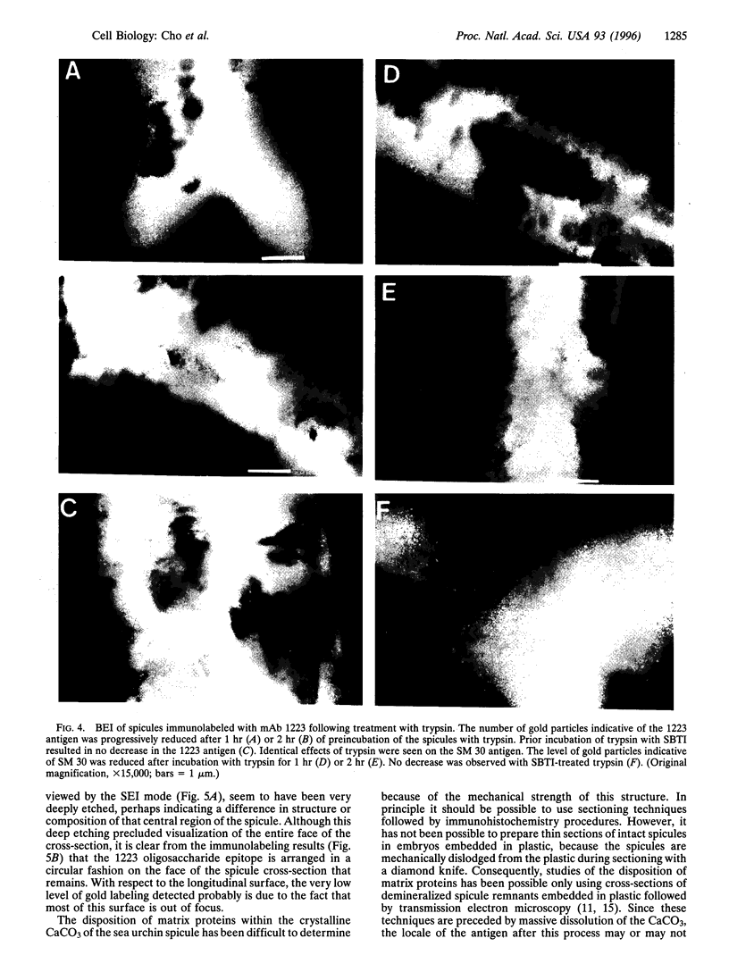
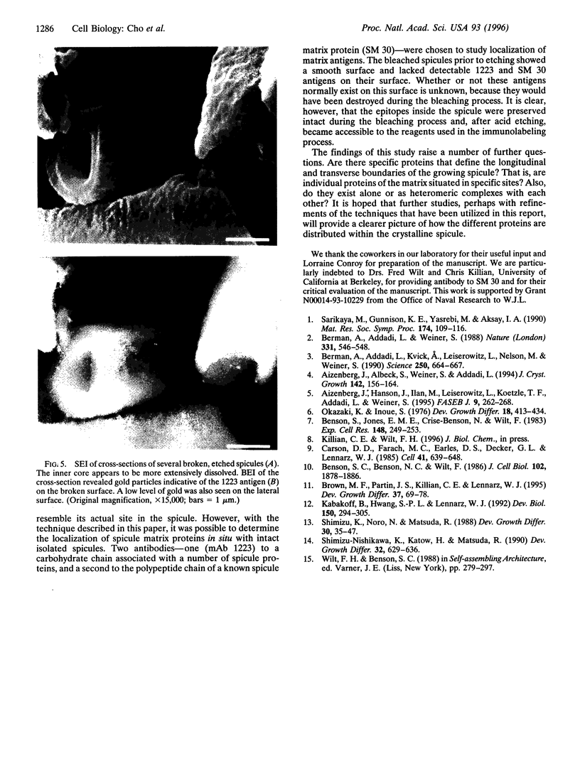
The presence of proteins associated with the CaCO3-containing biocrystals found in a wide variety of marine organisms is well established. In these organisms, including the primitive skeleton (spicule) of the sea urchin embryo, the structural and functional role of these proteins either in the biomineralization process or in control of the structural features of the biocrystals is unclear. Recently, one of the matrix proteins of the sea urchin spicule, SM 30, has been shown to contain a carbohydrate chain (the 1223 epitope) that has been implicated in the process whereby Ca2+ is deposited as CaCo3. Because an understanding of the localization of this protein, as well as other proteins found within the spicule, is central to understanding their function, we undertook to develop methods to localize spicule matrix proteins in intact spicules, using immunogold techniques and scanning electron microscopy. Gold particles indicative of this matrix glycoprotein could not be detected on the surface of spicules that had been isolated from embryo homogenates and treated with alkaline hypochlorite to remove any associated membranous material. However, when isolated spicules were etched for 2 min with dilute acetic acid (10 mM) to expose more internal regions of the crystal, SM 30 and perhaps other proteins bearing the 1223 carbohydrate epitope were detected in the calcite matrix. These results, indicating that these two antigens are widely distributed in the spicule, suggest that this technique should be applicable to any matrix protein for which antibodies are available.
Full text
PDF
Images in this article
Selected References
These references are in PubMed. This may not be the complete list of references from this article.
- Aizenberg J., Hanson J., Ilan M., Leiserowitz L., Koetzle T. F., Addadi L., Weiner S. Morphogenesis of calcitic sponge spicules: a role for specialized proteins interacting with growing crystals. FASEB J. 1995 Feb;9(2):262–268. doi: 10.1096/fasebj.9.2.7781928. [DOI] [PubMed] [Google Scholar]
- Benson S. C., Benson N. C., Wilt F. The organic matrix of the skeletal spicule of sea urchin embryos. J Cell Biol. 1986 May;102(5):1878–1886. doi: 10.1083/jcb.102.5.1878. [DOI] [PMC free article] [PubMed] [Google Scholar]
- Benson S., Jones E. M., Crise-Benson N., Wilt F. Morphology of the organic matrix of the spicule of the sea urchin larva. Exp Cell Res. 1983 Oct;148(1):249–253. doi: 10.1016/0014-4827(83)90204-5. [DOI] [PubMed] [Google Scholar]
- Berman A., Addadi L., Kvick A., Leiserowitz L., Nelson M., Weiner S. Intercalation of sea urchin proteins in calcite: study of a crystalline composite material. Science. 1990 Nov 2;250(4981):664–667. doi: 10.1126/science.250.4981.664. [DOI] [PubMed] [Google Scholar]
- Carson D. D., Farach M. C., Earles D. S., Decker G. L., Lennarz W. J. A monoclonal antibody inhibits calcium accumulation and skeleton formation in cultured embryonic cells of the sea urchin. Cell. 1985 Jun;41(2):639–648. doi: 10.1016/s0092-8674(85)80036-2. [DOI] [PubMed] [Google Scholar]
- Kabakoff B., Hwang S. P., Lennarz W. J. Characterization of post-translational modifications common to three primary mesenchyme cell-specific glycoproteins involved in sea urchin embryonic skeleton formation. Dev Biol. 1992 Apr;150(2):294–305. doi: 10.1016/0012-1606(92)90243-a. [DOI] [PubMed] [Google Scholar]