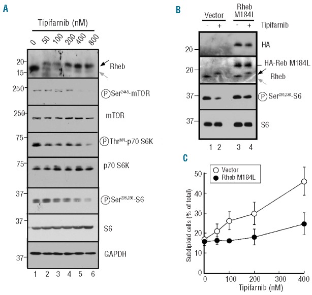
Figure 3.

Role of Rheb in tipifarnib-induced apoptosis. (A) After U937 cells were treated for 24 h with the indicated tipifarnib concentration, whole cell lysates were subjected to immunoblotting with antibodies that recognize the indicated antigens. GAPDH served as a loading control. In this and subsequent figures, gray arrow indicates farnesylated antigen and black arrow indicates unfarnesylated antigen. (B) U937 cells stably transduced with empty vector or HA-tagged Rheb M184L were treated for 24 h with diluent (0.1% DMSO) or 800 nM tipifarnib. Whole cell lysates were then probed with antibodies to the indicated antigen. Note that S6 phosphorylation diminishes less after tipifarnib treatment in cells transduced with HA-tagged Rheb M184L than in cells transduced with empty vector. (C) U937 cells stably transduced with empty vector or HA-tagged Rheb M184L were treated for 6 days with the indicated tipifarnib concentration, stained with propidium iodide and subjected to flow microfluorimetry as depicted in Online Supplementary Figure S2A.
