


Abstract
Neuropathy is the most common complication of diabetes. As a consequence of longstanding hyperglycemia, a downstream metabolic cascade leads to peripheral nerve injury through an increased flux of the polyol pathway, enhanced advanced glycation end‐products formation, excessive release of cytokines, activation of protein kinase C and exaggerated oxidative stress, as well as other confounding factors. Although these metabolic aberrations are deemed as the main stream for the pathogenesis of diabetic microvascular complications, organ‐specific histological and biochemical characteristics constitute distinct mechanistic processes of neuropathy different from retinopathy or nephropathy. Extremely long axons originating in the small neuronal body are vulnerable on the most distal side as a result of malnutritional axonal support or environmental insults. Sparse vascular supply with impaired autoregulation is likely to cause hypoxic damage in the nerve. Such dual influences exerted by long‐term hyperglycemia are critical for peripheral nerve damage, resulting in distal‐predominant nerve fiber degeneration. More recently, cellular factors derived from the bone marrow also appear to have a strong impact on the development of peripheral nerve pathology. As evident from such complicated processes, inhibition of single metabolic factors might not be sufficient for the treatment of neuropathy, but a combination of several inhibitors might be a promising approach to overcome this serious disorder. (J Diabetes Invest, doi: 10.1111/j.2040‐1124.2010.00070.x, 2010)
Keywords: Diabetic neuropathy, Novel treatment, Pathogenesis
Introduction
Peripheral neuropathy is the most common and intractable complication of diabetes1,2. It involves somatic sensory and motor nerves, as well as autonomic nerves. In fact, the prevalence of diabetic neuropathy ranges from 7% within 1 year of diagnosis to 50% for those with diabetes for >25 years3. If patients with subclinical levels of neuropathic disturbances are included, the prevalence might exceed 90%4. The presence of cardiovascular autonomic neuropathy dramatically shortens the patients’ longevity and increases the mortality5,6. Loss of feeling in the lower limbs is a high risk for limb amputation, which occurs in 1–2% of diabetic patients and necessitates extreme cost4,7.
Despite efforts to make an early diagnosis and to halt the progression of diabetic neuropathy, currently there is no effective treatment available at a global level, except for tight control of blood glucose. This might be as a result, at least in part, of insufficient clarification of the pathogenesis of diabetic neuropathy, complicated clinical pictures that do not necessarily reflect proper progression of the disease, or inadequate design of clinical trials. There might also be a possibility that the development of a candidate drug might not be based on genuine inciting factors. To overcome this serious disorder, it is therefore essential to explore the precise role of causative factors in nerve fiber dysfunction and fiber loss. The present review summarizes the most up‐to‐date considerations on the pathogenesis of diabetic neuropathy and discusses the direction of its treatment.
Risk factors for progression of neuropathy
The duration of diabetes and glycated hemoglobin levels have been well associated with a high incidence of neuropathy8,9. Classically, the Diabetes Control and Complications Trials (DCCT) confirmed the beneficial effects of meticulous control of blood glucose on the incidence of chronic complications in 1441 type 1 diabetic patients10. In that study, intensive insulin treatment for 6.5 years lowered HbA1c levels (average 7%) by 2% compared with a conventionally treated group (average 9%) and successfully decreased the incidence of neuropathy by 60% (13 vs 5%)10. More striking are the so‐called ‘legacy effects’ (glucose memory) of tight blood glucose control for the suppression of new development of neuropathy during a post‐trial observation period for 8 years11. In type 2 diabetic patients, the Kumamoto study showed that intensive insulin treatment for 7 years improved nerve conduction velocity (NCV) and the vibration perception threshold (VPT) compared with those conventionally treated12. In contrast, the UK prospective diabetes study (UKPDS) on 3867 type 2 diabetic patients did not find the effects of glucose control (to the extent of a 0.9% decrease in HbA1c) on the prevalence of neuropathy, whereas there was a significant reduction in the risk for retinopathy and nephropathy13. Tesfaye et al. in the EURO‐Diab group reported that blood glucose control, duration of diabetes, hypertension, hyperlipidemia and smoking were all significant risk factors for the development of neuropathy in type 1 diabetic patients14. The impact of hyperlipidemia has also been emphasized by a follow‐up study of the DCCT trial15. However, this trend is different in cohorts of other countries, because Japanese studies could not find a significant influence of the blood concentrations of triglyceride or cholesterol on the prevalence of neuropathy16. It is clear after all that high blood glucose leads to peripheral nerve injury through a downstream metabolic cascade. The following section will concentrate on how hyperglycemia leads to peripheral nerve injury.
Anatomy and vascular supply of peripheral nervous system
Anatomical characteristics of the peripheral nervous system might explain why the pathogenesis of neuropathy is distinct from other microvascular complications17,18. Peripheral nerves are covered by perineurium, where only a few transperineurial arterioles penetrate into the endoneurium (Figure 1). The vascular supply in peripheral nerves is sparse and blood flow is likely to be compromised and lacks autoregulation19. This system makes peripheral nerves vulnerable to ischemia. Endoneurial microvessels are tightly connected with endothelial cells on their inner surface, but when destroyed they are leaky and affect the endoneurial tissue components20. Leaky vessels are mainly located in the ganglion with fenestrated vessels, and nerve terminals on the distal side are directly exposed to environments not covered by perineurium and are susceptible to traumatic injury.
Figure 1.
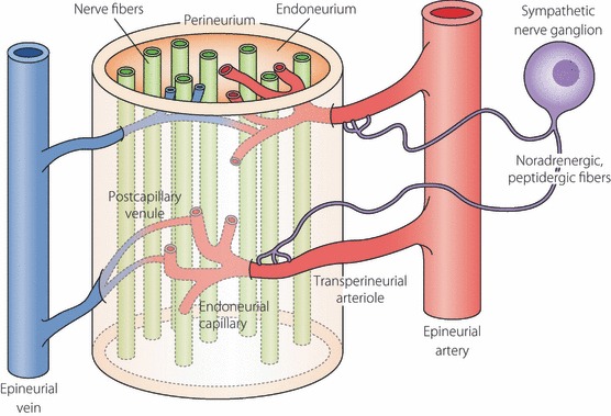
Vascular supply of the peripheral nervous system is sparse and transperineurial arteriole penetrates into endoneurium. Autonomic nerve endings contact with the wall of arterioles, but vascular autoregulation is lacking in peripheral nerves as a result of sparse innervations. In diabetes, autonomic nerve endings to the arteriole are likely to be lost and therefore vasoregulation is further impaired (modified from Pathology of Diabetes Mellitus for Clinicians by Soroku Yagihashi, Shindan‐to‐Chiryo Co., Tokyo, 2004, page 110).
Innervation of epineurial microvessels is involved in diabetes, resulting in impaired blood supply in diabetic nerves21,22. Endoneurial microvessels show thickened and multilayered basement membranes, cell debris of pericytes, as well as disrupted endothelial cells, and thus constitute salient structural changes in diabetic nerves.
Independent of vascular supply, three dimensions of neuronal architecture specific to the peripheral nervous system might account for the reason why the most distal side is susceptible in diabetes. Ganglion cells have extensively long axons covered by Schwann cells. The neuronal cell body is relatively small compared with the extremely long distance of axonal neurites, and thereby distal axons are innately too weak to support themselves for the long transport of nutrients, nerve trophic factors, as well as other signals.
Pathological background of neuropathy
Most characteristic findings of the peripheral nervous system in diabetic patients are distal and sensory predominant nerve fiber degeneration, axonal loss and endoneurial microangiopathy23,24. Both large and small caliber sizes of nerve fibers are affected. Based on this anatomical condition, Dyck et al. proposed that microvascular injury is the most probable factor for focal fiber loss and its summation appears to be the cause of diffuse fiber loss of distal predominant axonal neuropathy in diabetes25,26. However, this explanation is too simplistic and does not explain why hyperglycemia and duration of diabetes are crucial for its occurrence. There also emerges a controversy as to whether there is any predominance for the involvement of small fibers in early diabetic neuropathy. Questions on this issue were further raised by the report that the focality of nerve fiber loss was not universally demonstrated, indicating that microangiopathy does not always account for the fiber loss27. Nevertheless, vascular influence on the development of neuropathy was further supported by subsequent studies on humans. Malik et al. showed that patients who did not have clinically evident neuropathy at the time of nerve biopsy, but who showed high‐grade microangiopathic changes of endoneurial microvessels later, developed overt neuropathy, whereas the patients without microvessel changes did not develop neuropathy28. The extent of microangiopathic changes correlated well with subsequent nerve fiber loss in diabetic nerves29. We ourselves found a correlation between the thickness of the basement membrane of endoneurial microvessels and reduced myelinated fiber density30.
The most distal axons of small fibers distribute in the epidermis of the skin, sensing pain or pricking. Currently, punched skin biopsy immunostained with protein gene product (PGP)‐9.5 is widely used for the evaluation of peripheral neuropathy31. The method is simple and minimally invasive, but requires the equipment of confocal laser scan microscopy and skills for the staining and measurement. Usually, skin over the calf muscle is used, but other sites might also be added. In diabetes, the nerve fibers in the epidermis of the skin are significantly affected, resulting in distortion, twisting, focal swelling or beading, and finally, disappearance of nerve fibers32–34 (Figure 2). The reduction was found even in subjects of impaired glucose tolerance (IGT) and the extent of fiber loss was marked in established diabetic patients35,36. The nerve fiber loss in the skin was associated with fiber loss in the nerve trunk of the sural nerve, thus in keeping with the presence of clinically evident neuropathy32. In relation to the alteration of epidermal innervation, a non‐invasive method using corneal confocal microscopy has now been developed for the evaluation of neuropathy37,38. With this method, small nerve fibers distributed in the cornea can be observed without tissue sampling in live conditions38,39. Diabetic patients showed significant loss of nerve fibers, twisting and increased branching on the cornea38,39. Taking advantage of non‐invasiveness, it is easy to follow by repeated observations and to evaluate the treatment effects on neuropathy by this method. In fact, the recovery of nerve fibers by regeneration was detected in long‐standing type 1 diabetic patients 6 months after pancreas transplantation40. To understand the cause and the development of neuropathy, spatial and temporal changes of nerve pathology and their clinical significance should be explored in more detail.
Figure 2.

Epidermal innervation in diabetic patients as shown by immunostaining with PGP9.5. (a) In a normal subject (a 32‐year‐old man), small branching fibers (arrows) penetrating to basal lamina (arrowhead) derived from dermis distribute diffusely and end in the surface of the epidermis of the skin. (b) In contrast, in a type 2 diabetic subject with symptomatic neuropathy (a 52‐year‐old woman with 15 years duration of diabetes), fibers in the epidermis are completely lost. Only a few fibers are sparsely left in the dermis. Vascular systems also develop in the upper dermis (red color of tortuous structure). Bar, 100 mm.
To compensate for the paucity of information on human materials, animal models have served the basis of functional and biochemical changes that might be translated into human diabetic neuropathy. Unfortunately, diabetic animal models did not show the pathological features in the peripheral nerves trunk observed in human diabetic patients. However, recent studies have overcome this discrepancy by showing significant nerve fiber loss in the skin of diabetic animal models41,42. It is therefore now possible to search in more detail for the contribution of possible factors to the loss of nerve fibers of the skin by studying animal models. More importantly, it provides us a great tool for the exploration of effective compounds to inhibit nerve fiber loss and promote nerve fiber regeneration43,44.
Unlike human diabetic subjects, distinct pathological changes of endoneurial microvessels are not consistently shown in animal models, although reduced nerve blood flow is reproducibly shown45,46. In streptozotocin (STZ)‐induced diabetic rats, there was only a modest dilatation of vascular lumina, but no reduction of microvessel density or thickening of basement membranes in the peripheral nerve47–49. Although some studies reported reduced microvessel density in diabetic animals that reverted to normal by intervention with vascular endothelial growth factor (VEGF) or other angiotrophic factors, the recovery of nerve blood flow by these agents might be explained by functional improvement of endoneurial vessels rather than robust angiogenesis in the endoneurium.
How does hyperglycemia lead to peripheral nerve injury?
Polyol pathway
Increased polyol flux regulated by aldose reductase (AR) activation has been studied most extensively and there is no doubt that this metabolic cascade contributes to the development of neuropathy. With this premise, numerous AR inhibitors (ARI) have been developed, but clinical trials have mostly been unsuccessful, in part due to the adverse effects or insignificant improvement at the clinical end‐point. Currently, epalrestat (ONO2235) is the only one licensed in Japan. It was approved after a 3‐month double‐blinded trial50, which showed improvement of symptoms and nerve function. Further extended 3‐year double‐blinded randomized trials confirmed that ARI treatment significantly suppressed the progressive delay of nerve conduction51. The ARI effects were more marked in patients with early neuropathy and modestly elevated levels of glycated hemoglobin52. Another challenge of a new ARI will be expected to succeed in future trials, because other mechanisms do not amply replace the polyol pathway hypothesis53,54.
Despite a long history of preclinical studies, the detailed mechanism of how the polyol pathway is involved in neuropathy remains elusive. Earlier studies proposed the osmotic theory in which increased polyol flux caused intracellular hyperosmolarity by an accumulation of impermeable sorbitol in the cytoplasm, resulting in the expansion of cells and cell lysis55,56. Although this theory might be applied to the genesis of diabetic cataracts55,56, there is no consistent evidence of nerve edema or swollen cells in diabetic nerve tissues57. Following the osmotic hypothesis, Greene raised the poor energy utilization theory as the surrogate of osmotic theory58,59. With an accumulation of sorbitol, other osmolytes of myo‐inositol, taurine and adenosine were depleted in the cytoplasm. In turn, myo‐inositol deficiency caused phosphatidyl‐inositol depletion and then poor production of adenosine triphosphate (ATP), leading to reduced Na,K‐ATPase activity and protein kinase C (PKC) activity58,59. In this process, however, there is no confirmative data of myo‐inositol depletion in diabetic nerves60. In addition, clinical application of myo‐inositol was not successful61.
Consistent with the data from human IGT subjects, it was shown that ob/ob mice revealed neuropathic changes represented by NCV delay and increased oxidative stress‐induced damage62. High‐fat diet fed mice that showed typical glucose intolerance also showed neuropathic changes63. In these mice, postprandial hyperglycemia itself exerted increased flux of the polyol pathway in the peripheral nerve tissues.
The advent of transgenic technology has greatly advanced the polyol pathway story. Transgenic mice that overexpress human AR developed severe neuropathy when they were fed galactose, which is also the substrate of AR64. Thus, without hyperglycemia or insulin deficiency, increased flux of the polyol pathway in fact caused peripheral nerve dysfunction and myelinated fiber pathology, similar to those found in diabetic animal models64. The study was extended to the STZ‐induced diabetic condition in this model, which showed more severe NCV delay and reduced Na,K‐ATPase activity with an accumulation of sorbitol and fructose, compared with those in non‐transgenic diabetic mice, despite comparable levels of hyperglycemia65. The functional changes were accompanied by more severe structural changes in peripheral nerves and alterations of neuropeptide expressions in dorsal root ganglia (DRG)66. Concurrently, transgenic mice with hyperglycemia‐induced activation of the polyol pathway showed endoneurial reduction of PKC activity with decreased membranous expression of PKCα and a relative increase in PKCβ isoform (Figure 3). The neuropathic changes were improved by giving diabetic transgenic mice ARI. In contrast, studies using targeted mice lacking the AR gene showed that AR‐deficient mice were protective against neuropathy through the preservation of glutathione and nicotinamide adenine dinucleotide phosphate (NADPH)67.
Figure 3.

Tissue‐specific regulation of polyol pathway and its metabolic cascade to diabetic neuropathy. Major regulating enzymes of the polyol pathway are differentially expressed in the epineurial artery and endoneurial tissues. Aldose reductase (AR) is strongly expressed in both the endoneurium and the wall of the epineurial artery, whereas expression of sorbitol dehydrogenase (SDH) is equivocal in the endoneurium, but clearly positive for the wall of the epineurial artery (see reference 120, with kind permission from Springer Science + Business Media: Virchows Arch, Vol. 439, 2001, page 48. Enhanced in situ expression of aldose reductase in peripheral nerve and renal glomeruli in diabetic patients; Kasajima H, Yamagishi SI, Sugai S, Yagihashi N, Yagihashi S, Figure 2). Hence, hyperglycemia in nerve tissues exerts conversion from glucose to sorbitol by AR, thereby causing the depletion of reduced glutathione (GSH) and nitric oxide (NO) consequent from the overconsumption of nicotinamide adenine di‐nucleotide phosphate (NADPH). Concurrently, intracellular myo‐inositol is depleted to cause phosphatidylinositol (PI) depletion, which further suppresses diacylglycerol (DAG) production and finally protein kinase C (PKC) activity. As a consequence, Na,K‐ATPase activity will be reduced to result in functional and structural changes of neuropathy. In contrast, the second portion of the polyol pathway regulated by SDH is activated in the vascular wall in the hyperglycemic condition. As a result of redox changes of NAD/NADH, conversion from glyceraldehyde‐3‐phosphate (Glycer‐3P) to phosphatidic acid will be promoted. Then enhanced synthesis of DAG results in increased PKC activity. In our studies, major isoforms that underwent changes in the diabetic condition are PKCα in the nerve and PKCβ in the epineurial artery (reference 122).
Although these studies confirmed the critical role of AR in diabetic neuropathy, clinical experience of ARI trials50 showed that the polyol pathway cannot completely account for the development of neuropathy. Indeed, when blood glucose is poorly controlled, severe hyperglycemia can cause neuropathic changes, even in AR‐deficient diabetic mice68. A pathway independent of AR is yet to be determined and further studies are required for the complete prevention or intervention of the progression of diabetic neuropathy.
The implications of AR in ischemia/reperfusion injury have now revitalized the polyol pathway theory for vascular events, not only in diabetic patients but non‐diabetic patients as well (Figure 4). Ischemia/reperfusion causes polyol activation, leading to severe tissue injury against which ARI is preventive69–75. In experimental studies, ARI alleviated the pathological lesions in infarction of the brain, the heart, as well as the kidney or retina71,75,76. Because diabetic nerves are susceptible to ischemia/reperfusion injury, there emerges a new perspective that ischemia/reperfusion might be involved in the progression or exacerbation of neuropathy to which ARI is effective77,78.
Figure 4.
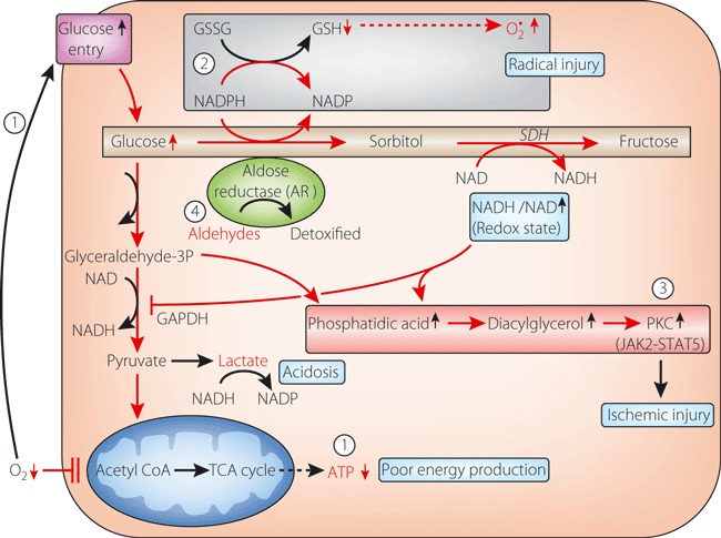
Implication of aldose reductase in ischemia/reperfusion injury. Recently, a new role of aldose reductase in ischemia/reperfusion and inflammatory injury was proposed. When a cell becomes ischemic, glucose uptake is enhanced to compensate energy depletion ( ). However, because mitochondria are impaired to produce ATP as a result of oxygen depletion, surplus glucose enters the collateral pathway to sorbitol and phosphatidic acid. From the former, aldose reductase is activated to cause glutathione deficiency and redox deviation, as in the hyperglycemic condition (
 ). As a result, free radical injury and protein kinase C (PKC) activation ensue to aggravate ischemic injury (
 ). Once reperfusion starts, oxygen radicals accumulate aldehydes, which are also substrates of aldose reductase, and enhance radical injury (
 ) (adapted from reference 69 and modified by the author).
Glycation and Advanced Glycation End‐products
Glycation has long been implicated in the pathogenesis of diabetic neuropathy30,79,80. Every component of nerve tissues can be excessively glycated in diabetic nerves. In fact, deposition of advanced glycation end‐products (AGE) was shown in human and animal diabetic nerves, in every component of peripheral nerve tissues30,80. The deposition was found in the stromal collagens, axoplasms of nerve fibers and Schwann cells, as well as endoneurial vessels81. The intensity of AGE deposition detected by carboxymethyllysine immunoreactions correlated well with reduced myelinated nerve fiber density81. Hence, AGE was considered to exert injurious processes in the endoneurium through direct toxicity to nerve tissues together with endoneurial microangiopathy (Figure 5). In vitro, Schwann cells underwent apoptotic processes with release of tumor necrosis factor (TNF)‐α, as well as other inflammatory cytokines, when exposed to a high AGE environment82. Axonal cytoskeletons of tubulin and neuro‐filaments were glycated to stagnate axonal transport, resulting in distal fiber degeneration30. Glycation of basement membrane collagen, laminin and fibronectin also caused impairment of regenerative efforts in diabetic nerves83,84.
Figure 5.
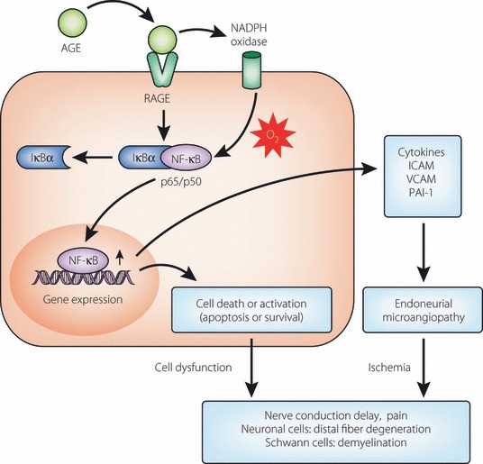
Advanced glycation end‐products (AGE) and receptor for AGE (RAGE) reactions in the pathogenesis of diabetic neuropathy. Nerve tissues, such as Schwann cells, nerve fibers and endothelial cells of vasa nervosum all express RAGE. When AGE bind with RAGE, the reaction generates oxidative stress mainly through the activation of NADPH oxidase. Complexes of IκBα‐nuclear factor‐(NF)‐κB will be separated into each fraction of IκBα and NFκB, the latter of which translocates into the nucleus as a transcription factor to activate genes related to cell death or survival. As a result, both microangiopathic processes and neural dysfunction ensue, resulting in the manifestation of pain or nerve conduction delay.
Transgenic mice with enhanced expression of the receptor for AGE (RAGE) in endothelial cells showed augmented neuropathic changes in the diabetic condition, exemplified by delayed NCV and more severe structural changes85. In this setting, it can be speculated that AGE exerts biological reactions after binding with RAGE expressed on endothelial cells and Schwann cells, leading to the functional and structural phenotype of neuropathy. During this process, intracellular oxidative stress mediated by NADPH oxidase activation might be elicited and then activate transcription of nuclear factor‐κB (NF‐κB)86,87. Bierhaus et al. reported that the activation of NF‐κB was associated with the alteration of pain sensation in STZ‐induced hyperglycemic mice88. Diabetic mice lacking the RAGE gene were protective against the induction of neuropathy89. Thus, these findings support the crucial role of AGE in the development of diabetic neuropathy.
Indirect evidence that suggests the role of AGE in neuropathy might be the effects of aminoguanidine on experimental diabetic neuropathy47,90–92. This compound was found to inhibit the formation of AGE, concurrently with the improvement of endoneurial blood flow90, NCV, Na,K‐ATPase activity and myelinated fiber structure91,92. It should be of note that aminoguanidine effects might also be mediated by its alternate action as an inducible nitric oxide synthase (iNOS) inhibitor or an anti‐oxidative function93.
In our most recent study, we showed that animals given AGE exogenously showed significant NCV delay resembling that found in experimental diabetic neuropathy (Figure 6)94. With delayed NCV, nerve Na,K‐ATPase activity was reduced and myelinated nerve fibers underwent reduction of fiber size. In this setting, vascular reactions in response to exogenous AGE elicited functional impairment of peripheral nervous systems. In fact, endothelial cells showed a high expression of NF‐κBp65 together with swollen and vacuolar changes at the ultrastructural levels. From these findings, AGE action mediated by binding with RAGE causes activation of NF‐κB and thereby its downstream signals88,95,96. Although preliminary clinical trials of anti‐glycation agent, benfotiamime, showed some efficacy for diabetic neuropathy97, there is still no effective compound that can suppress the AGE formation in vivo and improve diabetic neuropathy in humans.
Figure 6.
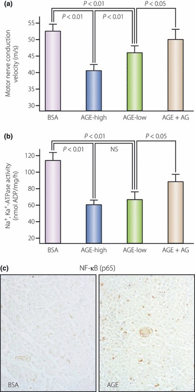
Neuropathy in normal rats given exogenous advanced glycation end‐products (AGE). When AGE were given exogenously, normal rats showed neuropathic changes, similar to those found in experimental diabetic animals. Rats given AGE showed (a) a significant delay of motor nerve conduction velocity and (b) suppression of nerve Na,K‐ATPase activity, whereas no effects were detected in bovine serum albumin (BSA)‐treated rats. Such suppression was corrected by co‐treatment with aminoguanidine, an inhibitor of glycation and nitric oxide. (c) On the sections, AGE‐treated rats showed strong expression of nuclear factor‐κB on the nuclei of endothelial cells of microvessels and Schwann cells (quoted from reference 94).
Oxidative Stress
As a cause of diabetic neuropathy, the generation of free radicals is proposed to be a major factor through increased glycolytic process98,99. In fact, there are numerous data that showed oxidative stress‐induced tissue injury in the peripheral nerve in experimental diabetes45,63,88,92,95,98. Based on this background, attempts have been made to inhibit neuropathy with anti‐oxidants100,101. In particular, α‐lipoic acid has been used for the suppression of oxidative stress in experimental diabetic rats and it was found that it improved NCV delay, nerve blood flow and nerve structure102–104.
Concurrent with the generation of free radicals during the glycolytic process, mitochondria have a crucial role in cellular death by activation of specific signals and the endonuclease system105,106. Hyperglycemia‐induced mitochondrial changes include the release of cytochrome C, activation of caspase 3, altered biogenesis and fission, resulting in programmed cell death105,107. Excessive entry of glucose causes surplus transport of electrons to generate oxidants in mitochondria, leading to reduced mitochondrial action potentials (MMP) with poor energy synthesis of ATP108,109. Neurotrophic support is also impaired by mitochondrial damage to cause reduced neurotrophin‐3 (NT‐3) and nerve growth factor (NGF)108. It is interesting that a small amount of insulin, that does not alter systemic blood glucose levels, was shown to improve the impaired mitochondrial membrane potential and delayed nerve conduction in STZ‐diabetic rats109.
As already alluded to, both the polyol pathway and AGE formation produce a large amount of oxidants, and ARI treatment suppresses the oxidative nerve injury110–112. In addition to mitochondria, other organelles, such as the Golgi apparatus and endoplasmic reticulum (ER), might also be regarded as an important source of free radicals, resulting in not only apoptosis, but cell death from autophagy113. Indeed, nitro‐oxidative stress in conjunction with hyperglycemia exerts poly ADP‐ribose polymerase (PARP) activation114, resulting in cellular dysfunction and cell death, which can be prevented by PARP inhibitor115. Serum from type 2 diabetic patients accelerates neuroblastoma cell death by increased autophagic processes with activation of cell death signals116. α‐Lipoic acid was found to be beneficial to some extent to alleviate neuropathic symptoms in diabetic patients117. However, to confirm whether this compound is in fact effective to inhibit the progression of the disease, further confirmation is required.
PKC Activity
PKC is central in nerve function and a key in the pathogenesis of diabetic neuropathy118,119. However, the alterations are complicated in nerve tissues and their supportive endoneurial vascular system, as the major enzymes of collateral glycolytic pathway are different between these two tissues120 (Figure 3). Such inhomogeneous tissue composition might explain the inconsistent findings on PKC activity in diabetic nerves. Nakamura et al. did not find any significant change of PKC activity in the homogenized whole peripheral nerve tissues in STZ diabetic rats, although PKC‐β specific inhibitor improved NCV delay and nerve blood flow121. In contrast, in our studies on STZ‐induced diabetic mice, we separated the tissues into endoneurium and epineurium for the measurement of PKC activity, the latter of which is rich in microvessels122. We found that the former showed decreased PKC activity with significantly decreased membranous expression of the PKC‐α isoform, as we already stated earlier about polyol pathway, whereas the latter showed increased PKC activity with enhanced expression of PKC‐β (Figure 3). The results of epineurial tissues were consistent with the changes in other systemic vascular tissues. In keeping with this finding, hyperglycemia caused reduced PKC activity in cultured Schwann cells exposed to high glucose123.
Hence, the application of PKC‐β‐specific inhibitor is expected to be useful for the treatment of diabetic vascular complications. Experimental studies showed beneficial effects of PKC‐β‐specific inhibitor on neuropathic changes in STZ‐induced diabetic rats121,124,125. Despite extensive efforts, however, clinical trials were not successful due, in part, to the high improvement rate in the placebo group126. Other isoforms of PKC were also implicated in the causation of diabetic neuropathy and inhibitors for these isoforms have been explored127,128.
Proinflammatory Processes
There is emerging evidence that nerve tissues in diabetes undergo a pro‐inflammatory process that presents symptoms and enhances the development of neuropathy129,130. Indeed, diabetic nerves contain macrophages, occasionally lymphocytes and release increased TNF‐α or interleukins (IL) in humans and animals129,131,132 (Figure 7). Inhibition of cytokine release or macrophage migration was associated with the improvement of NCV delay and structure in STZ‐diabetic rats treated with N‐acetylcysteine133 or pioglitazone134. The arachidonic acid pathway is activated to increase in cyclooxygenase (COX)‐2 concentrations in the peripheral nerves of STZ diabetic rats in which inhibition of COX‐2 corrected nerve blood flow and NCV delay135. To further confirm this data, COX‐2 gene‐deficient mice were protective for NCV delay and neuropathic deficits after STZ‐induced hyperglycemia136. The pro‐inflammatory condition activated the stress‐kinase, mitogen‐activated protein (MAP)‐kinase, in diabetic nerves, which was also suppressed by treatment with pioglitazone134. Thus, MAP‐kinase is considered to be a potential target for a new treatment of diabetic neuropathy137,138. In this process, NF‐κB is activated to lead the cell to cell death or proliferation139,140. Because a pro‐inflammatory reaction is induced by the polyol pathway hyperactivity or increased AGE formation as well, it should be clear to what extent the pro‐inflammatory process is a single initiating or influential factor for the development of neuropathy. Ischemia reperfusion might also accelerate the inflammatory processes to which diabetic nerves are susceptible77,78.
Figure 7.

Pro‐inflammatory reactions and experimental diabetic neuropathy. In the sciatic nerve of STZ‐induced diabetic rats, there were many macrophages stained positive for ED1 (upper center). Migration of macrophages was inhibited when diabetic rats were treated with pioglitazone (upper right). Pioglitazone treatment also corrected the delay of motor nerve conduction velocity (MNCV) and sensory nerve conduction velocity (SNCV), and activation of extracellular signal‐regulated kinase (ERK), one of mitogen activated protein kinases (MAPK) (adapted from reference 134).
With increasing information about the role of inflammation, approaches to suppress the pain symptoms or neuropathy itself are now carried out with the specific target of cytokines or cell signals141–143.
Cellular and Trophic Factors
The lack of neurotrophins plays an important role in the pathogenesis of diabetic neuropathy144–149. In fact, the production of NGF was suppressed in the skin and substitution of NGF ameliorated neuropathic changes of small fibers and autonomic pathology in diabetic animals150,152. NT‐3, brain‐derived neurotrophic factor (BDNF) and ciliary neurotrophic factor (CNTF) were also decreased in the muscle tissues in diabetic patients153. NT‐3 was shown to protect the NCV delay and perception threshold in diabetic animals154, but the results were not always positive155,156. Unfortunately, application of NGF in a clinical trial did not succeed in the correction of neuropathy, in part because of the emergence of pain157. Efforts have now been made to more efficiently deliver or produce trophic factors at the target tissues by introducing gene therapy or cell transplantations59,159–162.
Recent studies have shown a new insight into the pathogenesis of neuropathy. In diabetic nerves, there were chimeric cells that were a combination of resident Schwann cells or neuronal cells and migrated proinsulin‐producing cells derived from bone marrow163. Although the significance of such chimeric cells is yet to be known, they eventually undergo apoptotic cell death, thus injuring the constitutive cells, leading to neuropathic changes. Much remains to be further investigated to confirm such intriguing cells and to clarify their significance.
Direction of Treatment
Based on the proposed mechanisms of neuropathy so far (Figure 8), efforts have been continuously made to develop effective means for the treatment of neuropathy. However, to date, there are only a few agents available in limited countries; ARI (epalrestat) in Japan and α‐lipoic acid (thioctic acid) in Germany. Other agents, such as benfotiamine as an anti‐glycation agent, PKC‐β‐inhibitor (ruboxitaurine) or NGF were unsuccessful at the final stage of randomized clinical trials. Nevertheless, there are still ongoing trials that we hope will be successful in future. Very recently, it was shown that autonomic neuropathy in the bone marrow impaired activation and migration of endothelial precursor cells (EPC), which might determine the fate of vascular complications164. It also becomes clear that the vagus nerve conveys signals for regeneration of islet β‐cells165, which might be disturbed in diabetic patients. These novel findings reinforce the importance of diabetic neuropathy for patient care and direction of treatment in diabetes. In particular, early inhibition of causative factors is extremely important not only to halt, but to reverse, the lesions. However, once the lesions are developed, as stated earlier, a variety of factors are exerted to accelerate the neuropathy. In this setting, the combination of several inhibitors might be required.
Figure 8.
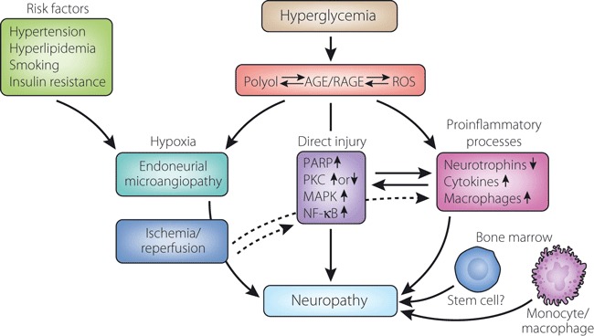
Summary of pathogenetic mechanisms of diabetic neuropathy. Long‐term hyperglycemia causes downstream metabolic cascades of polyol pathway hyperactivity, advanced glycation end‐products (AGE)/receptor for AGE (RAGE) reactions and increased reactive oxygen species (ROS). They compromise both endoneurial microvessels and neural tissues themselves through activation of poly‐ADP‐ribose polymerase (PARP), alterations of protein kinase C (PKC) and an increase in mitogen‐activated protein kinase (MAPK), as well as activation of nuclear factor‐(NF)‐κB, resulting in functional and structural changes of peripheral neuropathy. Metabolic aberrations in the nerve elicit pro‐inflammatory reactions, inducing release of cytokines, suppression of neurotrophins and migration of macrophages, and promote the development of neuropathy. Recently, cellular factors derived from the bone marrow were found to produce chimeric cells in peripheral nerves of diabetic animals to elicit nerve injury. There is also the possibility that other cellular components from the bone marrow have an influence on the nerve pathology in diabetes. In addition, ischemia/reperfusion might also accelerate nerve injury, in part mediated by inflammatory reactions. Risk factors represented by hypertension, hyperlipidemia, smoking and insulin resistance are also important contributors to the development of neuropathy.
Neuropathy has long been regarded merely as a disorder of the most distal portion of the body. Effects of hyperglycemia on the nervous system have now been shown to be a much more serious condition. Neuropathy itself is an important trigger for systemic abnormalities in diabetic patients. Much more investigation on the nerve changes in the pancreas, liver and related organs is required for a better understanding of the whole body in diabetic patients and to develop effective treatment of this disease.
Acknowledgements
The authors express sincere appreciation to colleagues who were involved in collaborative studies with our laboratory. The special technical assistance of Saori Ogasawara, Mari Tsujii, Hiroko Mori, Shiho Fujiwara are appreciated. Our studies quoted in this review were supported by the Japanese Ministry of Health and Welfare, Japanese Ministry of Science, Education, Culture and Sports, and the Juvenile Diabetes Research Foundation International. The authors declares no conflict of interest regarding this review.
References
- 1.Boulton AJ, Vinik AI, Arezzo JC, et al. Diabetic neuropathies: a statement by the American Diabetes Association. Diabetes Care 2005; 28: 956–962 [DOI] [PubMed] [Google Scholar]
- 2.Strotmeyer ES, de Rekeneire N, Schwartz AV, et al. Sensory and motor peripheral nerve function and lower‐extremity quadriceps strength: the health, aging and body composition study. J Am Geriatr Soc 2009; 57: 2004–2010 [DOI] [PMC free article] [PubMed] [Google Scholar]
- 3.Pirart J. Diabetes mellitus and its degenerative complications: a prospective study of 4400 patients observed. Diabetes Care 1978; 1: 168–188 [Google Scholar]
- 4.Vinik AI, Liuzze FJ, Holland MT, et al. Diabetic neuropathies. Diabetes Care 1992; 15: 1926–1975 [DOI] [PubMed] [Google Scholar]
- 5.Ewing DJ, Campbell IW, Clarke BF. Mortality in diabetic autonomic neuropathy. Lancet 1976; 1: 601–603 [DOI] [PubMed] [Google Scholar]
- 6.Ewing DJ, Campbell IW, Clarke BF. The natural history of diabetic autonomic neuropathy. Q J Med 1980; 49: 95–108 [PubMed] [Google Scholar]
- 7.Vinik AI, Ziegler D. Diabetic cardiovascular autonomic neuropathy. Circulation 2007; 115: 387–397 [DOI] [PubMed] [Google Scholar]
- 8.Sabanayagam C, Liew G, Tai ES, et al. Relationship between glycated haemoglobin and microvascular complications: is there a natural cut‐off point for the diagnosis of diabetes? Diabetologia 2009; 52: 1279–1289 [DOI] [PubMed] [Google Scholar]
- 9.Pop‐Busui R, Lu J, Lopes N, et al. Prevalence of diabetic peripheral neuropathy and relation to glycemic control therapies at baseline in the BARI 2D cohort. J Peripher Nerv Syst 2009; 14: 1–13 [DOI] [PMC free article] [PubMed] [Google Scholar]
- 10.Diabetes Control and Complications Trial Research Group: the effect of intensive treatment of diabetes on the development and progression of long‐term complications in insulin‐dependent diabetes mellitus. N Engl J Med 1993; 329: 977–986 [DOI] [PubMed] [Google Scholar]
- 11.Martin CL, Albers J, Herman WH, et al. Neuropathy among the diabetes control and complications trial cohort 8 years after trial completion. Diabetes Care 2006; 29: 340–344 [DOI] [PMC free article] [PubMed] [Google Scholar]
- 12.Ohkubo Y, Kishikawa H, Araki E, et al. Intensive insulin therapy prevents the progression of diabetic microvascular complications in Japanese patients with non‐insulin‐dependent diabetes mellitus: a randomized prospective 6‐year study. Diabetes Res Clin Pract 1995; 28: 103–117 [DOI] [PubMed] [Google Scholar]
- 13.UK ProspectiveDiabetesStudyGroup . Intensive blood‐glucose control with sulphonylureas or insulin compared with conventional treatment and risk of complications in patients with type 2 diabetes (UKPDS33). Lancet 1998; 352: 837–853 [PubMed] [Google Scholar]
- 14.Tesfaye S, Chaturvedi N, Eaton SE, et al. Vascular risk factors and diabetic neuropathy. N Engl J Med 2005; 352: 341–350 [DOI] [PubMed] [Google Scholar]
- 15.Vincent AM, Hinder LM, Pop‐Busui R, et al. Hyperlipidemia: a new therapeutic target for diabetic neuropathy. J Peripher Nerv Syst 2009; 14: 257–267 [DOI] [PMC free article] [PubMed] [Google Scholar]
- 16.Yagihashi S, Yasuda H, Deguchi T, et al. Clinical staging of diabetic polyneuropathy and its risk factors for the severity. 20th Neurodiab Meeting, Stockholm, Sept 17–20, 2010
- 17.Kihara M, Weerasuriya A, Low PA. Endoneurial blood flow in rat sciatic nerve during development. J Physiol 1991; 439: 351–360 [DOI] [PMC free article] [PubMed] [Google Scholar]
- 18.Sugimoto H, Monafo WW. Regional blood flow in sciatic nerve, biceps femoris muscle, and truncal skin in response to hemorrhagic hypotension. J Trauma 1987; 27: 1025–1030 [DOI] [PubMed] [Google Scholar]
- 19.Smith DR, Kobrine AI, Rizzoli HV. Absence of autoregulation in peripheral nerve blood flow. J Neurol Sci 1977; 33: 347–352 [DOI] [PubMed] [Google Scholar]
- 20.Sima AA, Nathaniel V, Prashar A, et al. Endoneurial microvessels in human diabetic neuropathy. Endothelial cell dysjunction and lack of treatment effect by aldose reductase inhibitor. Diabetes 1991; 40: 1090–1099 [DOI] [PubMed] [Google Scholar]
- 21.Grover‐Johnson NM, Baumann FG, Imparato AM, et al. Abnormal innervation of lower limb epineurial arterioles in human diabetes. Diabetologia 1981; 20: 31–38 [DOI] [PubMed] [Google Scholar]
- 22.Beggs J, Johnson PC, Olafsen A, et al. Innervation of the vasa nervorum: changes in human diabetics. J Neuropathol Exp Neurol 1992; 51: 612–629 [DOI] [PubMed] [Google Scholar]
- 23.Yagihashi S, Matsunaga M. Ultrastructural pathology of peripheral nerves in patients with diabetic neuropathy. Tohoku J Exp Med 1979; 129: 357–366 [DOI] [PubMed] [Google Scholar]
- 24.Dyck PJ, Giannini C. Pathologic alterations in the diabetic neuropathies of humans: a review. J Neuropathol Exp Neurol 1996; 55: 1181–1193 [DOI] [PubMed] [Google Scholar]
- 25.Dyck PJ, Karnes J, O’Brien P, et al. Spatial pattern of nerve fiber abnormality indicative of pathologic mechanism. Am J Pathol 1984; 117: 225–238 [PMC free article] [PubMed] [Google Scholar]
- 26.Dyck PJ, Karnes JL, O’Brien P, et al. The spatial distribution of fiber loss in diabetic polyneuropathy suggests ischemia. Ann Neurol 1986; 19: 440–449 [DOI] [PubMed] [Google Scholar]
- 27.Llewelyn JG, Thomas PK, Gilbey SG, et al. Pattern of myelinated fibre loss in the sural nerve in neuropathy related to type 1 (insulin‐dependent) diabetes. Diabetologia 1988; 31: 162–167 [DOI] [PubMed] [Google Scholar]
- 28.Thrainsdottir S, Malik RA, Dahlin LB, et al. Endoneurial capillary abnormalities presage deterioration of glucose tolerance and accompany peripheral neuropathy in man. Diabetes 2003; 52: 2615–2622 [DOI] [PubMed] [Google Scholar]
- 29.Malik RA, Tesfaye S, Newrick PG, et al. Sural nerve pathology in diabetic patients with minimal but progressive neuro‐pathy. Diabetologia 2005; 48: 578–585 [DOI] [PubMed] [Google Scholar]
- 30.Yagihashi S. The pathogenesis of diabetic neuropathy. Diabetes Metab Res Rev 1995; 11: 193–225 [DOI] [PubMed] [Google Scholar]
- 31.McCarthy BG, Hsieh ST, Stocks A, et al. Cutaneous innervation in sensory neuropathies: evaluation by skin biopsy. Neurology 1995; 45: 1848–1855 [DOI] [PubMed] [Google Scholar]
- 32.Kennedy WR, Wendelschafer‐Crabb G, Johnson T. Quantitation of epidermal nerves in diabetic neuropathy. Neurology 1996; 47: 1042–1048 [DOI] [PubMed] [Google Scholar]
- 33.Polydefkis M, Hauer P, Sheth S, et al. The time course of epidermal nerve fibre regeneration: studies in normal controls and in people with diabetes, with and without neuropathy. Brain 2004; 127: 1606–1615 [DOI] [PubMed] [Google Scholar]
- 34.Shun CT, Chang YC, Wu HP, et al. Skin denervation in type 2 diabetes: correlations with diabetic duration and functional impairments. Brain 2004; 127: 1593–1605 [DOI] [PubMed] [Google Scholar]
- 35.Smith AG, Ramachandran P, Tripp S, et al. Epidermal nerve innervations in impaired glucose tolerance and diabetes‐associated neuropathy. Neurology 2001; 57: 1701–1704 [DOI] [PubMed] [Google Scholar]
- 36.Sumner CJ, Sheth S, Griffin JW, et al. The spectrum of neuropathy in diabetes and impaired glucose tolerance. Neurology 2003; 60: 108–111 [DOI] [PubMed] [Google Scholar]
- 37.Malik RA, Kallinikos P, Abbott CA, et al. Corneal confocal microscopy: a non‐invasive surrogate of nerve fibre damage and repair in diabetic patients. Diabetologia 2003; 46: 683–638 [DOI] [PubMed] [Google Scholar]
- 38.Hossain P, Sachdev A, Malik RA. Early detection of diabetic peripheral neuropathy with corneal confocal microscopy. Lancet 2005; 366: 1340–1343 [DOI] [PubMed] [Google Scholar]
- 39.Quattrini C, Tavakoli M, Jeziorska M, et al. Surrogate markers of small fiber damage in human diabetic neuropathy. Diabetes 2007; 56: 2148–2154 [DOI] [PubMed] [Google Scholar]
- 40.Mehra S, Tavakoli M, Kallinikos PA, et al. Corneal confocal microscopy detects early nerve regeneration after pancreas transplantation in patients with type 1 diabetes. Diabetes Care 2007; 30: 2608–2612 [DOI] [PubMed] [Google Scholar]
- 41.Christianson JA, Riekhof JT, Wright DE. Restorative effects of neurotrophin treatment on diabetes‐induced cutaneous axon loss in mice. Exp Neurol 2003; 179: 188–199 [DOI] [PubMed] [Google Scholar]
- 42.Kennedy JM, Zochodne DW. Experimental diabetic neuropathy with spontaneous recovery: is there irreparable damage? Diabetes 2005; 54: 830–837 [DOI] [PubMed] [Google Scholar]
- 43.Chattopadhyay M, Mata M, Goss J, et al. Prolonged preservation of nerve function in diabetic neuropathy in mice by herpes simplex virus‐mediated gene transfer. Diabetologia 2007; 50: 1550–1558 [DOI] [PubMed] [Google Scholar]
- 44.Leonelli E, Bianchi R, Cavaletti G, et al. Progesterone and its derivatives are neuroprotective agents in experimental diabetic neuropathy: a multimodal analysis. Neuroscience 2007; 144: 1293–1304 [DOI] [PubMed] [Google Scholar]
- 45.Stevens MJ, Zhang W, Li F, et al. C‐peptide corrects endoneurial blood flow but not oxidative stress in type 1 BB/Wor rats. Am J Physiol Endocrinol Metab 2004; 287: E497–E505 [DOI] [PubMed] [Google Scholar]
- 46.Kuzumoto Y, Kusunoki S, Kato N, et al. Effect of the aldose reductase inhibitor fidarestat on experimental diabetic neuropathy in the rat. Diabetologia 2006; 49: 3085–3093 [DOI] [PubMed] [Google Scholar]
- 47.Sugimoto K, Yagihashi S. Effects of aminoguanidine on structural alterations of microvessels in peripheral nerve of streptozotocin diabetic rats. Microvasc Res 1997; 53: 105–112 [DOI] [PubMed] [Google Scholar]
- 48.Uehara K, Sugimoto K, Wada R, et al. Effects of cilostazol on the peripheral nerve function and structure in STZ‐induced diabetic rats. J Diabetes Complications 1997; 11: 194–202 [DOI] [PubMed] [Google Scholar]
- 49.Walker D, Carrington A, Cannan SA, et al. Structural abnormalities do not explain the early functional abnormalities in the peripheral nerves of the streptozotocin diabetic rat. J Anat 1999; 195: 419–427 [DOI] [PMC free article] [PubMed] [Google Scholar]
- 50.Goto Y, Hotta N, Shigeta Y, et al. Effects of an aldose reductase inhibitor, epalrestat, on diabetic neuropathy. Clinical benefit and indication for the drug assessed from the results of a placebo‐controlled double‐blind study. Biomed Pharmacother 1995; 49: 269–277 [DOI] [PubMed] [Google Scholar]
- 51.Hotta N, Akanuma Y, Kawamori R, et al. Long‐term clinical effects of epalrestat, an aldose reductase inhibitor, on diabetic peripheral neuropathy: the 3‐year, multicenter, comparative Aldose Reductase Inhibitor‐Diabetes Complications Trial. Diabetes Care 2006; 29: 1538–1544 [DOI] [PubMed] [Google Scholar]
- 52.Hotta N, Kawamori R, Atsumi Y, et al. Stratified analyses for selecting appropriate target patients with diabetic peripheral neuropathy for long‐term treatment with an aldose reductase inhibitor, epalrestat. Diabet Med 2008; 25: 818–825 [DOI] [PMC free article] [PubMed] [Google Scholar]
- 53.Pfeifer MA, Schumer MP, Gelber DA. Aldose reductase inhibitors: the end of an era or the need for different trial designs? Diabetes 1997; 46(Suppl 2): S82–S89 [DOI] [PubMed] [Google Scholar]
- 54.Ryan GJ. New pharmacologic approaches to treating diabetic retinopathy. Am J Health Syst Pharm 2007; 17(Suppl 12): S15–S21 [DOI] [PubMed] [Google Scholar]
- 55.Gabbay KH. Hyperglycemia, polyol metabolism, and complications of diabetes mellitus. Annu Rev Med 1975; 26: 521–536 [DOI] [PubMed] [Google Scholar]
- 56.Kinoshita JH. A thirty year journey in the polyol pathway. Exp Eye Res 1990; 50: 567–573 [DOI] [PubMed] [Google Scholar]
- 57.Jakobsen J, Sidenius P. Nerve morphology in experimental diabetes. Clin Physiol 1985; 5(Suppl 5): 9–13 [PubMed] [Google Scholar]
- 58.Greene DA, Lattimer SA, Sima AA. Sorbitol, phosphoinositides, and sodium‐potassium‐ATPase in the pathogenesis of diabetic complications. N Engl J Med 1987; 316: 599–606 [DOI] [PubMed] [Google Scholar]
- 59.Greene DA, Sima AA, Stevens MJ, et al. Complications: neuropathy, pathogenetic considerations. Diabetes Care 1992; 15: 1902–1925 [DOI] [PubMed] [Google Scholar]
- 60.Dyck PJ, Sherman WR, Hallcher LM, et al. Human diabetic endoneurial sorbitol, fructose, and myo‐inositol related to sural nerve morphometry. Ann Neurol 1980; 8: 590–596 [DOI] [PubMed] [Google Scholar]
- 61.Dyck PJ, Zimmerman BR, Vilen TH, et al. Nerve glucose, fructose, sorbitol, myo‐inositol, and fiber degeneration and regeneration in diabetic neuropathy. N Engl J Med 1988; 319: 542–548 [DOI] [PubMed] [Google Scholar]
- 62.Drel VR, Mashtalir N, Ilnytska O, et al. The leptin‐deficient (ob/ob) mouse: a new animal model of peripheral neuro‐pathy of type 2 diabetes and obesity. Diabetes 2006; 55: 3335–3343 [DOI] [PubMed] [Google Scholar]
- 63.Obrosova IG, Ilnytska O, Lyzogubov VV, et al. High‐fat diet induced neuropathy of pre‐diabetes and obesity: effects of “healthy” diet and aldose reductase inhibition. Diabetes 2007; 56: 2598–2608 [DOI] [PubMed] [Google Scholar]
- 64.Yagihashi S, Yamagishi S, Wada R, et al. Galactosemic neuropathy in transgenic mice for human aldose reductase. Diabetes 1996; 45: 56–59 [DOI] [PubMed] [Google Scholar]
- 65.Yagihashi S, Yamagishi SI, Wada R, et al. Neuropathy in diabetic mice overexpressing human aldose reductase and effects of aldose reductase inhibitor. Brain 2001; 124: 2448–2458 [DOI] [PubMed] [Google Scholar]
- 66.Uehara K, Yamagishi S, Otsuki S, et al. Effects of polyol pathway hyperactivity on protein kinase C activity, nociceptive peptide expression, and neuronal structure in dorsal root ganglia in diabetic mice. Diabetes 2004; 53: 3239–3247 [DOI] [PubMed] [Google Scholar]
- 67.Ho EC, Lam KS, Chen YS, et al. Aldose reductase‐deficient mice are protected from delayed motor nerve conduction velocity, increased c‐Jun NH2‐terminal kinase activation, depletion of reduced glutathione, increased superoxide accumulation, and DNA damage. Diabetes 2006; 55: 1946–1953 [DOI] [PubMed] [Google Scholar]
- 68.Yagihashi S, Yamagishi SI, Mizukami H, et al. Escape phenomenon from polyol pathway to other metabolic cascades may underlie nerve conduction delay in severely hyperglycemic AR‐deficient mice. 68th American Diabetes Association, San Francisco, USA, June 6–10, 2008 [Google Scholar]
- 69.Hwang YC, Shaw S, Kaneko M, et al. Aldose reductase pathway mediates JAK‐STAT signaling: a novel axis in myocardial ischemic injury. FASEB J 2005; 19: 795–797 [DOI] [PubMed] [Google Scholar]
- 70.Iwata K, Matsuno K, Nishinaka T, et al. Aldose reductase inhibitors improve myocardial reperfusion injury in mice by a dual mechanism. J Pharmacol Sci 2006; 102: 37–46 [DOI] [PubMed] [Google Scholar]
- 71.Cheung AK, Lo AC, So KF, et al. Gene deletion and pharmacological inhibition of aldose reductase protect against retinal ischemic injury. Exp Eye Res 2007; 85: 608–616 [DOI] [PubMed] [Google Scholar]
- 72.Yeung CM, Lo AC, Cheung AK, et al. More severe type 2 diabetes‐associated ischemic stroke injury is alleviated in aldose reductase‐deficient mice. J Neurosci Res 2010; 88: 2026–2034 [DOI] [PubMed] [Google Scholar]
- 73.Agardh CD, Agardh E, Obrosova IG, et al. The aldose reductase inhibitor fidarestat suppresses ischemia‐reperfusion‐induced inflammatory response in rat retina. Pharmacology 2009; 84: 257–263 [DOI] [PubMed] [Google Scholar]
- 74.Yagihashi S, Mizukami H, Ogasawara S, et al. The role of the polyol pathway in acute kidney injury caused by hindlimb ischaemia in mice. J Pathol 2010; 220: 530–541 [DOI] [PubMed] [Google Scholar]
- 75.Obrosova IG, Maksimchyk Y, Pacher P, et al. Evaluation of the aldose reductase inhibitor fidarestat on ischemia‐reperfusion injury in rat retina. Int J Mol Med 2010; 26: 135–142 [DOI] [PMC free article] [PubMed] [Google Scholar]
- 76.Ramasamy R, Goldberg IJ. Aldose reductase and cardiovascular diseases, creating human‐like diabetic complications in an experimental model. Circ Res 2010; 106: 1449–1458 [DOI] [PMC free article] [PubMed] [Google Scholar]
- 77.Nukada H, Lynch CD, McMorran PD. Aggravated reperfusion injury in STZ‐diabetic nerve. J Peripher Nerv Syst 2002; 7: 37–43 [DOI] [PubMed] [Google Scholar]
- 78.Baba M, Nukada H, McMorran D, et al. Prolonged ischemic conduction failure after reperfusion in diabetic nerve. Muscle Nerve 2006; 33: 350–355 [DOI] [PubMed] [Google Scholar]
- 79.Thornalley PJ. Glycation in diabetic neuropathy: characteristics, consequences, causes, and therapeutic options. Int Rev Neurobiol 2002; 50: 37–57 [DOI] [PubMed] [Google Scholar]
- 80.Sugimoto K, Yasujima M, Yagihashi S. Role of advanced glycation end products in diabetic neuropathy. Curr Pharm Des 2008; 14: 953–961 [DOI] [PubMed] [Google Scholar]
- 81.Sugimoto K, Nishizawa Y, Horiuchi S, et al. Localization in human diabetic peripheral nerve of N(epsilon)‐carboxymethyllysine‐protein adducts, an advanced glycation endproduct. Diabetologia 1997; 40: 1380–1387 [DOI] [PubMed] [Google Scholar]
- 82.Sekido H, Suzuki T, Jomori T, et al. Reduced cell replication and induction of apoptosis by advanced glycation end products in rat Schwann cells. Biochem Biophys Res Commun 2004; 320: 241–248 [DOI] [PubMed] [Google Scholar]
- 83.Ryle C, Leow CK, Donaghy M. Nonenzymatic glycation of peripheral and central nervous system proteins in experimental diabetes mellitus. Muscle Nerve 1997; 20: 577–584 [DOI] [PubMed] [Google Scholar]
- 84.Duran‐Jimenez B, Dobler D, Moffatt S, et al. Advanced glycation end products in extracellular matrix proteins contribute to the failure of sensory nerve regeneration in diabetes. Diabetes 2009; 58: 2893–2903 [DOI] [PMC free article] [PubMed] [Google Scholar]
- 85.Wada R, Yagihashi S. Role of advanced glycation end products and their receptors in development of diabetic neuropathy. Ann N Y Acad Sci 2005; 1043: 598–604 [DOI] [PubMed] [Google Scholar]
- 86.Haslbeck KM, Schleicher E, Bierhaus A, et al. The AGE/RAGE/NF‐(kappa)B pathway may contribute to the pathogenesis of polyneuropathy in impaired glucose tolerance (IGT). Exp Clin Endocrinol Diabetes 2005; 113: 288–291 [DOI] [PubMed] [Google Scholar]
- 87.Vincent AM, Perrone L, Sullivan KA, et al. Receptor for advanced glycation end products activation injures primary sensory neurons via oxidative stress. Endocrinology 2007; 148: 548–558 [DOI] [PubMed] [Google Scholar]
- 88.Bierhaus A, Haslbeck KM, Humpert PM, et al. Loss of pain perception in diabetes is dependent on a receptor of the immunoglobulin superfamily. J Clin Invest 2004; 114: 1741–1751 [DOI] [PMC free article] [PubMed] [Google Scholar]
- 89.Toth C, Rong LL, Yang C, et al. Receptor for advanced glycation end products (RAGEs) and experimental diabetic neuropathy. Diabetes 2008; 57: 1002–1017 [DOI] [PubMed] [Google Scholar]
- 90.Kihara M, Schmelzer JD, Poduslo JF, et al. Aminoguanidine effects on nerve blood flow, vascular permeability, electrophysiology, and oxygen free radicals. Proc Natl Acad Sci USA 1991; 88: 6107–6111 [DOI] [PMC free article] [PubMed] [Google Scholar]
- 91.Yagihashi S, Kamijo M, Baba M, et al. Effect of aminoguanidine on functional and structural abnormalities in peripheral nerve of STZ‐induced diabetic rats. Diabetes 1992; 41: 47–52 [DOI] [PubMed] [Google Scholar]
- 92.Wada R, Nishizawa Y, Yagihashi N, et al. Effects of OPB‐9195, anti‐glycation agent, on experimental diabetic neuropathy. Eur J Clin Invest 2001; 31: 513–520 [DOI] [PubMed] [Google Scholar]
- 93.Cameron NE, Gibson TM, Nangle MR, et al. Inhibitors of advanced glycation end product formation and neurovascular dysfunction in experimental diabetes. Ann N Y Acad Sci 2005; 1043: 784–792 [DOI] [PubMed] [Google Scholar]
- 94.Nishizawa Y, Wada R, Baba M, et al. Neuropathy induced by exogenously administered advanced glycation end‐products. J Diabetes Invest 2010; 1: 40–49 [DOI] [PMC free article] [PubMed] [Google Scholar]
- 95.Toth C, Martinez J, Zochodne DW. RAGE, diabetes, and the nervous system. Curr Mol Med 2007; 7: 766–776 [DOI] [PubMed] [Google Scholar]
- 96.Lukic IK, Humpert PM, Nawroth PP, et al. The RAGE pathway: activation and perpetuation in the pathogenesis of diabetic neuropathy. Ann N Y Acad Sci 2008; 1126: 76–80 [DOI] [PubMed] [Google Scholar]
- 97.Haupt E, Ledermann H, Köpcke W. Benfotiamine in the treatment of diabetic polyneuropathy – a three‐week randomized, controlled pilot study (BEDIP study). Int J Clin Pharmacol Ther 2005; 43: 71–77 [DOI] [PubMed] [Google Scholar]
- 98.Greene DA, Stevens MJ, Obrosova I, et al. Glucose‐induced oxidative stress and programmed cell death in diabetic neuropathy. Eur J Pharmacol 1999; 375: 217–223 [DOI] [PubMed] [Google Scholar]
- 99.Pop‐Busui R, Sima A, Stevens M. Diabetic neuropathy and oxidative stress. Diabetes Metab Res Rev 2006; 22: 257–273 [DOI] [PubMed] [Google Scholar]
- 100.Obrosova IG. Diabetes and the peripheral nerve. Biochim Biophys Acta 2009; 1792: 931–940 [DOI] [PubMed] [Google Scholar]
- 101.Cameron NE, Cotter MA. Potential therapeutic approaches to the treatment or prevention of diabetic neuropathy: evidence from experimental studies. Diabet Med 1993; 10: 593–605 [DOI] [PubMed] [Google Scholar]
- 102.Cameron NE, Cotter MA, Maxfield EK. Anti‐oxidant treatment prevents the development of peripheral nerve dysfunction in streptozotocin‐diabetic rats. Diabetologia 1993; 36: 299–304 [DOI] [PubMed] [Google Scholar]
- 103.Kishi Y, Schmelzer JD, Yao JK, et al. Alpha‐lipoic acid: effect on glucose uptake, sorbitol pathway, and energy metabolism in experimental diabetic neuropathy. Diabetes 1999; 48: 2045–2051 [DOI] [PubMed] [Google Scholar]
- 104.Stevens MJ, Obrosova I, Cao X, et al. Effects of DL‐alpha‐lipoic acid on peripheral nerve conduction, blood flow, energy metabolism, and oxidative stress in experimental diabetic neuropathy. Diabetes 2000; 49: 1006–1015 [DOI] [PubMed] [Google Scholar]
- 105.Leinninger GM, Edwards JL, Lipshaw MJ, et al. Mechanisms of disease: mitochondria as new therapeutic targets in diabetic neuropathy. Nat Clin Pract Neurol 2006; 2: 620–628 [DOI] [PubMed] [Google Scholar]
- 106.Edwards JL, Quattrini A, Lentz SI, et al. Diabetes regulates mitochondrial biogenesis and fission in mouse neurons. Diabetologia 2010; 53: 160–169 [DOI] [PMC free article] [PubMed] [Google Scholar]
- 107.Vincent AM, McLean LL, Backus C, et al. Short‐term hyperglycemia produces oxidative damage and apoptosis in neurons. FASEB J 2005; 19: 638–640 [DOI] [PubMed] [Google Scholar]
- 108.Zherebitskaya E, Akude E, Smith DR, et al. Development of selective axonopathy in adult sensory neurons isolated from diabetic rats: role of glucose‐induced oxidative stress. Diabetes 2009; 58: 1356–1364 [DOI] [PMC free article] [PubMed] [Google Scholar]
- 109.Huang TJ, Price SA, Chilton L, et al. Diabetes. Insulin prevents depolarization of the mitochondrial inner membrane in sensory neurons of type 1 diabetic rats in the presence of sustained hyperglycemia. Diabetes 2003; 52: 2129–2136 [DOI] [PubMed] [Google Scholar]
- 110.Drel VR, Pacher P, Stevens MJ, et al. Aldose reductase inhibition counteracts nitrosative stress and poly(ADP‐ribose) polymerase activation in diabetic rat kidney and high‐glucose‐exposed human mesangial cells. Free Radic Biol Med 2006; 40: 1454–1465 [DOI] [PMC free article] [PubMed] [Google Scholar]
- 111.Obrosova IG, Pacher P, Szabó C, et al. Aldose reductase inhibition counteracts oxidative‐nitrosative stress and poly(ADP‐ribose) polymerase activation in tissue sites for diabetes complications. Diabetes 2005; 54: 234–242 [DOI] [PMC free article] [PubMed] [Google Scholar]
- 112.Obrosova IG, Van Huysen C, Fathallah L, et al. An aldose reductase inhibitor reverses early diabetes‐induced changes in peripheral nerve function, metabolism, and antioxidative defense. FASEB J 2002; 16: 123–125 [DOI] [PubMed] [Google Scholar]
- 113.Kamiya H, Zhang W, Sima AA. Degeneration of the Golgi and neuronal loss in dorsal root ganglia in diabetic BioBreeding/Worcester rats. Diabetologia 2006; 49: 2763–2774 [DOI] [PubMed] [Google Scholar]
- 114.Obrosova IG, Drel VR, Oltman CL, et al. Role of nitrosative stress in early neuropathy and vascular dysfunction in streptozotocin‐diabetic rats. Am J Physiol Endocrinol Metab 2007; 293: E1645–E1655 [DOI] [PubMed] [Google Scholar]
- 115.Obrosova IG, Li F, Abatan OI, et al. Role of poly(ADP‐ribose) polymerase activation in diabetic neuropathy. Diabetes 2004; 53: 711–720 [DOI] [PubMed] [Google Scholar]
- 116.Towns R, Pietropaolo M, Wiley JW. Stimulation of autophagy by autoantibody‐mediated activation of death receptor cascades. Autophagy 2008; 4: 715–716 [DOI] [PubMed] [Google Scholar]
- 117.Ziegler D, Hanefeld M, Ruhnau KJ, et al. Treatment of symptomatic diabetic peripheral neuropathy with the anti‐oxidant alpha‐lipoic acid. A 3‐week multicentre randomized controlled trial (ALADIN Study). Diabetologia 1995; 38: 1425–1433 [DOI] [PubMed] [Google Scholar]
- 118.Way KJ, Katai N, King GL. Protein kinase C and the development of diabetic vascular complications. Diabet Med 2001; 18: 945–959 [DOI] [PubMed] [Google Scholar]
- 119.Geraldes P, King GL. Activation of protein kinase C isoforms and its impact on diabetic complications. Circ Res 2010; 106: 1319–1331 [DOI] [PMC free article] [PubMed] [Google Scholar]
- 120.Kasajima H, Yamagishi S, Sugai S, et al. Enhanced in situ expression of aldose reductase in peripheral nerve and renal glomeruli in diabetic patients. Virchows Arch 2001; 439: 46–54 [DOI] [PubMed] [Google Scholar]
- 121.Nakamura J, Kato K, Hamada Y, et al. A protein kinase C‐beta‐selective inhibitor ameliorates neural dysfunction in streptozotocin‐induced diabetic rats. Diabetes 1999; 48: 2090–2095 [DOI] [PubMed] [Google Scholar]
- 122.Yamagishi S, Uehara K, Otsuki S, et al. Differential influence of increased polyol pathway on protein kinase C expressions between endoneurial and epineurial tissues in diabetic mice. J Neurochem 2003; 87: 497–507 [DOI] [PubMed] [Google Scholar]
- 123.Kamiya H, Nakamura J, Hamada Y, et al. Polyol pathway and protein kinase C activity of rat Schwannoma cells. Diabetes Metab Res Rev 2003; 19: 131–139 [DOI] [PubMed] [Google Scholar]
- 124.Cameron NE, Cotter MA. Effects of protein kinase Cbeta inhibition on neurovascular dysfunction in diabetic rats: interaction with oxidative stress and essential fatty acid dysmetabolism. Diabetes Metab Res Rev 2002; 18: 315–323 [DOI] [PubMed] [Google Scholar]
- 125.Sasase T, Yamada H, Sakoda K, et al. Novel protein kinase C‐beta isoform selective inhibitor JTT‐010 ameliorates both hyper‐ and hypoalgesia in streptozotocin‐ induced diabetic rats. Diabetes Obes Metab 2005; 7: 586–594 [DOI] [PubMed] [Google Scholar]
- 126.Casellini CM, Barlow PM, Rice AL, et al. A 6‐month, randomized, double‐masked, placebo‐controlled study evaluating the effects of the protein kinase C‐beta inhibitor ruboxistaurin on skin microvascular blood flow and other measures of diabetic peripheral neuropathy. Diabetes Care 2007; 30: 896–902 [DOI] [PubMed] [Google Scholar]
- 127.Shangguan Y, Hall KE, Neubig RR, et al. Diabetic neuropathy: inhibitory G protein dysfunction involves PKC‐dependent phosphorylation of Goalpha. J Neurochem 2003; 86: 1006–1014 [DOI] [PubMed] [Google Scholar]
- 128.Sakaue Y, Sanada M, Sasaki T, et al. Amelioration of retarded neurite outgrowth of dorsal root ganglion neurons by overexpression of PKCdelta in diabetic rats. Neuroreport 2003; 14: 431–436 [DOI] [PubMed] [Google Scholar]
- 129.Younger DS, Rosoklija G, Hays AP, et al. Diabetic peripheral neuropathy: a clinicopathologic and immunohistochemical analysis of sural nerve biopsies. Muscle Nerve 1996; 19: 722–727 [DOI] [PubMed] [Google Scholar]
- 130.Satoh J, Yagihashi S, Toyota T. The possible role of tumor necrosis factor‐alpha in diabetic polyneuropathy. Exp Diabesity Res 2003; 4: 65–71 [DOI] [PMC free article] [PubMed] [Google Scholar]
- 131.Conti G, Scarpini E, Baron P, et al. Macrophage infiltration and death in the nerve during the early phases of experimental diabetic neuropathy: a process concomitant with endoneurial induction of IL‐1beta and p75NTR. J Neurol Sci 2002; 195: 35–40 [DOI] [PubMed] [Google Scholar]
- 132.Yagihashi S, Yamagishi S, Wada R. Pathology and pathogenetic mechanisms of diabetic neuropathy: correlation with clinical signs and symptoms. Diabetes Res Clin Pract 2007; 77(Suppl 1): S184–S189 [DOI] [PubMed] [Google Scholar]
- 133.Sagara M, Satoh J, Wada R, et al. Inhibition of development of peripheral neuropathy in streptozotocin‐induced diabetic rats with N‐acetylcysteine. Diabetologia 1996; 39: 263–269 [DOI] [PubMed] [Google Scholar]
- 134.Yamagishi S, Ogasawara S, Mizukami H, et al. Correction of protein kinase C activity and macrophage migration in peripheral nerve by pioglitazone, peroxisome proliferator activated‐gamma‐ligand, in insulin‐deficient diabetic rats. J Neurochem 2008; 104: 491–499 [DOI] [PubMed] [Google Scholar]
- 135.Pop‐Busui R, Marinescu V, Van Huysen C, et al. Dissection of metabolic, vascular, and nerve conduction interrelationships in experimental diabetic neuropathy by cyclooxygenase inhibition and acetyl‐L‐carnitine administration. Diabetes 2002; 51: 2619–2628 [DOI] [PubMed] [Google Scholar]
- 136.Kellogg AP, Converso K, Wiggin T, et al. Effects of cyclooxygenase‐2 gene inactivation on cardiac autonomic and left ventricular function in experimental diabetes. Am J Physiol Heart Circ Physiol 2009; 296: H453–H461 [DOI] [PMC free article] [PubMed] [Google Scholar]
- 137.Tomlinson DR. Mitogen‐activated protein kinases as glucose transducers for diabetic complications. Diabetologia 1999; 42: 1271–1281 [DOI] [PubMed] [Google Scholar]
- 138.Du Y, Tang J, Li G, et al. Effects of p38 MAPK inhibition on early stages of diabetic retinopathy and sensory nerve function. Invest Ophthalmol Vis Sci 2010; 51: 2158–2164 [DOI] [PMC free article] [PubMed] [Google Scholar]
- 139.Wang Y, Schmeichel AM, Iida H, et al. Enhanced inflam‐matory response via activation of NF‐kappaB in acute experimental diabetic neuropathy subjected to ischemia‐reperfusion injury. J Neurol Sci 2006; 247: 47–52 [DOI] [PubMed] [Google Scholar]
- 140.Kumar A, Sharma SS. NF‐kappaB inhibitory action of resveratrol: a probable mechanism of neuroprotection in experimental diabetic neuropathy. Biochem Biophys Res Commun 2010; 394: 360–365 [DOI] [PubMed] [Google Scholar]
- 141.Ramos KM, Jiang Y, Svensson CI, et al. Pathogenesis of spinally mediated hyperalgesia in diabetes. Diabetes 2007; 56: 1569–1576 [DOI] [PubMed] [Google Scholar]
- 142.Doupis J, Lyons TE, Wu S, et al. Microvascular reactivity and inflammatory cytokines in painful and painless peripheral diabetic neuropathy. J Clin Endocrinol Metab 2009; 94: 2157–2163 [DOI] [PMC free article] [PubMed] [Google Scholar]
- 143.Saini AK, Kumar HSA, Sharma SS. Preventive and curative effect of edaravone on nerve functions and oxidative stress in experimental diabetic neuropathy. Eur J Pharmacol 2007; 568: 164–172 [DOI] [PubMed] [Google Scholar]
- 144.Tomlinson DR, Fernyhough P, Diemel LT. Role of neurotrophins in diabetic neuropathy and treatment with nerve growth factors. Diabetes 1997; 46(Suppl 2): S43–S49 [DOI] [PubMed] [Google Scholar]
- 145.Zochodne DW. Neurotrophins and other growth factors in diabetic neuropathy. Semin Neurol 1996; 16: 153–161 [DOI] [PubMed] [Google Scholar]
- 146.Apfel SC. Neurotrophic factors in the therapy of diabetic neuropathy. Am J Med 1999; 107: 34S–42S [DOI] [PubMed] [Google Scholar]
- 147.Pittenger G, Vinik A. Nerve growth factor and diabetic neuropathy. Exp Diabesity Res 2003; 4: 271–285 [DOI] [PMC free article] [PubMed] [Google Scholar]
- 148.Hellweg R, Hartung HD. Endogenous levels of nerve growth factor (NGF) are altered in experimental diabetes mellitus: a possible role for NGF in the pathogenesis of diabetic neuropathy. J Neurosci Res 1990; 26: 258–267 [DOI] [PubMed] [Google Scholar]
- 149.Anand P, Terenghi G, Warner G, et al. The role of endogenous nerve growth factor in human diabetic neuropathy. Nat Med 1996; 2: 703–707 [DOI] [PubMed] [Google Scholar]
- 150.Apfel SC, Arezzo JC, Brownlee M, et al. Nerve growth factor administration protects against experimental diabetic sensory neuropathy. Brain Res 1994; 634: 7–12 [DOI] [PubMed] [Google Scholar]
- 151.Elias KA, Cronin MJ, Stewart TA, et al. Peripheral neuropathy in transgenic diabetic mice: restoration of C‐fiber function with human recombinant nerve growth factor. Diabetes 1998; 47: 1637–1642 [DOI] [PubMed] [Google Scholar]
- 152.Schmidt RE, Dorsey DA, Beaudet LN, et al. Effect of NGF and neurotrophin‐3 treatment on experimental diabetic autonomic neuropathy. J Neuropathol Exp Neurol 2001; 60: 263–273 [DOI] [PubMed] [Google Scholar]
- 153.Andreassen CS, Jakobsen J, Flyvbjerg A, et al. Expression of neurotrophic factors in diabetic muscle – relation to neuropathy and muscle strength. Brain 2009; 132: 2724–2733 [DOI] [PubMed] [Google Scholar]
- 154.Huang TJ, Sayers NM, Verkhratsky A, et al. Neurotrophin‐3 prevents mitochondrial dysfunction in sensory neurons of streptozotocin‐diabetic rats. Exp Neurol 2005; 194: 279–283 [DOI] [PubMed] [Google Scholar]
- 155.Middlemas A, Delcroix JD, Sayers NM, et al. Enhanced activation of axonally transported stress‐activated protein kinases in peripheral nerve in diabetic neuropathy is prevented by neurotrophin‐3. Brain 2003; 126: 1671–1682 [DOI] [PubMed] [Google Scholar]
- 156.Christianson JA, Ryals JM, Johnson MS, et al. Neurotrophic modulation of myelinated cutaneous innervation and mechanical sensory loss in diabetic mice. Neuroscience 2007; 145: 303–313 [DOI] [PMC free article] [PubMed] [Google Scholar]
- 157.Apfel SC, Kessler JA, Adornato BT, et al. Recombinant human nerve growth factor in the treatment of diabetic polyneuropathy. NGF Study Group. Neurology 1998; 51: 695–702 [DOI] [PubMed] [Google Scholar]
- 158.Price SA, Dent C, Duran‐Jimenez B, et al. Gene transfer of an engineered transcription factor promoting expression of VEGF‐A protects against experimental diabetic neuropathy. Diabetes 2006; 55: 1847–1854 [DOI] [PubMed] [Google Scholar]
- 159.Murakami T, Arai M, Sunada Y, et al. VEGF 164 gene transfer by electroporation improves diabetic sensory neuropathy in mice. J Gene Med 2006; 8: 773–781 [DOI] [PubMed] [Google Scholar]
- 160.Kim H, Park JS, Choi YJ, et al. Bone marrow mononuclear cells have neurovascular tropism and improve diabetic neuropathy. Stem Cells 2009; 27: 1686–1696 [DOI] [PMC free article] [PubMed] [Google Scholar]
- 161.Jeong JO, Kim MO, Kim H, et al. Dual angiogenic and neurotrophic effects of bone marrow‐derived endothelial progenitor cells on diabetic neuropathy. Circulation 2009; 119: 699–708 [DOI] [PMC free article] [PubMed] [Google Scholar]
- 162.Terashima T, Kojima H, Fujimiya M, et al. The fusion of bone‐marrow‐derived proinsulin‐expressing cells with nerve cells underlies diabetic neuropathy. Proc Natl Acad Sci USA 2005; 102: 12525–12530 [DOI] [PMC free article] [PubMed] [Google Scholar]
- 163.Busik JV, Tikhonenko M, Bhatwadekar A, et al. Diabetic neuropathy is associated with bone marrow neuropathy and a depressed peripheral clock. J Exp Med 2010; 206: 2897–2906 [DOI] [PMC free article] [PubMed] [Google Scholar]
- 164.Imai J, Katagiri H, Yamada T, et al. Regulation of pancreatic beta cell mass by neuronal signals from the liver. Science 2008; 322: 1250–1254 [DOI] [PubMed] [Google Scholar]