

Abstract
Fibroblast growth factor-1 (FGF-1) is a well characterized growth factor among the 22 members of the FGF superfamily in humans. It binds to all the four known FGF receptors and regulates a plethora of functions including cell growth, proliferation, migration, differentiation, and survival in different cell types. FGF-1 is involved in the regulation of diverse physiological processes such as development, angiogenesis, wound healing, adipogenesis, and neurogenesis. Deregulation of FGF-1 signaling is not only implicated in tumorigenesis but also is associated with tumor invasion and metastasis. Given the biomedical significance of FGFs and the fact that individual FGFs have different roles in diverse physiological processes, the analysis of signaling pathways induced by the binding of specific FGFs to their cognate receptors demands more focused efforts. Currently, there are no resources in the public domain that facilitate the analysis of signaling pathways induced by individual FGFs in the FGF/FGFR signaling system. Towards this, we have developed a resource of signaling reactions triggered by FGF-1/FGFR system in various cell types/tissues. The pathway data and the reaction map are made available for download in different community standard data exchange formats through NetPath and NetSlim signaling pathway resources.
1. Introduction
Fibroblast growth factor (FGF) superfamily consists of structurally related polypeptides most of which function through its high affinity fibroblast growth factor receptors (FGFRs). In addition to FGFRs, they also bind to heparan sulfate proteoglycans (HPSGs) and their analog, heparin. These interactions influence the stability of FGFs in the extracellular matrix and also regulate their binding and activation of FGFRs [1–9]. In humans, FGFs are encoded by 22 genes, FGF-1-14 and FGF-16-23, and are divided into 7 subfamilies. FGFs 1–10 and 16–23 are FGFR ligands, while FGFs 11–14 are intracellular FGF homologous factors which act in a receptor-independent fashion [10]. Knock-out mice of different FGFs exhibit diverse developmental and physiological disorders [11]. For instance, FGF-9 is involved in the development of lung and testes [12, 13], FGF-3 is critical for inner ear development [14], and FGF-18 is important in bone and lung development [15–17]. Moreover, knock-out of FGFs 4, 8, 9, 10, 15, 18, or 23 was found to be lethal in mice [18]. FGFs are also involved in wound healing, tissue repair [19, 20], and angiogenesis [21]. Facilitating cell proliferation, migration, and differentiation [16, 22–26], FGFs are implicated in diverse pathological conditions including cancer [27] as well as metabolic and developmental disorders [18].
Most FGFs have an N-terminal signal peptide and are thus secreted. FGFs 1, 2, 9, 16, and 20 do not have signal peptides. FGFs 9, 16, and 20 may be released through classical secretory pathway; however, FGF-1 and FGF-2 are released from damaged cells or through endoplasmic reticulum-golgi independent exocytotic pathway [10]. FGF-1 along with FGF-2 was initially isolated from bovine pituitary extracts based on their ability to induce proliferation in 3T3 fibroblasts [28, 29]. Also known as acidic FGF, FGF-1 is a 155 amino acid long non-glycosylated polypeptide. FGF-1 is not released from the cells under normal physiological conditions, but it was secreted in response to stress conditions such as heat shock, hypoxia [30, 31], serum starvation [32], and exposure to low-density lipoproteins [33]. Stress induces the release of inactive disulfide bond-linked homodimeric form of FGF-1, which is dependent on p40-Syt1, S100A13, and Cu2+ ions [34–37]. FGF-1 has been shown to reduce apoptosis in vascular injury [38–40]. Administration of FGF-1 has shown promise as a therapeutic strategy against human cervical spinal cord injury [41] and ischemic conditions [42–44]. Increased expression of FGF-1 was observed in ovarian [45] and prostate cancers [46]. Taken together, FGF1 is involved in different cellular functions that are mediated through its interaction with the four FGF receptors [47, 48]. A pathway resource representing these diverse functions and the underlying mechanisms that regulate these processes would be immensely useful.
Curated pathway maps are invaluable resources for scientific community. Such comprehensive pathway datasets are being increasingly used in bioinformatics efforts directed towards analysis of high-throughput datasets from various disease contexts. Repositories including Pathway Interaction Database of the National Cancer Institute (http://pid.nci.nih.gov/), Database of Cell Signaling (http://stke.sciencemag.org/cm/), KEGG Pathway Database (http://www.genome.jp/kegg/pathway.html), and INOH Pathway Database (http://inoh.org/) have cataloged basic components of FGF signaling. We have expanded the scope of this by providing a comprehensive representation of FGF1 signaling pathway and its diverse roles in regulating various cellular processes.
2. Methodology
Documentation of specific pathway reactions scattered in the literature into an organized, user-friendly, query-enabled platform is primary to the analysis of signaling pathways. We used NCBI PubMed database to carry out an extensive literature search to retrieve research articles where molecular events triggered by the FGF-1/FGFR signaling system were studied. Specific molecular events screened include (a) physical associations between proteins, (b) posttranslational modifications (PTMs), (c) change in subcellular localization of proteins, (d) activation or inhibition of specific proteins, and (e) regulation of gene expression. Relevant information from research articles were manually documented using the curation tool, PathBuilder. To streamline and organize data collection from literature, we followed the previously described criteria for the inclusion/exclusion of pathway specific reactions [49, 50]. The data accumulated was submitted to the NetPath signaling pathway resource developed by our group [51]. We then generated a signaling map for this pathway using PathVisio pathway visualization software. We also applied additional criteria to filter out low confidence reactions from the gathered data [52] and generated a NetSlim map. In addition to curation of molecular level information, we have also cataloged physiological effects brought about by FGF-1 in different cell types/tissues.
3. Results and Discussion
Canonical FGF/FGFR signaling reactions have been documented in a few public repositories and review articles. Vast amount of literature in the last few years have revealed several novel pathway intermediates of FGF/FGFR signaling system. In order to generate a comprehensive view of FGF/FGFR signaling pathway, we carried out extensive literature search on PubMed for articles pertaining to FGF-1 signaling. Of a total of 3275 articles that were screened, 237 of them had molecular reactions reported downstream of FGF-1 in various cell types/tissues. Manual curation from these research articles revealed 109 molecules involved in FGF-1 induced physical associations, modulation by PTMs, activity, and subcellular or cell surface translocation events. Of the 42 physical associations that were cataloged, 29 were “binary” and 13 were “complex” interactions inclusive of the ligand/receptor interactors. We could record a total of 87 catalysis events, 15 activation/inhibition, and 21 translocation events. The 87 catalysis events include 19 events, where the enzymes directly catalyzing the reactions were studied and reported, and 68 events for which the enzymes which post-translationally modified the proteins are not studied under FGF-1 stimulation. Apart from these molecular reactions, we have also cataloged 117 genes whose expression is reported to be either upregulated or downregulated by FGF-1 treatment. However, only a total of 25 genes were reported to be differentially regulated at mRNA level by FGF-1 stimulation in different human cell types. A list of genes reported to be regulated by FGF-1 in different mammalian systems at the mRNA and/or the protein level is provided in Table 1. After the annotation process, all the entries were reviewed and approved by internal reviewers. Internally reviewed pathways were further reviewed and approved by an external pathway authority (LC, who is an author in this paper).
Table 1.
List of genes that are reported to be transcriptionally and translationally regulated by FGF-1 in humans and other mammals.
| Gene symbol | Up-/down regulation | mRNA/Protein | Experiment | Organism | Tissue/cell line/type | PubMed ID | Transcriptional regulator | Regulator Gene ID | PubMed ID | |
|---|---|---|---|---|---|---|---|---|---|---|
| 1 | APOE | Up | mRNA and protein | RT-PCR, Western blot | Rat | Astrocytes | 18216067, 19229075, 17548887, 15627653 | |||
| 2 | BAMBI | Down | mRNA and protein | RT-PCR, Western blot | Human | Preadipocytes | 22187378 | |||
| 3 | CCND1 | Up | mRNA and protein | Gene chip array, Western blot | Human, rat | MG63 osteoblastic cells, Rat Wister bladder tumor cells (NBT-II) | 15572039, 18189245 | |||
| 4 | CDK5R1 | Up | mRNA and protein | Q-PCR, Western blot | Rat | PC12 cells | 19249349 | |||
| 5 | CDKN1A | Up | mRNA and protein | RT-PCR, Western blot | Human, mouse, rat | Chondrocytes, REtsAF cells | 16091747, 16153144, 11779141, 10364154 | STAT1 | 6772 | 11779141, 10364154 |
| 6 | CEBPA | Up | mRNA and protein | RT-PCR, Western blot | Human, mouse | Preadipocytes, 3T3-L1 cells | 17068114 | |||
| 7 | CEBPB | Up | mRNA and protein | RT-PCR, Western blot | Human, mouse | Preadipocytes, 3T3-L1 cells | 17068114 | |||
| 8 | COX2 | Up | mRNA and protein | Northern blot, ELISA | Human, rabbit | Cardiac muscle microvessel endothelial cells | 8790580, 2107185 | |||
| 9 | EGR1 | Up | mRNA and protein | Q-PCR, Western blot | Mouse, rat | PC12 cells, Hippocampal neuronal cell line HT22, human periodontal ligament cells | 19249349, 20649566, 18179472, 24396070 | STAT3, SP1 | 6774, 6667 | 24396070 |
| 10 | FOS | Up | mRNA and protein | RT-PCR, northern blot (mouse and rat), Immunohistochemistry, Western blot | Mouse, rat, human | 3T3 cells, Adipocytes, ENU1564 cell, Astrocytes of periventricular zone of third ventricle, SUM-52PE cells | 16309174, 2507555, 18041768, 11172932, 20388777 | |||
| 11 | JUN | Up | mRNA and protein | RT-PCR, Western blot | Rat | ENU1564 cells | 18041768 | |||
| 12 | JUNB | Up | mRNA and protein | Gene chip array (Rat), Western blot | Rat, human | Rat Wister bladder tumor cells (NBT-II), SUM-52PE cells | 18189245, 20388777 | |||
| 13 | MDM2 | Up | mRNA and protein | RT-PCR, Western blot | Rat | REtsAF cells | 16091747 | |||
| 14 | MMP14 | Up | mRNA and protein | Northern blot, Gene chip array, Western blot | Human, rat | Prostate cancer cell line, LNCaP, Rat Wister bladder tumor cells (NBT-II) | 14673954, 18189245 | STAT3 | 6774 | 14673954 |
| 15 | MMP9 | Up | mRNA and protein | RT-PCR, Gene chip array, Western blot | Rat | ENU1564 cells, Rat Wister bladder tumor cells (NBT-II) | 18041768, 18189245 | RELA, JUN, FOS | 5970, 3725, 2353 | 18041768 |
| 16 | MYC | Up | mRNA and protein | Northern blot (Mouse), Western blot | Mouse, human | 3T3 cells, SUM-52PE cells | 16309174, 20388777 | |||
| 17 | NOS2 | Up | mRNA and protein | RT-PCR, Western blot | Rat | Astrocytes | 16524372 | |||
| 18 | PLAU | Up | mRNA and protein | RT-PCR, ELISA | Human | Fibroblasts | 12008951 | |||
| 19 | PPARG | Up | mRNA and protein | RT-PCR, Western blot | Human, mouse | Preadipocytes, 3T3-L1 cells | 17068114, 22187378 | |||
| 20 | SLC2A4 | Up | mRNA and protein | RT-PCR, Western blot | Human, mouse | Preadipocytes, 3T3-L1 cells | 22187378, 17068114 | |||
| 21 | THY1 | Up | mRNA and protein | Northern blot, Western blot | Rat | PC12 cell lines | 11084019 | |||
| 22 | TNFRSF12A | Up | mRNA and protein | RT-PCR, Immunoblot | Rat | Cardiomyocytes | 19629561 | |||
| 23 | NGF | Up | mRNA and Protein | RT-PCR, Enzyme Immuno assay | Rat | Hippocampal astrocytes, skin fibroblasts, Primary spinal cord astrocyte | 1377078, 15773903 | |||
| 24 | VEGFA | Up | mRNA and protein | Real time PCR, ELISA | Human | Primary human airway smooth muscle cells | 22205500 | |||
| 25 | ACPL2 | Down | mRNA | Microarray | Mouse | Osteoblast cells | 18505824 | |||
| 26 | ARG1 | Up | mRNA | Gene chip array, Q-PCR | Rat | Rat Wister bladder tumor cells (NBT-II) | 18189245 | |||
| 27 | ATP2A2 | Up | mRNA | RNA gel blot | Mouse | NIH 3T3 cells | 7506544 | |||
| 28 | AXIN2 | Down | mRNA | Microarray | Mouse | Osteoblast cells | 18505824 | |||
| 29 | BGLAP | Up | mRNA | in situ hybridization | Mouse | Mouse calvaria cells (coronal sutures) | 12674336 | |||
| 30 | CTSC | Up | mRNA | Gene chip array | Rat | Rat wister bladder tumor cells (NBT-II) | 18189245 | |||
| 31 | DKK3 | Down | mRNA | Microarray | Mouse | Osteoblast cells | 18505824 | |||
| 32 | DLL1 | Down | mRNA | Northern blot | Mouse | Neuroepithelial precursor (E10) | 11466430 | |||
| 33 | DUSP1 | Up | mRNA | Gene chip array | Rat | Rat Wister bladder tumor cells (NBT-II) | 18189245 | |||
| 34 | DYNC2LI1 | Up | mRNA | Gene chip array | Rat | Rat Wister bladder tumor cells (NBT-II) | 18189245 | |||
| 35 | EDNRA | Up | mRNA | Northern blot | Rat | Arterial smooth muscle cells | 12851419 | |||
| 36 | EFNB1 | Up | mRNA | Gene chip array | Rat | Rat Wister bladder tumor cells (NBT-II) | 18189245 | |||
| 37 | ELF4 | Up | mRNA | Gene chip array | Rat | Rat Wister bladder tumor cells (NBT-II) | 18189245 | |||
| 38 | FASN | Up | mRNA | RNA gel blot | Mouse | NIH 3T3 cells | 7506544 | |||
| 39 | FGF1 | Up | mRNA | RT-PCR | Rat | Pheochromocytoma cells | 8576258 | |||
| 40 | FGF7 | Up | mRNA | RT-PCR | Mouse | Embryonic lung mesenchymal cells | 10446271 | |||
| 41 | FN1 | Up | mRNA | Gene chip array | Rat | Rat Wister bladder tumor cells (NBT-II) | 18189245 | |||
| 42 | FZD1 | Down | mRNA | Microarray | Mouse | Osteoblast cells | 18505824 | |||
| 43 | FZD2 | Down | mRNA | Microarray | Mouse | Osteoblast cells | 18505824 | |||
| 44 | FZD7 | Down | mRNA | Microarray | Mouse | Osteoblast cells | 18505824 | |||
| 45 | FZD8 | Down | mRNA | Microarray | Mouse | Osteoblast cells | 18505824 | |||
| 46 | F3 | Down | mRNA | Northern blot | Human | Human umbilical vein endothelial cells | 9157959 | |||
| 47 | GADD45A | Down | mRNA | Microarray | Mouse | Osteoblast cells | 18505824 | |||
| 48 | HBEGF | Up | mRNA | Gene chip array | Rat | Rat Wister bladder tumor cells (NBT-II) | 18189245 | |||
| 49 | HMGA2 | Down | mRNA | Northern blot | Rat | 3T3-L1 cells | 10490844 | |||
| 50 | IBSP | Up | mRNA | in situ hybridization | Mouse | Mouse calvaria cells (coronal sutures) | 12674336 | |||
| 51 | IGF1 | Down | mRNA | RT-PCR | Human | Fibroblasts | 12008951 | |||
| 52 | IGF2 | Down | mRNA | RT-PCR | Human | Fibroblasts | 12008951 | |||
| 53 | IGF1R | Down | mRNA | RT-PCR | Human | Fibroblasts | 12008951 | |||
| 54 | IGF2R | Down | mRNA | RT-PCR | Human | Fibroblasts | 12008951 | |||
| 55 | IGFBP4 | Down | mRNA | RT-PCR | Human | Fibroblasts | 12008951 | |||
| 56 | IL4 | Up | mRNA | Q-PCR | Rat | Transected spinal cord tissue | 21411654 | |||
| 57 | IRS1 | Down | mRNA | Microarray | Mouse | Osteoblast cells | 18505824 | |||
| 58 | LAMA3 | Up | mRNA | Gene chip array | Rat | Rat Wister bladder tumor cells (NBT-II) | 18189245 | |||
| 59 | LRRC17 | Down | mRNA | Microarray | Mouse | Osteoblast cells | 18505824 | |||
| 60 | MITF | Up | mRNA | Microarray | Mouse | Osteoblast cells | 18505824 | |||
| 61 | MMP13 | Up | mRNA | Gene chip array, Q-PCR | Rat | Rat Wister bladder tumor cells (NBT-II) | 18189245 | |||
| 62 | MMP3 | Up | mRNA | Northern blot | Rat | PC12 cell lines | 11084019 | |||
| 63 | MSH6 | Up | mRNA | RNA gel blot | Mouse | NIH 3T3 cells | 8870641 | |||
| 64 | MSX2 | Up | mRNA | in situ hybridization | Mouse | Mouse calvaria cells | 12674336 | |||
| 65 | NID2 | Up | mRNA | Gene chip array | Rat | Rat Wister bladder tumor cells (NBT-II) | 18189245 | |||
| 66 | NOTCH1 | Up | mRNA | Northern blot, Gene chip array, Q-PCR | Mouse, rat | Neuroepithelial precursor (E10), bladder tumor cells (NBT-II) | 11466430, 18189245 | |||
| 67 | NR1H3 | Up | mRNA | RT-PCR | Rat | Astrocytes | 19229075 | |||
| 68 | ODC1 | Up | mRNA | Northern blot | Mouse | NIH 3T3 cells | 9223379 | |||
| 69 | PDGFA | Up | mRNA | RNA gel blot | Human | HUVE cells | 1689299 | |||
| 70 | PFKL | Up | mRNA | RNA gel blot | Mouse | NIH 3T3 cells | 7506544 | |||
| 71 | PLAT | Up | mRNA | RT-PCR | Human | Fibroblasts | 12008951 | |||
| 72 | PLAUR | Up | mRNA | RT-PCR | Human | Fibroblasts | 12008951 | |||
| 73 | PLF | Up | mRNA | Northern blot | Mouse | NIH 3T3 cells | 9223379 | |||
| 74 | PMEPA1 | Down | mRNA | Microarray | Mouse | Osteoblast cells | 18505824 | |||
| 75 | PNRC1 | Up | mRNA | Gene chip array | Rat | Rat Wister bladder tumor cells (NBT-II) | 18189245 | |||
| 76 | POSTN | Up | mRNA | Northern blot | Rat | Pulmonary arterial smooth muscle cells | 15121739 | |||
| 77 | PPIA | Up | mRNA | Northern blot | Rat | PC12 cell lines | 11084019 | |||
| 78 | PRICKLE1 | Down | mRNA | Microarray | Mouse | Osteoblast cells | 18505824 | |||
| 79 | PRPH | Up | mRNA | Northern blot | Rat | PC12 cell lines | 11084019 | |||
| 80 | PTPRE | Up | mRNA | Gene chip array | Rat | Rat Wister bladder tumor cells (NBT-II) | 18189245 | |||
| 81 | RUNX2 | Up | mRNA | in situ hybridization | Mouse | Mouse calvaria cells (coronal sutures) | 12674336 | |||
| 82 | SCGB1A1 | Up | mRNA | RT-PCR | Mouse | Mouse lung epithelium | 12242715 | |||
| 83 | SDC1 | Up | mRNA | Gene chip array | Rat | Rat Wister bladder tumor cells (NBT-II) | 18189245 | |||
| 84 | SERPINB1 | Down | mRNA | Microarray | Mouse | Osteoblast cells | 18505824 | |||
| 85 | SERPINB2 | Up | mRNA | RT-PCR | Human | Fibroblasts | 12008951 | |||
| 86 | SERPINE1 | Up | mRNA | RT-PCR | Human | Fibroblasts | 12008951 | |||
| 87 | SFRP1 | Down | mRNA | Microarray | Mouse | Osteoblast cells | 18505824 | |||
| 88 | SFTPC | Up | mRNA | RT-PCR | Mouse | Mouse lung epithelium, Embryonic stem cell (mESC) line E14-Tg2a | 12242715, 20497026 | |||
| 89 | SOCS1 | Up | mRNA | Northern blot | Rat | Mouse lens epithelium | 14985304 | |||
| 90 | SOCS3 | Up | mRNA | Northern blot | Rat | Mouse lens epithelium | 14985304 | |||
| 91 | SOX2 | Up | mRNA | Microarray | Mouse | Osteoblast cells | 18505824 | |||
| 92 | SPP1 | Up | mRNA | Quantitative northern blot | Rat | Pulmonary arterial smooth muscle cells | 15121739 | |||
| 93 | SPRY1 | Up | mRNA | RNA gel blot | Mouse | MC3T3-E1 osteoblasts | 16604287 | |||
| 94 | SPRY2 | Up | mRNA | RNA gel blot | Mouse | MC3T3-E1 osteoblasts | 16604287 | |||
| 95 | SPRY4 | Up | mRNA | RNA gel blot | Mouse | MC3T3-E1 osteoblasts | 16604287 | |||
| 96 | S1PR3 | Up | mRNA | Northern blot | Human | Human umbilical vein endothelial cells | 9315732 | |||
| 97 | TCF3 | Down | mRNA | Microarray | Mouse | Osteoblast cells | 18505824 | |||
| 98 | TCF4 | Down | mRNA | RT-PCR | Human | Preadipocytes | 22187378 | |||
| 99 | TGFA | Up | mRNA | Northern blot | Mouse | Cultured keratinocytes | 7535082 | |||
| 100 | TGFB2 | Down | mRNA | Microarray | Mouse | Osteoblast cells | 18505824 | |||
| 101 | TGFBR3 | Down | mRNA | Microarray | Mouse | Osteoblast cells | 18505824 | |||
| 102 | THBS1 | Down | mRNA | Microarray | Mouse | Osteoblast cells | 18505824 | |||
| 103 | THBS1 | Up | mRNA | Northern blot | Mouse | NIH 3T3 cells | 9223379 | |||
| 104 | TIMP1 | Up | mRNA | Gene chip array | Rat | Rat Wister bladder tumor cells (NBT-II) | 18189245 | |||
| 105 | TIMP3 | Down | mRNA | Microarray | Mouse | Osteoblast cells | 18505824 | |||
| 106 | VIM | Up | mRNA | Gene chip array | Rat | Rat Wister bladder tumor cells (NBT-II) | 18189245 | |||
| 107 | ADIPOQ | Up | Protein | Radioimmunoassay | Human | Preadipocytes | 17068114 | |||
| 108 | CCNE1 | Up | Protein | Western blot | Human | MG63 osteoblastic cells | 15572039 | |||
| 109 | CTNNB1 | Down | Protein | Western blot | Human | Simpson Golabi Behmel syndrome (SGBS), Preadipocytes | 22187378 | |||
| 110 | HMOX1 | Up | Protein | Western blot | Human | Spinal cord astrocytes | 16524372 | |||
| 111 | MMP7 | Up | Protein | ELISA | Human | LNCaP cells | 11922392 | STAT3 | 6774 | 11922392 |
| 112 | PKMYT1 | Up | Protein | Immunoblot | Rat | Chondrosarcoma cells | 21051949 | |||
| 113 | PLIN1 | Up | Protein | Western blot | Human, mouse | Preadipocytes, 3T3-L1 cells | 17068114 | |||
| 114 | PTGIS | Down | Protein | ELISA | Human | Endothelial cells | 2107185 | |||
| 115 | PTGS2 | Down | Protein | ELISA | Human | Endothelial cells | 2107185 | |||
| 116 | RELA | Up | Protein | Western blot | Rat | ENU1564 cells | 18041768 | |||
| 117 | RHOA | Up | Protein | Immunoblot | Rat | Cardiomyocytes | 19629561 | |||
| 118 | SOX9 | Up | Protein | Western blot | Mouse | Costal chondrocytes | 10655493 | |||
| 119 | WEE1 | Up | Protein | Immunoblot | Rat | Chondrosarcoma cells | 21051949 | |||
| 120 | CDH2 | Up | Protein | Western blot | Rat | PC12 cells | 24396070 | STAT3, SP1 | 6774, 6667 | 24396070 |
| 121 | GAP43 | Up | Protein | Western blot | Rat | PC12 cells | 24396070 | STAT3 | 6774 | 24396070 |
3.1. Signaling Modules Activated by FGF-1
Signaling modules comprise a well-characterized group of molecules and their interactions downstream of activation of a receptor. We documented the following signaling modules to be activated upon stimulation with FGF-1.
3.1.1. Ras/Raf/Mek/Erk Pathway
The Ras/Raf/Mek/Erk pathway has been implicated in cellular processes including cell growth, proliferation, and migration. Stimulation of different cell types with FGF-1 resulted in the formation of multiple complexes involving FRS2, GAB1, SOS1, PTPN11, SHC1, SH2B1, and GRB2 [53–60]. These complexes are critical to the subsequent activation of Ras [53, 56]. Association of Ras with Raf kinase [53] induces autophosphorylation and activation of Raf. Activation of Raf leads to phosphorylation dependent activation of Map kinases 1/2 (MAP2K1/2) and subsequently Erk2/1 (MAPK1/3) [60–62]. In the context of FGF-1 signaling, this module was reported to be involved in a number of processes including neurogenesis, adipocyte differentiation, cell proliferation, cholesterogenesis, cardioprotection, and tumor invasion and metastasis [62–67].
3.1.2. Pi3k/Akt Pathway
The complexes mentioned above also lead to the activation of Pi3k/Akt pathway, another signaling module that regulates various processes including cell growth, survival, cell proliferation, and cell migration [68]. A number of studies have shown FGF-1 induced phosphorylation of Akt [63, 64, 69]. Pi3k inhibitor-based functional assays also proved the involvement of FGF-1 pathway in diverse physiological conditions including angiogenesis [70], lung development [71], maintenance of neuronal phenotype [72], neuroprotection [73], and ApoE-HDL secretion [69].
3.1.3. Jnk and p38 Mapk Pathway
The c-jun N-terminal kinase (Jnk) pathway is implicated in the regulation of cell cycle, cell survival and apoptosis. FGF-1 stimulates the phosphorylation of p38 Mapk (MAPK14) as well as Jnk1/2 (MAPK8/9). The Jnk1/2 was also found to be crucial to neurogenesis and vascular remodeling [63, 74]. The specific functions of FGF-1 signaling mediated by p38 Mapk include growth arrest, promotion of apoptosis in response to oxidative stress, and formation of actin stress fibers [75–77].
3.1.4. STAT3 and Nf-kb Pathway
FGF-1 also stimulates STATs (STAT1 and STAT3) and Nf-kB signaling modules. FGFR signaling is reported to be regulated through several downstream molecules including JAK2, SRC, SH2B1, MAPK1/3, MAPK8/9, and STAT3. This signaling axis is known to regulate various cellular processes including neurite outgrowth, cell proliferation, and increased cancer cell invasion [78–80]. In addition, FGF-1 is also reported to induce MMP9 expression in mammary adenocarcinoma cells through the Nf-kb pathway [81].
3.2. Physiological Effects Mediated by FGF-1
FGF-1 was found to be involved in a number of biological processes. It is associated with the development of heart [82], lens [83], lung, and liver [84–86]. Its crucial roles in neurogenesis as well as adipogenesis [65, 87, 88] have also been reported. FGF-1 induces growth arrest and differentiation in chondrocytes [89–92]. It is implicated in angiogenesis [93–95] and wound healing [95–99]. Multiple studies have also shown the role of FGF-1 in cardioprotection [99–101] and neuroprotection [22, 102]. FGF-1 also induces migration [103–105] and proliferation [106–108] in different types of cancer cells. It is also involved in the regulation of epithelial-to-mesenchymal transition [109, 110], and tumorigenesis [111] as well as invasion and metastasis [64, 112]. A list of functional effects of FGF-1 studied in different cell types/tissues is provided in Table 2.
Table 2.
Functions of FGF-1 identified in diverse cell/tissue types of human and other mammalian origins.
| Function | PubMed ID | Cell type/tissue | Organism |
|---|---|---|---|
| Adipogenesis | 22187378, 17068114 | Preadipocytes | Human |
| Apoptosis | 20657013 | Hepatoma cells, HEK293 cells | Human |
| 15773903 | Motor neuron | Rat | |
| 9681989 | Peroxynitrite-induced apoptosis in PC12 cells | Rat | |
| Cell cycle arrest | 16153144 | cells | Human |
| Cell migration | 9108375 | Skin fibroblasts | Human |
| 11019781 | Fibroblasts | Mouse | |
| Cell proliferation | 9182757 | Embryo fibroblasts | Rat |
| 2441696 | Arterial smooth muscle cells | Human | |
| 14966081 | AT2 alveolar cells | Human | |
| 15094393 | Human long-bone growth plate chondrocytes | Human | |
| 1699952 | Umbilical vein endothelial ceils | Human | |
| 15767480 | Y79 cells | Human | |
| 2303528 | Epidermal keratinocytes (BALB-MK1) | Mouse | |
| 2303528 | Keratinocytes (BALB/MK-1) | Mouse | |
| 2383402 | Leydig cells (TM3) | Mouse | |
| 1379845 | Megakaryocyte progenitor cells | Mouse | |
| 1379845 | Megakaryocytes | Mouse | |
| 14985304 | Murine lens epithelial cell lines CRLE2, 1AMLE6, TN4-1 and NKR11 | Mouse | |
| 15574884 | NIH-3T3 cells | Mouse | |
| 3272188 | Adrenal chromaffin cells | Rat | |
| 2566605 | Astroblasts | Rat | |
| 1377078 | Hippocampal astrocytes | Rat | |
| 2153969 | Rat bladder carcinoma cell line (NBT-II) | Rat | |
| 8622701 | PC12 cells | Rat | |
| 8732667 | Prostate cancer cells | Rat | |
| 1638984 | Retinal cells | Rat | |
| 1377078 | Skin fibroblasts | Rat | |
| 12907464 | Aortic smooth muscle cells | Human, rat | |
| 1638984 | Retinal cells | Rats | |
| 22108586 | Periodontal fibroblasts | Rat | |
| 3272188 | Adrenal chromaffin cells | Rat | |
| 22108586 | Periodontal ligament fibroblasts | Rat | |
| 20388777 | SUM-52PE cells | Human | |
| Cell rounding, growth inhibition | 11779141 | ATDC5 cells, chondroprogenitor cell lines | Mouse |
| Cholesterol biosynthesis | 19713443 | Mouse fibroblasts and rat astrocytes | Mouse, rat |
| 19229075 | Astrocytes | Rat | |
| 18216067 | Astrocytes | Rat | |
| 17548887 | Astrocytes | Rat | |
| Differentiation | 20497026 | Embryonic stem cell (mESC) line E14-Tg2a | Mouse |
| Epithelial-mesenchymal transition | 2153969 | NBT-II cells (Rat bladder carcinoma cell line) | Rat |
| 7593195 | NBT-II | Rat | |
| 2153969 | NBT-II | Rat | |
| Fiber cell differentiation | 7539358 | Lens epithelial cells | Mouse |
| G0/G1 arrest | 21051949 | Chondrosarcoma cells | Rat |
| G2 arrest | 21051949 | Chondrosarcoma cells | Rat |
| G2/M transition | 20044603 | Breast cancer cells | Human |
| Growth arrest | 14593093 | Rat chondrosarcoma (RCS) cells | Rat |
| Inhibition of apoptosis | 16524372 | Astrocytes | Rat |
| 17473910, 16091747 | PC12 and RetsAF cells | Rat | |
| Inhibition of cell growth | 17363592 | TAKA-1 cells | Hamster |
| Inhibition of neurogenesis | 11466430 | NEP cells | Mouse |
| Inhibition of proliferation | 10364154 | Chondrosarcoma cells (RCS) | Rat |
| Membrane ruffling | 7534069 | Human ductal breast epithelial tumor cell line (T47D) | Human |
| Neurite outgrowth | 20175207 | TREX 293 cells | Human |
| 3272188 | Adrenal chromaffin cells | Rat | |
| 8764646 | PC12 cells | Rat | |
| 19249349 | PC12 cells | Rat | |
| 3316527, 8576258 | PC12 cells | Rat | |
| 12127979, 9182757, 2157719 | PC12 cells | Rat | |
| Neuronal differentiation | 16716298 | Primary astrocyte from human fetal brain | Human |
| 7514169, 8622701, 2157719 | PC12 cells | Rat | |
| Osteoblast proliferation | 18041768 | ENU1564 cells | Rat |
| Osteoblast differentiation | 18505824 | Osteoblasts | Mouse |
| Osteogenic differentiation | 12674336 | Sutural mesenchyme in mouse calvaria | Mouse |
| Protection from apoptosis | 19765618, 8576258 | PC12 cells | Rat |
| Repression of myogenic differentiation | 1379245 | Skeletal muscle myoblasts (MM14) | Mouse |
| Retinal cell proliferation | 15978261 | Retinal cells | Mouse |
| Skeletal muscle development | 8601591 | Skeletal muscle myoblasts (MM14) | Mouse |
| Synaptic plasticity | 20649566 | Hippocampal neuronal cell line HT22 | Mouse |
| Tumorigenesis | 20889570 | JMSU1 urothelial carcinoma cell lines | Human |
| 9038374 | NBD-II | Rat | |
| Vascular remodeling | 15121739 | Pulmonary arterial smooth muscle cells (PASMCs) | Rat |
| 22205500 | ASM (Airway Smooth Muscle cells) | Human | |
| Regeneration | 3353388 | Retinal ganglion cells | Rat |
| Astrocyte activation | 15773903 | Primary spinal cord astrocyte | Rat |
| Neurogenesis | 20429889 | Embryonic stem cells | Mouse |
| Wound healing | 9036931 | Mouse | |
| Cord Formation | 16631103 | Rat | |
| Decrease in food intake | 7692459 | Rat | |
| Facilitation of memory | 7692459 | Rat | |
| Increase in sleep duration | 8985960 | Rabbit | |
| Maintenance of the integrity of the organ of corti, initiation of protective recovery and repair processes following damaging auditory stimuli | 7568115 | Rat | |
| Arteriole dilation | 8853345 | Rat | |
| Feeding suppressor function | 11172932 | Rat | |
| Hair-cell innervation during the terminal development of the sensory epithelium | 12792312 | Rat | |
| Lens regeneration | 3792708 | Bovine | |
| Lung morphogenesis and differentiation | 12242715 | Rat | |
| Metastasis | 1707175 | Rat | |
| Muscle regeneration | 1384586 | Mouse | |
| Myocardial remodeling | 19629561 | Rat | |
| Neuroprotection | 12095987 | Rat | |
| Prevention of premature angiogenesis and inflammatory responses | 17643421 | Mouse | |
| Protection against hypoxic-ischemic injury | 16635575 | Rat | |
| Spinal cord injury repair | 21411654 | Rat | |
| Cardioprotection | 15337227, 12176126 | Mouse |
3.3. Pathway Visualization, Data Formats, and Availability
User-friendly visualization of pathways is an important aspect to provide a concise view. A number of tools are available for visualization and analysis of pathway data including Cytoscape [113], ChisioBioPAX Editor (ChiBE) [114], visualization and layout services for BioPAX pathway models (VISIBIOweb) [115], and ingenuity pathway analysis. These tools use pathway and molecular interaction data in different XML-based community standard data exchange formats as input. These standard formats, which include Proteomics Standards Initiative for Molecular Interaction (PSI-MI version 2.5), Biological Pathway eXchange (BioPAX level 3), and Systems Biology Markup Language (SBML version 2.1), enable easy data exchange and interoperability with multiple software. We have provided the annotated pathway data in the standard formats mentioned above. This data can be downloaded and used from NetPath [51], an open source resource for signal transduction pathways developed by our group (http://www.netpath.org/index.html). Additionally, we have drawn a map of FGF-1/FGFR signaling using the data accumulated in NetPath. This network map represents the molecules and their reactions organized by topology and excludes the molecules identified through phosphoproteomics approaches for which topology could not be assigned (Figure 1). The map was manually drawn using freely available software, PathVisio [116]. The topology of the molecules and their reactions in the pathway was arranged based on (i) inhibitor-based assays, (ii) mutation-based assays, (iii) knock-out studies, (iv) prior knowledge of canonical modules, and/or (v) with reference to multiple review articles. Another map, which incorporated high confidence reactions in accordance with NetSlim criteria [52], is submitted to the NetSlim database. These maps can be visualized and downloaded in gpml, GenMAPP, png, and pdf formats from http://www.netpath.org/netslim/FGF-1_pathway.html. Each node in the map is linked to their molecule page in NetPath, thereby to other pathways in NetPath, and to HPRD [117] and RefSeq protein accessions. In the “map with citation” option, the edges connecting the nodes are linked to the corresponding articles in PubMed that report the FGF-1 stimulated reaction(s). Direct reactions are represented by solid edges. Indirect reactions are represented with dashed edges. The edges which represent the protein-protein interactions, enzyme-substrate reactions and translocation events are distinguished by different colors.
Figure 1.
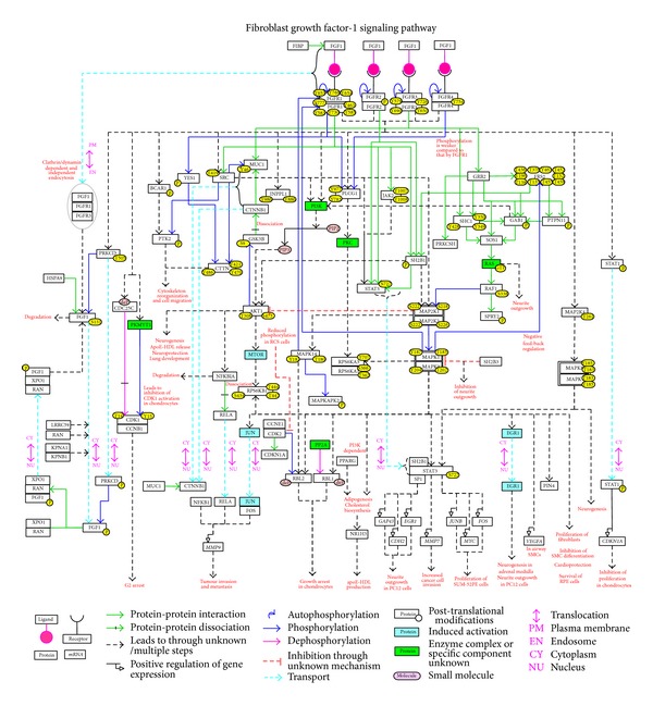
Network map of FGF-1 signaling. This map manually drawn using PathVisio [112] represents the reactions induced by FGF-1 through their receptors. Each node represents the molecules and the post-translationally modified states of proteins are also represented. Distinguished by color and continuous/dashed lines, the edges represent the specific information such as protein-protein interactions, enzyme-substrate reactions, reactions mediated through unknown/multiple steps, and protein translocations as provided in the legend. The biological processes that FGF-1 regulates through multiple signaling modules are also represented. A NetSlim [52] version of this map can be obtained from http://www.netpath.org/netslim/FGF-1_pathway.html.
4. Conclusions
Availability of specific ligand-receptor mediated signaling data in community approved formats is crucial to the understanding of proteins and their reactions in diverse biological processes. Analysis of high-throughput data obtained from microarray- and mass spectrometry-based platforms essentially relies on enrichment of biological function or signaling pathways available in databases to obtain insights into their physiological functions. Although some resources have cataloged FGF signaling in general, this is the first attempt to provide a comprehensive view of FGF-1 signaling. This will be extended to other FGF ligands and/or specific FGFRs in the future to facilitate the analysis of differences between different FGFs and/or FGFRs. The pathway information has been made available through NetPath and NetSlim resources in multiple community standard data formats. The FGF-1 signaling pathway data will be periodically updated in NetPath. We have cataloged multiple signaling modules that are activated upon activation of FGFR and their implications in diverse physiological and pathophysiological processes. We believe that the data presented here will boost further research in this area and will help identify novel therapeutically important molecules that could be targeted in pathological conditions involving aberrant FGF-1 signaling.
Acknowledgments
The authors thank the Department of Biotechnology (DBT), Government of India, for research support to the Institute of Bioinformatics, Bangalore. Shyam Mohan Palapetta is supported by a Senior Research Fellowship from the Council of Scientific and Industrial Research (CSIR), India. Varot K. Sandhya is a recipient of Inspire Fellowship from the Department of Science and Technology (DST), Government of India. Harsha Gowda is a Wellcome Trust/DBT India Alliance Early Career Fellow.
Abbreviations
- S100A13:
S100 calcium binding protein A13
- FRS2:
Fibroblast growth factor receptor substrate 2
- GAB1:
GRB2-associated binding protein 1
- SOS1:
Son of sevenless homolog 1
- PTPN11:
Protein tyrosine phosphatase, non-receptor type 11
- SHC1:
Src homology 2 domain containing transforming protein 1
- GRB2:
Growth factor receptor-bound protein 2
- Mapk:
Mitogen activated protein kinase
- Pi3k:
Phosphatidylinositide 3-kinase
- Akt:
v-akt murine thymoma viral oncogene homolog
- HDL:
High density lipoprotein
- Jnk:
Jun N-terminal kinase
- STAT3:
Signal transducer and activator of transcription 3.
Conflict of Interests
The authors have no conflict of interests.
Authors' Contribution
Shyam Mohan Palapetta, Varot K. Sandhya, and Apeksha Sahu contributed equally to the paper.
References
- 1.Harmer NJ. Insights into the role of heparan sulphate in fibroblast growth factor signalling. Biochemical Society Transactions. 2006;34(3):442–445. doi: 10.1042/BST0340442. [DOI] [PubMed] [Google Scholar]
- 2.Ibrahimi OA, Zhang F, Hrstka SCL, Mohammadi M, Linhardt RJ. Kinetic model for FGF, FGFR, and proteoglycan signal transduction complex assembly. Biochemistry. 2004;43(16):4724–4730. doi: 10.1021/bi0352320. [DOI] [PubMed] [Google Scholar]
- 3.Ornitz DM, Herr AB, Nilsson M, Westman J, Svahn C-M, Waksman G. FGF binding and FGF receptor activation by synthetic heparan-derived di- and trisaccharides. Science. 1995;268(5209):432–436. doi: 10.1126/science.7536345. [DOI] [PubMed] [Google Scholar]
- 4.Pantoliano MW. Multivalent ligand-receptor binding interactions in the fibroblast growth factor system produce a cooperative growth factor and heparin mechanism for receptor dimerization. Biochemistry. 1994;33(34):10229–10248. doi: 10.1021/bi00200a003. [DOI] [PubMed] [Google Scholar]
- 5.Ornitz DM, Leder P. Ligand specificity and heparin dependence of fibroblast growth factor receptors 1 and 3. The Journal of Biological Chemistry. 1992;267(23):16305–16311. [PubMed] [Google Scholar]
- 6.Ornitz DM, Yayon A, Flanagan JG, Svahn CM, Levi E, Leder P. Heparin is required for cell-free binding of basic fibroblast growth factor to a soluble receptor and for mitogenesis in whole cells. Molecular and Cellular Biology. 1992;12(1):240–247. doi: 10.1128/mcb.12.1.240. [DOI] [PMC free article] [PubMed] [Google Scholar]
- 7.Yayon A, Klagsbrun M, Esko JD, Leder P, Ornitz DM. Cell surface, heparin-like molecules are required for binding of basic fibroblast growth factor to its high affinity receptor. Cell. 1991;64(4):841–848. doi: 10.1016/0092-8674(91)90512-w. [DOI] [PubMed] [Google Scholar]
- 8.Saksela O, Moscatelli D, Sommer A, Rifkin DB. Endothelial cell-derived heparan sulfate binds basic fibroblast growth factor and protects it from proteolytic degradation. Journal of Cell Biology. 1988;107(2):743–751. doi: 10.1083/jcb.107.2.743. [DOI] [PMC free article] [PubMed] [Google Scholar]
- 9.Gospodarowicz D, Cheng J. Heparin protects basic and acidic FGF from inactivation. Journal of Cellular Physiology. 1986;128(3):475–484. doi: 10.1002/jcp.1041280317. [DOI] [PubMed] [Google Scholar]
- 10.Itoh N, Ornitz DM. Evolution of the Fgf and Fgfr gene families. Trends in Genetics. 2004;20(11):563–569. doi: 10.1016/j.tig.2004.08.007. [DOI] [PubMed] [Google Scholar]
- 11.Beenken A, Mohammadi M. The FGF family: biology, pathophysiology and therapy. Nature Reviews Drug Discovery. 2009;8(3):235–253. doi: 10.1038/nrd2792. [DOI] [PMC free article] [PubMed] [Google Scholar]
- 12.Colvin JS, white AC, Pratt SJ, Ornitz DM. Lung hypoplasia and neonatal death in Fgf9-null mice identify this gene as an essential regulator of lung mesenchyme. Development. 2001;128(11):2095–2106. doi: 10.1242/dev.128.11.2095. [DOI] [PubMed] [Google Scholar]
- 13.Colvin JS, Green RP, Schmahl J, Capel B, Ornitz DM. Male-to-female sex reversal in mice lacking fibroblast growth factor 9. Cell. 2001;104(6):875–889. doi: 10.1016/s0092-8674(01)00284-7. [DOI] [PubMed] [Google Scholar]
- 14.Tekin M, Hişmi BÖ, Fitoz S, et al. Homozygous mutations in fibroblast growth factor 3 are associated with a new form of syndromic deafness characterized by inner ear agenesis, microtia, and microdontia. American Journal of Human Genetics. 2007;80(2):338–344. doi: 10.1086/510920. [DOI] [PMC free article] [PubMed] [Google Scholar]
- 15.Usui H, Shibayama M, Ohbayashi N, Konishi M, Takada S, Itoh N. Fgf18 is required for embryonic lung alveolar development. Biochemical and Biophysical Research Communications. 2004;322(3):887–892. doi: 10.1016/j.bbrc.2004.07.198. [DOI] [PubMed] [Google Scholar]
- 16.Ohbayashi N, Shibayama M, Kurotaki Y, et al. FGF18 is required for normal cell proliferation and differentiation during osteogenesis and chondrogenesis. Genes and Development. 2002;16(7):870–879. doi: 10.1101/gad.965702. [DOI] [PMC free article] [PubMed] [Google Scholar]
- 17.Liu Z, Xu J, Colvin JS, Ornitz DM. Coordination of chondrogenesis and osteogenesis by fibroblast growth factor 18. Genes and Development. 2002;16(7):859–869. doi: 10.1101/gad.965602. [DOI] [PMC free article] [PubMed] [Google Scholar]
- 18.Itoh N, Ornitz DM. Fibroblast growth factors: from molecular evolution to roles in development, metabolism and disease. Journal of Biochemistry. 2011;149(2):121–130. doi: 10.1093/jb/mvq121. [DOI] [PMC free article] [PubMed] [Google Scholar]
- 19.Werner S, Peters KG, Longaker MT, Fuller-Pace F, Banda MJ, Williams LT. Large induction of keratinocyte growth factor expression in the dermis during wound healing. Proceedings of the National Academy of Sciences of the United States of America. 1992;89(15):6896–6900. doi: 10.1073/pnas.89.15.6896. [DOI] [PMC free article] [PubMed] [Google Scholar]
- 20.Rieck P, Assouline M, Savoldelli M, et al. Recombinant human basic fibroblast growth factor (Rh-bFGF) in three different wound models in rabbits: corneal wound healing effect and pharmacology. Experimental Eye Research. 1992;54(6):987–998. doi: 10.1016/0014-4835(92)90163-m. [DOI] [PubMed] [Google Scholar]
- 21.Slavin J. Fibroblast growth factors: at the heart of angiogenesis. Cell Biology International. 1995;19(5):431–444. doi: 10.1006/cbir.1995.1087. [DOI] [PubMed] [Google Scholar]
- 22.Hossain WA, Morest DK. Fibroblast growth factors (FGF-1, FGF-2) promote migration and neurite growth of mouse cochlear ganglion cells in vitro: immunohistochemistry and antibody perturbation. Journal of Neuroscience Research. 2000;62(1):40–55. doi: 10.1002/1097-4547(20001001)62:1<40::AID-JNR5>3.0.CO;2-L. [DOI] [PubMed] [Google Scholar]
- 23.Tanaka S, Kunath T, Hadjantonakis A-K, Nagy A, Rossant J. Promotion to trophoblast stem cell proliferation by FGF4. Science. 1998;282(5396):2072–2075. doi: 10.1126/science.282.5396.2072. [DOI] [PubMed] [Google Scholar]
- 24.Webb SE, Lee KK, Tang MK, et al. Fibroblast growth factors 2 and 4 stimulate migration of mouse embryonic limb myogenic cells. Developmental Dynamics. 1997;209(2):206–216. doi: 10.1002/(SICI)1097-0177(199706)209:2<206::AID-AJA6>3.0.CO;2-M. [DOI] [PubMed] [Google Scholar]
- 25.Werner S, Weinberg W, Liao X, et al. Targeted expression of a dominant-negative FGF receptor mutant in the epidermis of transgenic mice reveals a role of FGF in keratinocyte organization and differentiation. EMBO Journal. 1993;12(7):2635–2643. doi: 10.1002/j.1460-2075.1993.tb05924.x. [DOI] [PMC free article] [PubMed] [Google Scholar]
- 26.Murphy M, Drago J, Bartlett PF. Fibroblast growth factor stimulates the proliferation and differentiation of neural precursor cells in vitro. Journal of Neuroscience Research. 1990;25(4):463–475. doi: 10.1002/jnr.490250404. [DOI] [PubMed] [Google Scholar]
- 27.Turner N, Grose R. Fibroblast growth factor signalling: from development to cancer. Nature Reviews Cancer. 2010;10(2):116–129. doi: 10.1038/nrc2780. [DOI] [PubMed] [Google Scholar]
- 28.Gospodarowicz D. Localisation of a fibroblast growth factor and its effect along and with hydrocortisone on 3T3 cell growth. Nature. 1974;249(5453):123–127. doi: 10.1038/249123a0. [DOI] [PubMed] [Google Scholar]
- 29.Armelin HA. Pituitary extracts and steroid hormones in the control of 3T3 cell growth. Proceedings of the National Academy of Sciences of the United States of America. 1973;70(9):2702–2706. doi: 10.1073/pnas.70.9.2702. [DOI] [PMC free article] [PubMed] [Google Scholar]
- 30.Carreira CM, Landriscina M, Bellum S, Prudovsky I, Maciag T. The comparative release of FGF1 by hypoxia and temperature stress. Growth Factors. 2001;18(4):277–285. doi: 10.3109/08977190109029116. [DOI] [PubMed] [Google Scholar]
- 31.Jackson A, Friedman S, Zhan X, Engleka KA, Forough R, Maciag T. Heat shock induces the release of fibroblast growth factor 1 from NIH 3T3 cells. Proceedings of the National Academy of Sciences of the United States of America. 1992;89(22):10691–10695. doi: 10.1073/pnas.89.22.10691. [DOI] [PMC free article] [PubMed] [Google Scholar]
- 32.Shin JT, Opalenik SR, Wehby JN, et al. Serum-starvation induces the extracellular appearance of FGF-1. Biochimica et Biophysica Acta, Molecular Cell Research. 1996;1312(1):27–38. doi: 10.1016/0167-4889(96)00013-4. [DOI] [PubMed] [Google Scholar]
- 33.Ananyeva NM, Tjurmin AV, Berliner JA, et al. Oxidized LDL mediates the release of fibroblast growth factor-1. Arteriosclerosis, Thrombosis, and Vascular Biology. 1997;17(3):445–453. doi: 10.1161/01.atv.17.3.445. [DOI] [PubMed] [Google Scholar]
- 34.Mohan SK, Rani SG, Kumar SM, Yu C. S100A13-C2A binary complex structure-a key component in the acidic fibroblast growth factor for the non-classical pathway. Biochemical and Biophysical Research Communications. 2009;380(3):514–519. doi: 10.1016/j.bbrc.2009.01.143. [DOI] [PubMed] [Google Scholar]
- 35.Landriscina M, Bagalá C, Mandinova A, et al. Copper induces the assembly of a multiprotein aggregate implicated in the release of fibroblast growth factor 1 in response to stress. The Journal of Biological Chemistry. 2001;276(27):25549–25557. doi: 10.1074/jbc.M102925200. [DOI] [PubMed] [Google Scholar]
- 36.Carreira CM, LaVallee TM, Tarantini F, et al. S100A13 is involved in the regulation of fibroblast growth factor-1 and p40 synaptotagmin-1 release in vitro. The Journal of Biological Chemistry. 1998;273(35):22224–22231. doi: 10.1074/jbc.273.35.22224. [DOI] [PubMed] [Google Scholar]
- 37.Tarantini F, Lavallee T, Jackson A, et al. The extravesicular domain of synaptotagmin-1 is released with the latent fibroblast growth factor-1 homodimer in response to heat shock. The Journal of Biological Chemistry. 1998;273(35):22209–22216. doi: 10.1074/jbc.273.35.22209. [DOI] [PubMed] [Google Scholar]
- 38.Uriel S, Brey EM, Greisler HP. Sustained low levels of fibroblast growth factor-1 promote persistent microvascular network formation. American Journal of Surgery. 2006;192(5):604–609. doi: 10.1016/j.amjsurg.2006.08.012. [DOI] [PubMed] [Google Scholar]
- 39.Iwakura A, Fujita M, Ikemoto M, et al. Myocardial ischemia enhances the expression of acidic fibroblast growth factor in human pericardial fluid. Heart and Vessels. 2000;15(3):112–116. doi: 10.1007/pl00007264. [DOI] [PubMed] [Google Scholar]
- 40.Cuevas P, Reimers D, Carceller F, et al. Fibroblast growth factor-1 prevents myocardial apoptosis triggered by ischemia reperfusion injury. European Journal of Medical Research. 1997;2(11):465–468. [PubMed] [Google Scholar]
- 41.Wu J-C, Huang W-C, Tsai Y-A, Chen Y-C, Cheng H. Nerve repair using acidic fibroblast growth factor in human cervical spinal cord injury: a preliminary Phase I clinical study. Journal of Neurosurgery: Spine. 2008;8(3):208–214. doi: 10.3171/SPI/2008/8/3/208. [DOI] [PubMed] [Google Scholar]
- 42.Xia X, Babcock JP, Blaber SI, et al. Pharmacokinetic properties of 2nd-generation fibroblast growth factor-1 mutants for therapeutic application. PLoS ONE. 2012;7(11) doi: 10.1371/journal.pone.0048210.e48210 [DOI] [PMC free article] [PubMed] [Google Scholar]
- 43.Belch J, Hiatt WR, Baumgartner I, et al. Effect of fibroblast growth factor NV1FGF on amputation and death: a randomised placebo-controlled trial of gene therapy in critical limb ischaemia. The Lancet. 2011;377(9781):1929–1937. doi: 10.1016/S0140-6736(11)60394-2. [DOI] [PubMed] [Google Scholar]
- 44.Comerota AJ, Throm RC, Miller KA, et al. Naked plasmid DNA encoding fibroblast growth factor type 1 for the treatment of end-stage unreconstructible lower extremity ischemia: preliminary results of a phase I trial. Journal of Vascular Surgery. 2002;35(5):930–936. doi: 10.1067/mva.2002.123677. [DOI] [PubMed] [Google Scholar]
- 45.Birrer B. Whole genome oligonucleotide-based array comparative genomic hybridization analysis identified fibroblast growth factor 1 as a prognostic marker for advanced-stage serous ovarian adenocarcinomas. Journal of Clinical Oncology. 2007;25(21):p. 3184. doi: 10.1200/JCO.2006.09.0795. [DOI] [PubMed] [Google Scholar]
- 46.Kwabi-Addo B, Ozen M, Ittmann M. The role of fibroblast growth factors and their receptors in prostate cancer. Endocrine-Related Cancer. 2004;11(4):709–724. doi: 10.1677/erc.1.00535. [DOI] [PubMed] [Google Scholar]
- 47.Eswarakumar VP, Lax I, Schlessinger J. Cellular signaling by fibroblast growth factor receptors. Cytokine and Growth Factor Reviews. 2005;16(2):139–149. doi: 10.1016/j.cytogfr.2005.01.001. [DOI] [PubMed] [Google Scholar]
- 48.Ornitz DM, Itoh N. Fibroblast growth factors. Genome Biology. 2001;2(3, article 3005) doi: 10.1186/gb-2001-2-3-reviews3005. [DOI] [PMC free article] [PubMed] [Google Scholar]
- 49.Bhattacharjee M, Raju R, Radhakrishnan A, et al. A bioinformatics resource for TWEAK-Fn14 signaling pathway. Journal of Signal Transduction. 2012;2012:10 pages. doi: 10.1155/2012/376470.376470 [DOI] [PMC free article] [PubMed] [Google Scholar]
- 50.Telikicherla D, Ambekar A, Palapetta S, et al. A comprehensive curated resource for follicle stimulating hormone signaling. BMC Research Notes. 2011;4, article 408 doi: 10.1186/1756-0500-4-408. [DOI] [PMC free article] [PubMed] [Google Scholar]
- 51.Kandasamy K, Sujatha Mohan S, Raju R, et al. NetPath: A public resource of curated signal transduction pathways. Genome Biology. 2010;11(1, article r3) doi: 10.1186/gb-2010-11-1-r3. [DOI] [PMC free article] [PubMed] [Google Scholar]
- 52.Raju R, Nanjappa V, Balakrishnan L, et al. NetSlim: high-confidence curated signaling maps. The Journal of Biological Databases and Curation. 2011;2011:p. bar032. doi: 10.1093/database/bar032. [DOI] [PMC free article] [PubMed] [Google Scholar]
- 53.Manuvakhova M, Thottassery JV, Hays S, et al. Expression of the SNT-1/FRS2 phosphotyrosine binding domain inhibits activation of MAP kinase and PI3-kinase pathways and antiestrogen resistant growth induced by FGF-1 in human breast carcinoma cells. Oncogene. 2006;25(44):6003–6014. doi: 10.1038/sj.onc.1209592. [DOI] [PubMed] [Google Scholar]
- 54.Ong SH, Hadari YR, Gotoh N, Guy GR, Schlessinger J, Lax I. Stimulation of phosphatidylinositol 3-kinase by fibroblast growth factor receptors is mediated by coordinated recruitment of multiple docking proteins. Proceedings of the National Academy of Sciences of the United States of America. 2001;98(11):6074–6079. doi: 10.1073/pnas.111114298. [DOI] [PMC free article] [PubMed] [Google Scholar]
- 55.Hadari YR, Kouhara H, Lax I, Schlessinger J. Binding of Shp2 tyrosine phosphatase to FRS2 is essential for fibroblast growth factor-induced PC12 cell differentiation. Molecular and Cellular Biology. 1998;18(7):3966–3973. doi: 10.1128/mcb.18.7.3966. [DOI] [PMC free article] [PubMed] [Google Scholar]
- 56.Kouhara H, Hadari YR, Spivak-Kroizman T, et al. A lipid-anchored Grb2-binding protein that links FGF-receptor activation to the Ras/MAPK signaling pathway. Cell. 1997;89(5):693–702. doi: 10.1016/s0092-8674(00)80252-4. [DOI] [PubMed] [Google Scholar]
- 57.Kanai M, Göke M, Tsunekawa S, Podolsky DK. Signal transduction pathway of human fibroblast growth factor receptor 3. Identification of a novel 66-kDa phosphoprotein. The Journal of Biological Chemistry. 1997;272(10):6621–6628. doi: 10.1074/jbc.272.10.6621. [DOI] [PubMed] [Google Scholar]
- 58.Hadari YR, Kouhara H, Lax I, Schlessinger J. Binding of Shp2 tyrosine phosphatase to FRS2 is essential for fibroblast growth factor-induced PC12 cell differentiation. Molecular and Cellular Biology. 1998;18(7):3966–3973. doi: 10.1128/mcb.18.7.3966. [DOI] [PMC free article] [PubMed] [Google Scholar]
- 59.Lin W-F, Chen C-J, Chang Y-J, Chen S-L, Chiu I-M, Chen L. SH2B1β enhances fibroblast growth factor 1 (FGF1)-induced neurite outgrowth through MEK-ERK1/2-STAT3-Egr1 pathway. Cellular Signalling. 2009;21(7):1060–1072. doi: 10.1016/j.cellsig.2009.02.009. [DOI] [PubMed] [Google Scholar]
- 60.Mohammadi M, Dikic I, Sorokin A, Burgess WH, Jaye M, Schlessinger J. Identification of six novel autophosphorylation sites on fibroblast growth factor receptor 1 and elucidation of their importance in receptor activation and signal transduction. Molecular and Cellular Biology. 1996;16(3):977–989. doi: 10.1128/mcb.16.3.977. [DOI] [PMC free article] [PubMed] [Google Scholar]
- 61.Willems-Widyastuti A, Vanaudenaerde BM, Vos R, et al. Azithromycin attenuates fibroblast growth factors induced vascular endothelial growth factor Via p38MAPK signaling in human airway smooth muscle cells. Cell Biochemistry and Biophysics. 2013;67(2):331–339. doi: 10.1007/s12013-011-9331-0. [DOI] [PubMed] [Google Scholar]
- 62.Nishida T, Ito J-I, Nagayasu Y, Yokoyama S. FGF-1-induced reactions for biogenesis of apoE-HDL are mediated by Src in rat astrocytes. Journal of Biochemistry. 2009;146(6):881–886. doi: 10.1093/jb/mvp135. [DOI] [PubMed] [Google Scholar]
- 63.Chen CW, Liu CS, Chiu IM, et al. The signals of FGFs on the neurogenesis of embryonic stem cells. Journal of Biomedical Science. 2010;17:p. 33. doi: 10.1186/1423-0127-17-33. [DOI] [PMC free article] [PubMed] [Google Scholar]
- 64.Lungu G, Covaleda L, Mendes O, Martini-Stoica H, Stoica G. FGF-1-induced matrix metalloproteinase-9 expression in breast cancer cells is mediated by increased activities of NF-kappa;B and activating protein-1. Molecular Carcinogenesis. 2008;47(6):424–435. doi: 10.1002/mc.20398. [DOI] [PubMed] [Google Scholar]
- 65.Newell FS, Su H, Tornqvist H, Whitehead JP, Prins JB, Hutley LJ. Characterization of the transcriptional and functional effects of fibroblast growth factor-1 on human preadipocyte differentiation. The FASEB Journal: Official Publication of the Federation of American Societies for Experimental Biology. 2006;20(14):2615–2617. doi: 10.1096/fj.05-5710fje. [DOI] [PubMed] [Google Scholar]
- 66.Newman DR, Li C-M, Simmons R, Khosla J, Sannes PL. Heparin affects signaling pathways stimulated by fibroblast growth factor-1 and -2 in type II cells. American Journal of Physiology, Lung Cellular and Molecular Physiology. 2004;287(1):L191–L200. doi: 10.1152/ajplung.00284.2003. [DOI] [PubMed] [Google Scholar]
- 67.Buehlera A, Martirea A, Strohma C, et al. Angiogenesis-independent cardioprotection in FGF-1 transgenic mice. Cardiovascular Research. 2002;55(4):768–777. doi: 10.1016/s0008-6363(02)00494-7. [DOI] [PubMed] [Google Scholar]
- 68.Castaneda CA, Cortes-Funes H, Gomez HL, Ciruelos EM. The phosphatidyl inositol 3-kinase/AKT signaling pathway in breast cancer. Cancer Metastasis Reviews. 2010;29(4):751–759. doi: 10.1007/s10555-010-9261-0. [DOI] [PubMed] [Google Scholar]
- 69.Ito J-I, Nagayasu Y, Okumura-Noji K, et al. Mechanism for FGF-1 to regulate biogenesis of apoE-HDL in astrocytes. Journal of Lipid Research. 2007;48(9):2020–2027. doi: 10.1194/jlr.M700188-JLR200. [DOI] [PubMed] [Google Scholar]
- 70.Forough R, Weylie B, Patel C, Ambrus S, Singh US, Zhu J. Role of AKT/PKB signaling in fibroblast growth factor-1 (FGF-1)-induced angiogenesis in the chicken chorioallantoic membrane (CAM) Journal of Cellular Biochemistry. 2005;94(1):109–116. doi: 10.1002/jcb.20274. [DOI] [PubMed] [Google Scholar]
- 71.Wang J, Ito T, Udaka N, Okudela K, Yazawa T, Kitamura H. PI3K-AKT pathway mediates growth and survival signals during development of fetal mouse lung. Tissue and Cell. 2005;37(1):25–35. doi: 10.1016/j.tice.2004.09.002. [DOI] [PubMed] [Google Scholar]
- 72.Lin W-F, Chen C-J, Chang Y-J, Chen S-L, Chiu I-M, Chen L. SH2B1β enhances fibroblast growth factor 1 (FGF1)-induced neurite outgrowth through MEK-ERK1/2-STAT3-Egr1 pathway. Cellular Signalling. 2009;21(7):1060–1072. doi: 10.1016/j.cellsig.2009.02.009. [DOI] [PubMed] [Google Scholar]
- 73.Hossain MA, Russell JC, Gomes R, Laterra J. Neuroprotection by scatter factor/hepatocyte growth factor and FGF-1 in cerebellar granule neurons is phosphatidylinositol 3-kinase/Akt-dependent and MAPK/CREB-independent. Journal of Neurochemistry. 2002;81(2):365–378. doi: 10.1046/j.1471-4159.2002.00837.x. [DOI] [PubMed] [Google Scholar]
- 74.Li P, Oparil S, Feng W, Chen Y-F. Hypoxia-responsive growth factors upregulate periostin and osteopontin expression via distinct signaling pathways in rat pulmonary arterial smooth muscle cells. Journal of Applied Physiology. 2004;97(4):1550–1558. doi: 10.1152/japplphysiol.01311.2003. [DOI] [PubMed] [Google Scholar]
- 75.Raucci A, Laplantine E, Mansukhani A, Basilico C. Activation of the ERK1/2 and p38 mitogen-activated protein kinase pathways mediates fibroblast growth factor-induced growth arrest of chondrocytes. The Journal of Biological Chemistry. 2004;279(3):1747–1756. doi: 10.1074/jbc.M310384200. [DOI] [PubMed] [Google Scholar]
- 76.Jiao J, Greendorfer JS, Zhang P, Zinn KR, Diglio CA, Thompson JA. Alternatively spliced FGFR-1 isoform signaling differentially modulates endothelial cell responses to peroxynitrite. Archives of Biochemistry and Biophysics. 2003;410(2):187–200. doi: 10.1016/s0003-9861(02)00681-1. [DOI] [PubMed] [Google Scholar]
- 77.Mehta PB, Robson CN, Neal DE, Leung HY. Keratinocyte growth factor activates p38 MAPK to induce stress fibre formation in human prostate DU145 cells. Oncogene. 2001;20(38):5359–5365. doi: 10.1038/sj.onc.1204688. [DOI] [PubMed] [Google Scholar]
- 78.Chang YJ, Chen KW, Chen CJ, et al. SH2B1β interacts with STAT3 and enhances fibroblast growth factor 1-induced gene expression during neuronal differentiation. Molecular and Cellular Biology. 2014;34(6):1003–1019. doi: 10.1128/MCB.00940-13. [DOI] [PMC free article] [PubMed] [Google Scholar]
- 79.Dudka AA, Sweet SMM, Heath JK. Signal transducers and activators of transcription-3 binding to the fibroblast growth factor receptor is activated by receptor amplification. Cancer Research. 2010;70(8):3391–3401. doi: 10.1158/0008-5472.CAN-09-3033. [DOI] [PMC free article] [PubMed] [Google Scholar]
- 80.Lin W-F, Chen C-J, Chang Y-J, Chen S-L, Chiu I-M, Chen L. SH2B1β enhances fibroblast growth factor 1 (FGF1)-induced neurite outgrowth through MEK-ERK1/2-STAT3-Egr1 pathway. Cellular Signalling. 2009;21(7):1060–1072. doi: 10.1016/j.cellsig.2009.02.009. [DOI] [PubMed] [Google Scholar]
- 81.Lungu G, Covaleda L, Mendes O, Martini-Stoica H, Stoica G. FGF-1-induced matrix metalloproteinase-9 expression in breast cancer cells is mediated by increased activities of NF-kappa;B and activating protein-1. Molecular Carcinogenesis. 2008;47(6):424–435. doi: 10.1002/mc.20398. [DOI] [PubMed] [Google Scholar]
- 82.Zhu X, Sasse J, McAllister D, et al. Evidence that fibroblast growth factors 1 and 4 participate in regulation of cardiogenesis. Developmental Dynamics. 1996;207(4):429–438. doi: 10.1002/(SICI)1097-0177(199612)207:4<429::AID-AJA7>3.0.CO;2-J. [DOI] [PubMed] [Google Scholar]
- 83.Qu X, Hertzler K, Pan Y, Grobe K, Robinson ML, Zhang X. Genetic epistasis between heparan sulfate and FGF-Ras signaling controls lens development. Developmental Biology. 2011;355(1):12–20. doi: 10.1016/j.ydbio.2011.04.007. [DOI] [PMC free article] [PubMed] [Google Scholar]
- 84.Serls AE, Doherty S, Parvatiyar P, Wells JM, Deutsch GH. Different thresholds of fibroblast growth factors pattern the ventral foregut into liver and lung. Development. 2005;132(1):35–47. doi: 10.1242/dev.01570. [DOI] [PubMed] [Google Scholar]
- 85.Lebeche D, Malpel S, Cardoso WV. Fibroblast growth factor interactions in the developing lung. Mechanisms of Development. 1999;86(1-2):125–136. doi: 10.1016/s0925-4773(99)00124-0. [DOI] [PubMed] [Google Scholar]
- 86.Jung J, Zheng M, Goldfarb M, Zaret KS. Initiation of mammalian liver development from endoderm by fibroblast growth factors. Science. 1999;284(5422):1998–2003. doi: 10.1126/science.284.5422.1998. [DOI] [PubMed] [Google Scholar]
- 87.Luo X, Hutley LJ, Webster JA, et al. Identification of BMP and activin membrane-bound inhibitor (BAMBI) as a potent negative regulator of adipogenesis and modulator of autocrine/paracrine adipogenic factors. Diabetes. 2012;61(1):124–136. doi: 10.2337/db11-0998. [DOI] [PMC free article] [PubMed] [Google Scholar]
- 88.Hutley L, Shurety W, Newell F, et al. Fibroblast growth factor 1: a key regulator of human adipogenesis. Diabetes. 2004;53(12):3097–3106. doi: 10.2337/diabetes.53.12.3097. [DOI] [PubMed] [Google Scholar]
- 89.Tran T, Kolupaeva V, Basilico C. FGF inhibits the activity of the cyclin B1/CDK1 kinase to induce a transient G2 arrest in RCS chondrocytes. Cell Cycle. 2010;9(21):4379–4386. doi: 10.4161/cc.9.21.13671. [DOI] [PMC free article] [PubMed] [Google Scholar]
- 90.Kolupaeva V, Laplantine E, Basilico C. PP2A-mediated dephosphorylation of p107 plays a critical role in chondrocyte cell cycle arrest by FGF. PLoS ONE. 2008;3(10) doi: 10.1371/journal.pone.0003447.e3447 [DOI] [PMC free article] [PubMed] [Google Scholar]
- 91.Priore R, Dailey L, Basilico C. Downregulation of Akt activity contributes to the growth arrest induced by FGF in chondrocytes. Journal of Cellular Physiology. 2006;207(3):800–808. doi: 10.1002/jcp.20620. [DOI] [PubMed] [Google Scholar]
- 92.Dailey L, Laplantine E, Priore R, Basilico C. A network of transcriptional and signaling events is activated by FGF to induce chondrocyte growth arrest and differentiation. Journal of Cell Biology. 2003;161(6):1053–1066. doi: 10.1083/jcb.200302075. [DOI] [PMC free article] [PubMed] [Google Scholar]
- 93.Zhang P, Greendorfer JS, Jiao J, Kelpke SC, Thompson JA. Alternatively spliced FGFR-1 isoforms differentially modulate endothelial cell activation of c-YES. Archives of Biochemistry and Biophysics. 2006;450(1):50–62. doi: 10.1016/j.abb.2006.03.017. [DOI] [PubMed] [Google Scholar]
- 94.Fernandez B, Buehler A, Wolfram S, et al. Transgenic myocardial overexpression of fibroblast growth factor-1 increases coronary artery density and branching. Circulation Research. 2000;87(3):207–213. doi: 10.1161/01.res.87.3.207. [DOI] [PubMed] [Google Scholar]
- 95.Schumacher B, Peecher P, Von Specht BU, Stegmann T. Induction of neoangiogenesis in ischemic myocardium by human growth factors: first clinical results of a new treatment of coronary heart disease. Circulation. 1998;97(7):645–650. doi: 10.1161/01.cir.97.7.645. [DOI] [PubMed] [Google Scholar]
- 96.Ito J-I, Nagayasu Y, Lu R, Kheirollah A, Hayashi M, Yokoyama S. Astrocytes produce and secrete FGF-1, which promotes the production of apoE-HDL in a manner of autocrine action. Journal of Lipid Research. 2005;46(4):679–686. doi: 10.1194/jlr.M400313-JLR200. [DOI] [PubMed] [Google Scholar]
- 97.Sun L, Xu L, Chang H, et al. Transfection with aFGF cDNA improves wound healing. Journal of Investigative Dermatology. 1997;108(3):313–318. doi: 10.1111/1523-1747.ep12286471. [DOI] [PubMed] [Google Scholar]
- 98.Pirvola U, Cao Y, Oellig C, Suoqiang Z, Pettersson RF, Ylikoski J. The site of action of neuronal acidic fibroblast growth factor is the organ of corti of the rat cochlea. Proceedings of the National Academy of Sciences of the United States of America. 1995;92(20):9269–9273. doi: 10.1073/pnas.92.20.9269. [DOI] [PMC free article] [PubMed] [Google Scholar]
- 99.Palmen M, Daemen MJAP, De Windt LJ, et al. Fibroblast growth factor-1 improves cardiac functional recovery and enhances cell survival after ischemia and reperfusion: a fibroblast growth factor receptor, protein kinase C, and tyrosine kinase-dependent mechanism. Journal of the American College of Cardiology. 2004;44(5):1113–1123. doi: 10.1016/j.jacc.2004.05.067. [DOI] [PubMed] [Google Scholar]
- 100.Buehler A, Martire A, Strohm C, et al. Angiogenesis-independent cardioprotection in FGF-1 transgenic mice. Cardiovascular Research. 2002;55(4):768–777. doi: 10.1016/s0008-6363(02)00494-7. [DOI] [PubMed] [Google Scholar]
- 101.Htun P, Ito WD, Hoefer IE, Schaper J, Schaper W. Intramyocardial infusion of FGF-1 mimics ischemic preconditioning in pig myocardium. Journal of Molecular and Cellular Cardiology. 1998;30(4):867–877. doi: 10.1006/jmcc.1998.0654. [DOI] [PubMed] [Google Scholar]
- 102.Hashimoto M, Sagara Y, Langford D, et al. Fibroblast growth factor 1 regulates signaling via the glycogen synthase kinase-3β pathway. Implications for neuroprotection. The Journal of Biological Chemistry. 2002;277(36):32985–32991. doi: 10.1074/jbc.M202803200. [DOI] [PubMed] [Google Scholar]
- 103.Taeger J, Moser C, Hellerbrand C, et al. Targeting FGFR/PDGFR/VEGFR impairs tumor growth, angiogenesis, and metastasis by effects on tumor cells, endothelial cells, and pericytes in pancreatic cancer. Molecular Cancer Therapeutics. 2011;10(11):2157–2167. doi: 10.1158/1535-7163.MCT-11-0312. [DOI] [PubMed] [Google Scholar]
- 104.Haugsten EM, Zakrzewska M, Brech A, et al. Clathrin- and dynamin-independent endocytosis of fgfr3—implications for signalling. PLoS ONE. 2011;6(7) doi: 10.1371/journal.pone.0021708.e21708 [DOI] [PMC free article] [PubMed] [Google Scholar]
- 105.Bonneton C, Sibarita JB, Thiery JP. Relationship between cell migration and cell cycle during the initiation of epithelial to fibroblastoid transition. Cell Motility and the Cytoskeleton. 1999;43(4):288–295. doi: 10.1002/(SICI)1097-0169(1999)43:4<288::AID-CM2>3.0.CO;2-Y. [DOI] [PubMed] [Google Scholar]
- 106.Liu Z, Hartman YE, Warram JM, et al. Fibroblast growth factor receptor mediates fibroblast-dependent growth in EMMPRIN-depleted head and neck cancer tumor cells. Molecular Cancer Research. 2011;9(8):1008–1017. doi: 10.1158/1541-7786.MCR-11-0043. [DOI] [PMC free article] [PubMed] [Google Scholar]
- 107.Estes NR, II, Thottassery JV, Westbrook L, Kern FG. MEK ablation in MCF-7 cells blocks DNA synthesis induced by serum, but not by estradiol or growth factors. International Journal of Oncology. 2006;29(6):1573–1580. [PubMed] [Google Scholar]
- 108.Klingenberg O, Wiçdłocha A, Rapak A, Muñoz R, Falnes PØ, Olsnes S. Inability of the acidic fibroblast growth factor mutant K132E to stimulate DNA synthesis after translocation into cells. The Journal of Biological Chemistry. 1998;273(18):11164–11172. doi: 10.1074/jbc.273.18.11164. [DOI] [PubMed] [Google Scholar]
- 109.Rodier J-M, Valles AM, Denoyelle M, Thiery JP, Boyer B. pp60(c-src) Is a positive regulator of growth factor-induced cell scattering in a rat bladder carcinoma cell line. Journal of Cell Biology. 1995;131(3):761–773. doi: 10.1083/jcb.131.3.761. [DOI] [PMC free article] [PubMed] [Google Scholar]
- 110.Valles AM, Boyer B, Badet J, Tucker GC, Barritault D, Thiery JP. Acidic fibroblast growth factor is a modulator of epithelial plasticity in a rat bladder carcinoma cell line. Proceedings of the National Academy of Sciences of the United States of America. 1990;87(3):1124–1128. doi: 10.1073/pnas.87.3.1124. [DOI] [PMC free article] [PubMed] [Google Scholar]
- 111.Jouanneau J, Plouet J, Moens G, Thiery JP. FGF-2 and FGF-1 expressed in rat bladder carcinoma cells have similar angiogenic potential but different tumorigenic properties in vivo. Oncogene. 1997;14(6):671–676. doi: 10.1038/sj.onc.1200883. [DOI] [PubMed] [Google Scholar]
- 112.Jouanneau J, Gavrilovic J, Caruelle D, et al. Secreted or nonsecreted forms of acidic fibroblast growth factor produced by transfected epithelial cells influence cell morphology, motility, and invasive potential. Proceedings of the National Academy of Sciences of the United States of America. 1991;88(7):2893–2897. doi: 10.1073/pnas.88.7.2893. [DOI] [PMC free article] [PubMed] [Google Scholar]
- 113.Shannon P, Markiel A, Ozier O, et al. Cytoscape: a software environment for integrated models of biomolecular interaction networks. Genome Research. 2003;13(11):2498–2504. doi: 10.1101/gr.1239303. [DOI] [PMC free article] [PubMed] [Google Scholar]
- 114.Babur O, Dogrusoz U, Demir E, Sander C. ChiBE: interactive visualization and manipulation of BioPAX pathway models. Bioinformatics. 2010;26(3):429–431. doi: 10.1093/bioinformatics/btp665. [DOI] [PMC free article] [PubMed] [Google Scholar]
- 115.Dilek A, Belviranli ME, Dogrusoz U. VISIBIOweb: visualization and layout services for BioPAX pathway models. Nucleic Acids Research. 2010;38(2):W150–W154. doi: 10.1093/nar/gkq352.gkq352 [DOI] [PMC free article] [PubMed] [Google Scholar]
- 116.van Iersel MP, Kelder T, Pico AR, et al. Presenting and exploring biological pathways with PathVisio. BMC Bioinformatics. 2008;9, article 399 doi: 10.1186/1471-2105-9-399. [DOI] [PMC free article] [PubMed] [Google Scholar]
- 117.Prasad TSK, Goel R, Kandasamy K, et al. Human protein reference database—2009 update. Nucleic Acids Research. 2009;37(1):D767–D772. doi: 10.1093/nar/gkn892. [DOI] [PMC free article] [PubMed] [Google Scholar]