Inhibition of T reg cells by type I IFNs is necessary for the generation of optimal antiviral T cell responses during acute LCMV infection.
Abstract
Regulatory T (T reg) cells play an essential role in preventing autoimmunity but can also impair clearance of foreign pathogens. Paradoxically, signals known to promote T reg cell function are abundant during infection and could inappropriately enhance T reg cell activity. How T reg cell function is restrained during infection to allow the generation of effective antiviral responses remains largely unclear. We demonstrate that the potent antiviral type I interferons (IFNs) directly inhibit co-stimulation–dependent T reg cell activation and proliferation, both in vitro and in vivo during acute infection with lymphocytic choriomeningitis virus (LCMV). Loss of the type I IFN receptor specifically in T reg cells results in functional impairment of virus-specific CD8+ and CD4+ T cells and inefficient viral clearance. Together, these data demonstrate that inhibition of T reg cells by IFNs is necessary for the generation of optimal antiviral T cell responses during acute LCMV infection.
CD4+ regulatory T (T reg) cells expressing the transcription factor Foxp3 are potent anti-inflammatory cells capable of restraining immune responses to both self- and foreign antigens (Sakaguchi et al., 2008). In addition to preventing autoimmunity and immunopathology, T reg cells can also inhibit immune responses during viral, bacterial, and parasitic infections (Belkaid and Tarbell, 2009). Although this activity is beneficial to the host in some instances (Lund et al., 2008), T reg cell–mediated suppression can also impair clearance of harmful pathogens. Enhanced T reg cell numbers, for example, are associated with higher viral burden and exaggerated liver pathology after infection with hepatitis C virus (Cabrera et al., 2004; Bolacchi et al., 2006), and T reg cell depletion protects mice infected with Plasmodium yoelii from death by restoring anti-parasite effector responses (Hisaeda et al., 2004). These studies highlight the need to tightly regulate T reg cell activity in different immune contexts to prevent autoimmunity while allowing protective immune responses to harmful pathogens.
Of the factors known to control T reg cell abundance and function in the periphery, the role of the cytokine IL-2 and antigen recognition are best understood. T reg cells constitutively express the IL-2 receptor component CD25, and because T reg cells are thought to be largely self-reactive their abundance is also influenced by TCR signaling. Indeed, changes in the availability of IL-2 or the activity of antigen-presenting DCs alter T reg cell abundance (Boyman et al., 2006; Darrasse-Jèze et al., 2009), and mutations in IL-2, CD25, or molecules important for T cell activation via the TCR, such as Zap70 or the costimulatory receptors CD28 and ICOS, all result in impaired T reg cell homeostasis and render mice susceptible to autoimmunity (Tang et al., 2003; Herman et al., 2004; Tanaka et al., 2010). Paradoxically, these signals that drive T reg cell proliferation are also abundant during infection when T reg cell activity may need to be curbed. IL-2 is produced by activated pathogen-specific CD4+ T cells (Long and Adler, 2006), and recognition of pathogen-associated molecular patterns drives dendritic cell activation, resulting in increased antigen presentation and expression of MHC class II and co-stimulatory ligands. Although this is essential for priming of pathogen-specific T cells, it could also lead to enhanced T reg cell activation, which could dampen protective T cell responses.
The type I IFNs are a family of cytokines that are essential for antiviral immunity in both mice and humans (Theofilopoulos et al., 2005). These cytokines signal through the heterodimeric type I IFN receptor (IFNαR), leading to phosphorylation and activation of STAT1 and STAT2, and induction of hundreds of IFN-stimulated genes. The IFNαR is expressed by nearly all nucleated cells, and type I IFNs can induce apoptosis, block translation, and inhibit cellular proliferation of many cell types. This helps limit viral spread and has made type I IFNs clinically useful in the treatment of chronic viral infection and certain types of leukemia (Trinchieri, 2010). Additionally, IFNs activate cytotoxic function in NK cells (Nguyen et al., 2002), enhance antigen-presentation and production of pro-inflammatory cytokines in DCs (Luft et al., 1998), and are required for the clonal expansion of virus-specific CD8+ and CD4+ T cells during murine infection with lymphocytic choriomeningitis virus (LCMV; Kolumam et al., 2005; Havenar-Daughton et al., 2006). Previous studies have provided conflicting results regarding the impact of type I IFNs on T reg cells (Golding et al., 2010; Namdar et al., 2010; Pace et al., 2010; Riley et al., 2011; Mozzillo and Ascierto, 2012) and have generally not used experimental systems to examine the direct effects of IFNs on T reg cell homeostasis and function. Thus, the influence of type I IFN signaling on T reg cell function and the importance of this for the generation of effective antiviral immune responses remain poorly understood.
Here, we demonstrate that type I IFNs directly inhibit co-stimulation–dependent T reg cell proliferation and activation both in vitro and in vivo during acute infection with LCMV. This inhibition is cell-intrinsic and preferentially targets CD62LloCD44hi effector/memory T reg cells that potently inhibit effector T cell responses. Selective loss of IFNαR expression on T reg cells during LCMV infection results in functional impairment of virus-specific CD8+ and CD4+ T cells and inefficient viral clearance. Together, these data indicate that transient inhibition of T reg cells by IFNs during acute viral infection is necessary for the development of optimal antiviral T cell responses.
RESULTS
T reg cells are dynamically regulated during acute viral infection
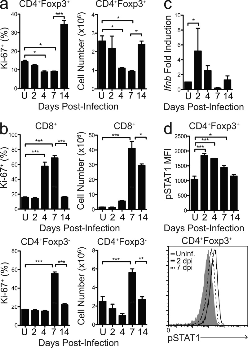
In C57BL/6 mice, infection with the Armstrong strain of LCMV causes acute viremia and induces potent CD4+ and CD8+ T cell responses that result in viral clearance by ∼8 d post infection (dpi). To determine how T reg cell activity is modulated during viral infection, we first monitored T reg cell proliferation and abundance over the course of LCMV-Armstrong infection. Whereas at 2 dpi there was little change in T reg cell abundance and phenotype, T reg cell numbers dropped progressively in the spleens of infected mice by 4 and 7 dpi (Fig. 1 a). This correlated with a significant decline in T reg cell proliferation at these time points, as measured by the percentage of cells expressing the cell cycle–associated nuclear antigen Ki-67, a common marker of cell proliferation which is expressed by cells in the G1, S, G2, and M phases of the cell cycle but not by quiescent cells in G0 (Fig. 1 a).
Figure 1.
T reg cells are dynamically regulated during LCMV infection. (a) Proportion of Ki-67+ cells among CD4+Foxp3+ T reg cells (left) and absolute number of CD4+Foxp3+ T reg cells (right) in spleens of mice left uninfected (U) or at various dpi with LCMV-Armstrong. (b) Proportion of Ki-67+ cells (left) and absolute number (right) of CD4+Foxp3− T cells (bottom) and CD8+ T cells (top) in spleens of mice infected with LCMV-Armstrong. (c) Fold induction of Ifnb mRNA from whole spleens of mice infected with LCMV-Armstrong relative to uninfected controls. Ifnb mRNA expression was normalized to Gapdh expression. (d) Median fluorescence intensity (MFI; top) and representative staining (bottom) of phospho-STAT1 in gated CD4+Foxp3+ T reg cells directly ex vivo from spleens of mice infected with LCMV-Armstrong. For all panels, n = 3–4 mice per group. Statistical significance was determined using one-way ANOVA with Tukey’s post-test (a–d). *, P < 0.05; **, P < 0.005; ***, P < 0.0001. Data are representative of two independent experiments with 3–4 mice per time point. All data are presented as the mean values ± SEM.
This reduction in T reg cell abundance correlated inversely with CD4+Foxp3− and CD8+ T effector cell responses, which peaked in proliferation and number at 7 dpi, consistent with published findings (Fig. 1 b; Murali-Krishna et al., 1998; Homann et al., 2001). After viral eradication, T reg cells displayed a proliferative burst at 14 dpi, which correlated with a significant recovery in T reg cell numbers and a reduction in the proliferation and number of CD4+Foxp3− and CD8+ T cells (Fig. 1, a and b).
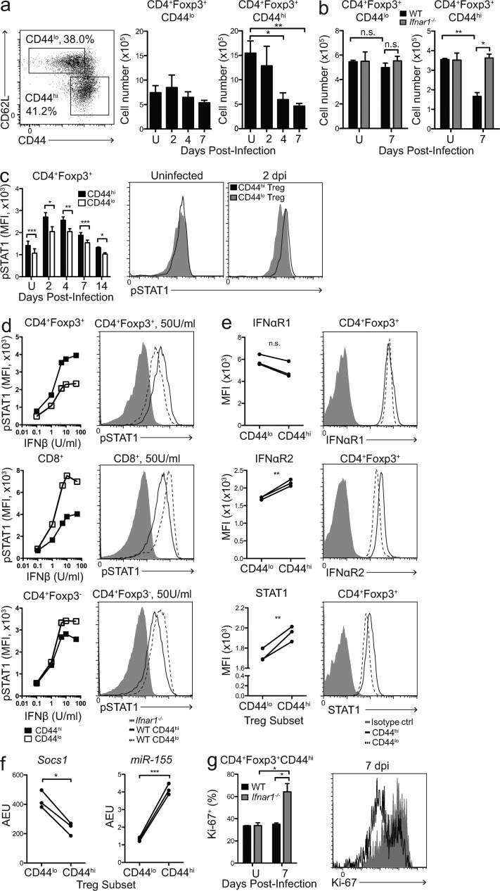
LCMV provokes a robust type I IFN response that peaks at ∼2 dpi and is required for antigen-specific T cell expansion and for viral clearance (Fig. 1 c; Müller et al., 1994). T reg cells analyzed directly ex vivo from infected spleens showed increased phosphorylation of STAT1 that peaked by 2 dpi and gradually declined in intensity, closely mimicking the kinetics of IFN-β induction in infected spleens (Fig. 1 d). Interestingly, STAT1 phosphorylation in T reg cells preceded the decline in T reg cell number and proliferation, suggesting that type I IFN signaling may play a role in inhibiting T reg cells during infection.
IFN-β directly inhibits T reg cell proliferation in vitro
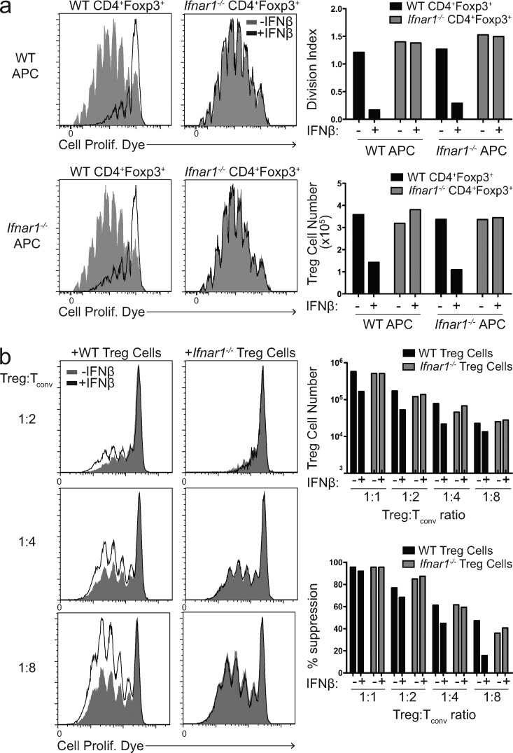
To determine whether type I IFNs could be responsible for the attrition of T reg cells we observed during acute LCMV infection, we tested the effect of IFN-β on T reg cell proliferation in vitro. For this, we isolated CD4+CD25+ T reg cells from WT or Ifnar1−/− mice that lack functional IFNαR expression, labeled them with CFSE, and cultured them with Ifnar1−/− APCs, soluble anti-CD3, and IL-2 in the presence or absence of recombinant IFN-β. After 72 h of culture, both WT and Ifnar1−/− T reg cells proliferated robustly in the absence of IFN-β. However, increasing concentrations of IFN-β substantially reduced the proliferation of WT but not Ifnar1−/− T reg cells (Fig. 2 a and not depicted). Under physiological conditions, however, both APCs and T reg cells will be exposed to IFNs during infection, and the sum of IFN’s effects on both cell populations may affect T reg proliferation differently. Thus, to determine how IFNs affect T reg proliferation in this setting, we cultured WT or Ifnar1−/− T reg cells with WT APCs in the presence or absence of IFN-β for 72 h. Interestingly, IFN-β inhibited the proliferation of WT T reg cells but not Ifnar1−/− T reg cells to the same extent whether in the presence of WT or Ifnar1−/− APCs, suggesting that inhibition was due to a direct effect of IFN-β on T reg cells and not due to an indirect effect on APCs (Fig. 2 a).
Figure 2.
Type I IFNs directly inhibit T reg cell proliferation and activity in vitro. (a, Left) T reg cell proliferation assay showing CPDye dilution of gated WT (left) or Ifnar1−/− (right) CD4+Foxp3+ T reg cells cultured with WT (top) or Ifnar1−/− APCs (bottom) in the absence (gray) or presence (black) of 50 U/ml IFN-β after 72 h of culture with soluble CD3 and IL-2. (a, Right) Division index (top) and absolute number (bottom) of WT (black) or Ifnar1−/− (gray) T reg cells after 72 h of culture in the indicated conditions. (b, Left) In vitro suppression assay showing CPDye dilution of Ifnar1−/− CD4+Foxp3− Tconv cells in the presence of WT or Ifnar1−/− T reg cells at the indicated T reg/Tconv cell ratios in the absence (gray) or presence (black) of 50 U/ml IFN-β after 72 h of culture with soluble CD3 and irradiated Ifnar1−/− APCs. (b, Right) Absolute number of and percent suppression by WT (black) or Ifnar1−/− (gray) T reg cells at decreasing T reg/Tconv cell ratios after 72 h culture in the absence (−) or presence (+) of 50 U/ml IFN-β. Data are representative of 3 independent experiments.
Next, we asked if addition of IFN-β could impair T reg cell function in a standard in vitro suppression assay. To ensure that the effects of IFN-β were restricted to T reg cells, we isolated CD4+CD25− conventional T cells (Tconv cells) and APCs from Ifnar1−/− mice. In the absence of added IFN-β, Tconv cell proliferation was suppressed equivalently by the addition of sorted WT or Ifnar1−/−CD4+Foxp3gfp+ T reg cells. However, addition of IFN-β substantially inhibited suppression by WT T reg cells but not by Ifnar1−/− T reg cells (Fig. 2 b). This inhibitory effect was magnified at decreasing T reg/Tconv cell ratios, suggesting that the effect of IFNs is most apparent when T reg cell activity is already limited and may be due to the impaired proliferation of T reg cells in these cultures. In fact, IFN-β–treated WT T reg cells showed a substantial reduction in cell number at the end of the culture period, whereas Ifnar1−/− T reg numbers were unaffected by the addition of IFN-β, suggesting that decreased T reg cell numbers may underlie the decline in suppression of Tconv cell proliferation (Fig. 2 b). Consistent with this, culture conditions with similar numbers of WT T reg cells (e.g., 1:4 with IFN compared with 1:8 without IFN) demonstrated similar suppression of Tconv cell proliferation (∼40%), regardless of the presence of IFN-β (Fig. 2 b). This suggests that reduced suppression was due to a decline in T reg cell numbers rather than an inhibition in T reg cell activity or function on a per-cell basis.
Type I IFNs directly inhibit T reg cell proliferation and activation during LCMV infection
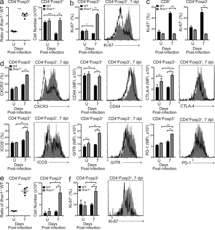
To determine if type I IFNs act directly on T reg cells to inhibit their activation during viral infection, we set up mixed bone marrow chimeras by transferring a 1:1 mixture of bone marrow from CD45.2+ Ifnar1−/− and CD45.1+ WT donor mice into irradiated T cell–deficient TCR-βδ−/− recipients. After full hematopoietic reconstitution, WT and Ifnar1−/− T reg cells were present at equal numbers and were phenotypically similar, with ∼15–20% of each population Ki-67+, indicating that Ifnar1−/− T reg cells were not intrinsically different from their WT counterparts (Fig. 3, a and b). However, at both 4 and 7 dpi, there was a dramatic decline in the number of WT T reg cells but not Ifnar1−/− T reg cells (Fig. 3 a and not depicted), consistent with our in vitro results (Fig. 2 a). Additionally, at both 4 and 7 dpi there was a dramatic increase in Ki-67 expression among Ifnar1−/− T reg cells compared with WT T reg cells in the same animals (Fig. 3 b and not depicted). This pattern was unique to T reg cells, as Ifnar1−/− effector CD8+ and CD4+Foxp3− T cells examined in the same mixed bone marrow chimeric mice showed impaired proliferation compared with their WT counterparts, consistent with previous studies demonstrating that type I IFNs are required for the expansion of virus-specific T cells (Fig. 3 c; Kolumam et al., 2005; Havenar-Daughton et al., 2006). Moreover, Ifnar1−/− T reg cells displayed a more activated phenotype, with a greater proportion expressing the chemokine receptor CXCR3, and elevated surface expression of the activation markers CD44 and ICOS (Fig. 3 d). Expression of inhibitory receptors important for T reg cell suppressive function, such as CTLA-4, PD-1, and GITR, was also increased on Ifnar1−/− T reg cells (Fig. 3 d), altogether indicating that type I IFNs directly inhibit T reg cell proliferation, activation, and function during LCMV infection.
Figure 3.
Type I IFNs directly inhibit T reg cell proliferation and activity during LCMV infection. (a, Left) Ratio of Ifnar1−/−/WT CD4+Foxp3+ T reg cells in spleens of mixed bone marrow chimeric mice left uninfected or 7 dpi with LCMV Armstrong. (a, Right) Absolute number of WT (black) and Ifnar1−/− CD4+Foxp3+ T reg cells in spleens of mixed bone marrow chimeric mice left uninfected or 7 dpi. (b, Left) Summary of Ki-67 expression by WT (black) and Ifnar1−/− (gray) CD4+Foxp3+ T reg cells from spleens of mixed bone marrow chimeric mice left uninfected or 7 dpi. (b, Right) Representative flow cytometry analysis of Ki-67 expression by gated CD45.1+ WT (black) and CD45.2+ Ifnar1−/− (gray) CD4+Foxp3+ T reg cells in spleens of mixed bone marrow chimeric mice 7 dpi. (c) Summary of Ki-67 expression by WT (black) and Ifnar1−/− (gray) CD8+ and CD4+Foxp3− T cells from spleens of mixed bone marrow chimeric mice left uninfected or 7 dpi. (d) Representative flow cytometry analysis and summaries of the indicated markers expressed by WT (black) and Ifnar1−/− (gray) CD4+Foxp3+ T reg cells in spleens of mixed bone marrow chimeric mice left uninfected or 7 dpi with LCMV-Armstrong. (e, Left) Ratio of Ifnar1−/−/WT CD4+Foxp3+ T reg cells in small intestinal LP (SI-LP) of mixed bone marrow chimeric mice left uninfected or 7 dpi with LCMV Armstrong. (e, Right) Absolute number of WT (black) and Ifnar1−/− CD4+Foxp3+ T reg cells in SI-LP of mixed bone marrow chimeric mice left uninfected or 7 dpi. (f, Left) Summary of Ki-67 expression by WT (black) and Ifnar1−/− (gray) CD4+Foxp3+ T reg cells from SI-LP of mixed bone marrow chimeric mice left uninfected or 7 dpi. (f, Right) Representative flow cytometry analysis of Ki-67 expression by gated CD45.1+ WT (black) and CD45.2+ Ifnar1−/− (gray) CD4+Foxp3+ T reg cells in SI-LP of mixed bone marrow chimeric mice 7 dpi. Statistical significance was determined using two-tailed paired t test when comparing WT and Ifnar1−/− cells within the same chimeric mouse. Two-tailed unpaired Student’s t test was used when comparing cells from different mice. n = 4 per group. Data are representative of 6 (a–d) or 2 (e and f) independent experiments with 3–4 mice per group. *, P < 0.05; **, P < 0.005; ***, P < 0.0001. All data are presented as the mean values ± SEM.
The decline in the number of WT T reg cells in the spleens of infected mice could reflect a selective redistribution of T reg cells to nonlymphoid tissues. Specifically, recent studies have posited a role for IFNs in the distribution of T reg cells in the mucosa during inflammatory colitis (Lee et al., 2012). However, at 7 dpi we did not detect any change in the number or proliferation of WT T reg cells in the intestinal lamina propria (LP; Fig. 3, e and f), suggesting that WT T reg cells from the spleen are not redistributing to the gut. Rather, there was a massive and significant increase in the proliferation and number of Ifnar1−/− T reg cells in the LP, and the ratio of Ifnar1−/−/WT T reg cells approached 6:1 in the LP, compared with 3:1 in the spleens of the same animals (Fig. 3, a, e, and f). Thus, these data suggest that IFNs do not simply induce T reg cells to redistribute to nonlymphoid/mucosal tissues, but that they directly inhibit T reg cell proliferation and accumulation in both lymphoid and nonlymphoid tissues during LCMV infection.
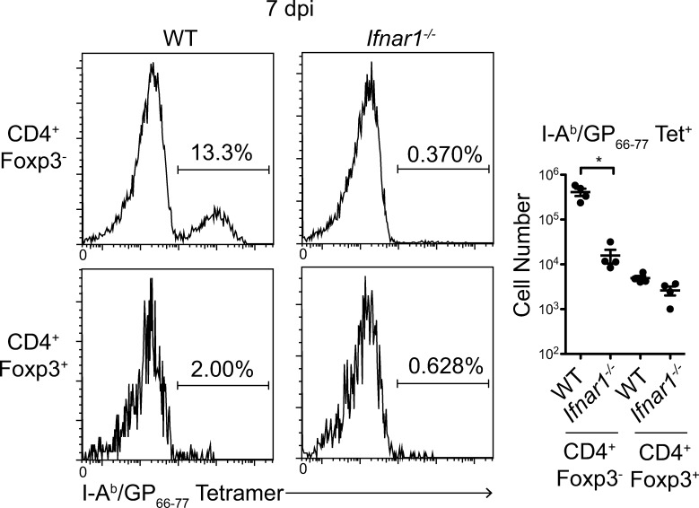
Recent studies have demonstrated that pathogen-specific natural T reg cells are potent suppressors of effector responses and can delay or prevent pathogen clearance (Zhao et al., 2011; Shafiani et al., 2013). To determine if loss of type I IFN responsiveness allowed for the expansion of LCMV-specific T reg cells, we used tetramer staining to track cells specific for the immunodominant I-Ab/GP66-77 epitope in our mixed bone marrow chimeric mice. LCMV infection provoked a robust response to the GP66-77 peptide only among WT-derived CD4+Foxp3− effector T cells (Fig. 4). As expected, GP66-77-specific cells were largely absent from the population of Ifnar1−/− CD4+Foxp3− T cells, consistent with the requirement for IFN in the expansion of antigen-specific effector CD4+ T cells during LCMV infection (Havenar-Daughton et al., 2006). However, only ∼1–2% of either WT or Ifnar1−/− T reg cells were I-Ab/GP66-77-specific, indicating that the majority of T reg cells during LCMV infection are not specific for the immunodominant I-Ab/GP66-77 epitope. Moreover, although Ifnar1−/− T reg cells are hyperproliferative, activated, and accumulate during LCMV infection, this was not due to an expansion of T reg cells specific for the I-Ab/GP66-77 epitope.
Figure 4.
Neither WT nor Ifnar1−/− T reg cells are GP66-77 specific during LCMV infection. Left: representative histograms showing I-Ab/GP66-77 tetramer staining among gated WT (left) or Ifnar1−/− (right) CD4+Foxp3− T cells (top) and CD4+Foxp3+ T cells (bottom) from spleens of the same mixed bone marrow chimeric mouse 7 dpi with LCMV-Armstrong. Numbers represent frequency of tetramer-positive cells among the indicated populations. Right: absolute number of the indicated tetramer-positive populations in spleens of chimeric mice 7 dpi with LCMV-Armstrong. Statistical significance was determined using two-tailed paired Student’s t test. n = 4 per group. Data are representative of 2 independent experiments with 3–4 mice per group. *, P < 0.05, P < 0.0001. All data are presented as the mean values ± SEM.
Type I IFNs preferentially inhibit CD62LloCD44hi effector T reg cells
Like effector T cells, T reg cells can be divided into distinct subsets based on their expression of CD44 and CD62L (Fig. 5 a; Huehn et al., 2004; Min et al., 2007). CD62LhiCD44lo (“CD44lo”) T reg cells have a quiescent phenotype, undergo minimal homeostatic proliferation, and recirculate through secondary lymphoid tissues. In contrast, CD62LloCD44hi (“CD44hi”) effector T reg cells are highly proliferative and express high levels of functional immunosuppressive molecules such as IL-10, GITR, and CTLA-4 (Cretney et al., 2013). Interestingly, the reduction in T reg cell number we observed during LCMV infection was restricted primarily to the CD44hi T reg cell subset, whereas the number of CD44lo T reg cells did not significantly change (Fig. 5 a). The selective decline in CD44hi T reg cells was IFN-dependent, as the number of CD44hi T reg cells was reduced among WT but not Ifnar1−/− T reg cells in mixed bone marrow chimeric mice 7 dpi, whereas the number of CD44lo T reg cells remained unchanged among both WT and Ifnar1−/− T reg cells (Fig. 5 b), suggesting that type I IFNs preferentially act on the effector T reg cell population. Consistent with this hypothesis, CD44hi T reg cells showed a significantly greater degree of STAT1 phosphorylation than CD44lo T reg cells in vivo during LCMV infection and in vitro after stimulation with IFN-β (Fig. 5, c and d). In contrast, STAT1 phosphorylation was reduced in CD44hi effector CD8+ and CD4+Foxp3− T cells (Fig. 5 d). Although both CD44lo and CD44hi T reg cells showed comparable expression of IFNαR1, CD44hi T reg cells expressed consistently higher amounts of IFNαR2 and total STAT1 protein (Fig. 5 e). Additionally, CD44hi T reg cells expressed lower amounts of Socs1 mRNA, a negative regulator of type I IFN signaling, as well as higher amounts of miR-155, a negative regulator of Socs1 (Fig. 5 f). Collectively, these changes in the IFNαR signaling machinery likely account for the higher responsiveness of CD44hi T reg cells to type I IFNs.
Figure 5.
Type I IFNs preferentially inhibit CD62LloCD44hi effector/memory T reg cells. (a, Left) Representative gating of CD44lo and CD44hi T reg cells. (a, Right) Absolute number of CD44lo and CD44hi CD4+Foxp3+ T reg cells in spleens of mice infected with LCMV-Armstrong. Data are representative of two independent experiments. n = 4 mice per group. Statistical significance was determined using one-way ANOVA with Tukey’s post-test. (b) Absolute number of CD44lo (left) and CD44hi (right) CD4+Foxp3+ T reg cells in mixed bone marrow chimeric mice left uninfected or 7 dpi. Data are representative of 6 independent experiments with 3–4 mice per group. Statistical significance was determined by two-tailed paired Student’s t test. Two-tailed unpaired Student’s t test was used when comparing cells from different mice. (c) Representative histograms (right) and MFI summary (left) of phospho-STAT1 in CD44lo (white) and CD44hi (black) T reg cells in spleens of mice infected with LCMV-Armstrong. Data are representative of two independent experiments. n = 4 mice per group. Statistical significance was determined using two-tailed paired Student’s t test. (d) Representative histograms (right) and MFI summary (left) of phospho-STAT1 in Ifnar1−/−, WT CD44lo and WT CD44hi T reg cells, CD8+, and CD4+Foxp3− T cells stimulated for 30 min with IFN-β. Data are representative of 3 independent experiments. (e) Representative histograms (right) and MFI summary (left) of IFNαR1, IFNαR2, and STAT1 in CD44lo and CD44hi CD4+Foxp3+ T reg cells determined by flow cytometry. n = 3 mice per group. Data are representative of three independent experiments. Statistical significance was determined using two-tailed paired Student’s t test. (f) miR-155 microRNA and Socs1 mRNA expression in sorted CD44lo and CD44hi CD4+Foxp3+ T reg cells, expressed in arbitrary expression units (AEUs) normalized to U6 and Gapdh expression, respectively. Data are representative of two independent experiments with three mice each. Statistical significance was determined using two-tailed paired Student’s t test. (g Left) Summary of Ki67 expression by WT and Ifnar1−/− CD4+Foxp3+CD62LloCD44hi (CD44hi) T reg cells from mixed bone marrow chimeric mice left uninfected (black) or infected with LCMV for 7 d (gray). (g, Right) Representative histogram showing Ki-67 expression by WT (black) and Ifnar1−/− (gray) CD4+Foxp3+CD62LloCD44hi T reg cells in mixed bone marrow chimeric mice 7 dpi with LCMV-Armstrong. n = 4 per group. Data are representative of 6 independent experiments. Statistical significance was determined by two-tailed paired Student’s t test. Two-tailed unpaired Student’s t test was used when comparing cells from different mice. *, P < 0.05; **, P < 0.005; ***, P < 0.0001. All data are presented as the mean values ± SEM.
On most cells, type I IFNs are pro-apoptotic and anti-proliferative, and this is thought to underlie much of their antiviral effects. To determine if IFNαR signaling results in attrition of CD44hi T reg cells during infection, we asked how LCMV infection impacted the recovery of sorted CD44hi WT and Ifnar1−/− T reg cells after adoptive transfer into congenically marked recipients. Indeed, we recovered significantly fewer of the transferred CD44hi WT T reg cells from infected mice, whereas the number of Ifnar1−/− CD44hi T reg cells did not change after infection (unpublished data). These results were consistent with the IFNαR-dependent loss of CD44hi T reg cells we observed in our mixed bone marrow chimeric mice upon LCMV infection (Fig. 5 b) and indicate that CD44hi T reg cells undergo IFN-dependent attrition during LCMV infection. Moreover, the proliferation of WT CD44hi T reg cells was substantially lower than that of Ifnar1−/− CD44hi T reg cells in LCMV-infected mixed bone marrow chimeras (Fig. 5 g), indicating that IFNs also have direct anti-proliferative effects on these cells. Thus, a combination of IFN’s anti-proliferative and pro-apoptotic effects on CD44hi effector T reg cells likely accounts for the loss of T reg cells observed during LCMV infection.
Enhanced proliferation of Ifnar1−/− T reg cells during LCMV infection is ICOSL- and CD28-dependent
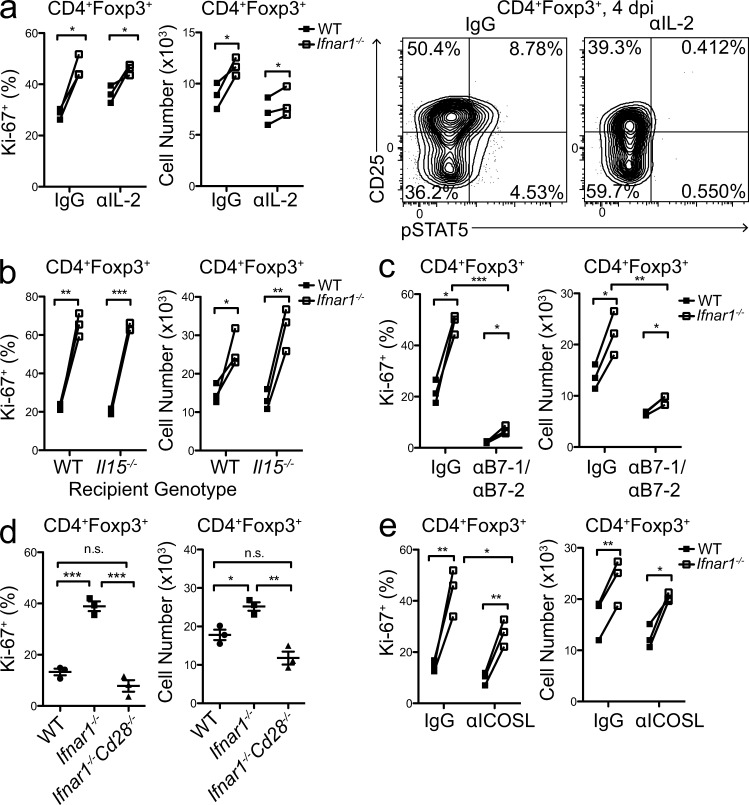
To better define the mechanism of IFN’s anti-proliferative effects on T reg cells, we determined whether IFNs blocked T reg cell proliferation in response to different activating signals. IL-2 and the closely related cytokine IL-15 can drive T reg cell proliferation both in vitro and in vivo (Boyman et al., 2006; Clark and Kupper, 2007; Xia et al., 2010), and both are expressed abundantly during LCMV infection by effector T cells and as an IFN-stimulated gene, respectively (Zhang et al., 1998; Williams et al., 2006). However, pretreatment of T reg cells for 30 min with IFN-β did not affect IL-2–induced phosphorylation of STAT5 (unpublished data). Additionally, using blocking antibodies and genetically deficient mice, we determined that neither IL-2 nor IL-15 was required for the enhanced proliferation of Ifnar1−/− T reg cells during LCMV infection (Fig. 6, a and b).
Figure 6.
Enhanced proliferation of Ifnar1−/− T reg cells is ICOSL- and CD28-dependent. (a, Left) Summary of Ki-67 expression and absolute number of transferred WT (black) and Ifnar1−/− (open squares) CD4+Foxp3+ T reg cells in WT mice 4 dpi with LCMV-Armstrong and treated with IgG or blocking anti–IL-2 antibody (αIL-2). (a, Right) Representative flow cytometric analysis showing pSTAT5 and CD25 expression by total CD4+Foxp3+ T reg cells in spleens of mice 4 dpi treated with IgG or αIL-2. Numbers represent frequency of the indicated quadrant among total CD4+Foxp3+ T reg cells. (b) Summary of Ki-67 expression and absolute number of transferred WT (black) and Ifnar1−/− (open squares) CD4+Foxp3+ T reg cells in WT or Il15−/− recipient mice 4 dpi with LCMV-Armstrong. (c) summary of Ki-67 expression and absolute number of transferred WT (black) and Ifnar1−/− (open squares) CD4+Foxp3+ T reg cells in WT mice 4 dpi with LCMV-Armstrong and treated with IgG or blocking anti–B7-1/B7-2 antibodies (αB7-1/B7-2). (d) Summary of Ki-67 expression and absolute number of transferred WT, Ifnar1−/−, or Ifnar1−/−Cd28−/− T reg cells in spleens of WT mice 4 dpi with LCMV-Armstrong. (e) Summary of Ki-67 expression and absolute number of transferred WT (black) and Ifnar1−/− (open squares) CD4+Foxp3+ T reg cells in WT mice 4 dpi treated with IgG or blocking anti-ICOSL antibody (αICOSL). a–c, e: statistical significance was determined using paired two-tailed Student’s t test when comparing WT and Ifnar1−/− T reg cells in the same recipient mice or unpaired two-tailed Student’s t test when comparing T reg cells in different mice. d: statistical significance was determined using one-way ANOVA with Tukey’s post-test. For all panels, n = 3 per group. Results are representative of 2 independent experiments. *, P < 0.05; **, P < 0.005; ***, P < 0.0001. All data are presented as the mean values ± SEM.
In addition to cytokine-driven proliferation/survival, recognition of self-antigen via stimulation of the TCR and co-receptors like CD28 and ICOS is also known to positively regulate T reg cell homeostasis (Tang et al., 2003; Darrasse-Jèze et al., 2009). Therefore, we hypothesized that during infection increased antigen presentation and expression of co-stimulatory ligands by pathogen-activated DCs drives the activation and proliferation of Ifnar1−/− T reg cells. Indeed, the number and proliferation of Ifnar1−/− T reg cells was significantly reduced 7 dpi after B7-1/B7-2 blockade compared with IgG-treated controls (Fig. 6 c). This inhibition was not secondary to impaired IL-2 signaling caused by reduced activation of effector T cells, as STAT5 phosphorylation in T reg cells was not decreased after B7-1/B7-2 blockade (unpublished data). To confirm that enhanced proliferation of Ifnar1−/− T reg cells required T reg cell–intrinsic B7/CD28 co-stimulation, we compared the proliferation of Ifnar1−/− and Ifnar1−/−Cd28−/− T reg cells transferred into WT recipients 4 dpi. Similar to our results with B7 blockade, loss of CD28 expression completely blocked the enhanced proliferation and accumulation of Ifnar1−/− T reg cells during infection (Fig. 6 d), indicating that the hyperproliferation of Ifnar1−/− T reg cells during LCMV infection is dependent on CD28 co-stimulation. However, Ifnar1−/− T reg cells still proliferated roughly twofold more and were present at higher number than WT T reg cells in both control and B7-1/B7-2–blocked mice, indicating that the ability of type I IFNs to inhibit T reg cell proliferation is independent of any direct effects on CD28 signaling. Consistent with this, IFN-β had no effect on CD3/28-dependent phosphorylation of the downstream PI3K/Akt substrate S6 in T reg cells (unpublished data), indicating that type I IFNs do not inhibit T reg cells by directly modifying this important costimulatory signaling pathway.
Stimulation through the TCR and CD28 leads to up-regulation of the inducible costimulatory molecule ICOS, which can assume many of the functions of CD28 in promoting T cell expansion/survival (Simpson et al., 2010). As ICOS expression was significantly up-regulated on Ifnar1−/− T reg cells during infection (Fig. 3 d), we also asked if enhanced ICOS–ICOSL interactions contributed to the hyperproliferation of Ifnar1−/− T reg cells by transferring WT and Ifnar1−/− T reg cells into mice, treating them with the α-ICOSL blocking antibody HK5.3 (Iwai et al., 2003; Choi et al., 2011) and infecting them with LCMV. Blockade of ICOS signaling partially inhibited the enhanced proliferation of Ifnar1−/− T reg cells, indicating that up-regulation of ICOS contributes to CD28-dependent activation of Ifnar1−/− T reg cells during infection (Fig. 6 e). Together, these data demonstrate that although LCMV infection results in a co-stimulatory environment highly favorable for T reg cell expansion, this is prevented by the direct action of type I IFNs on T reg cells.
IFN-mediated inhibition of T reg cells is necessary for optimal antiviral T cell responses
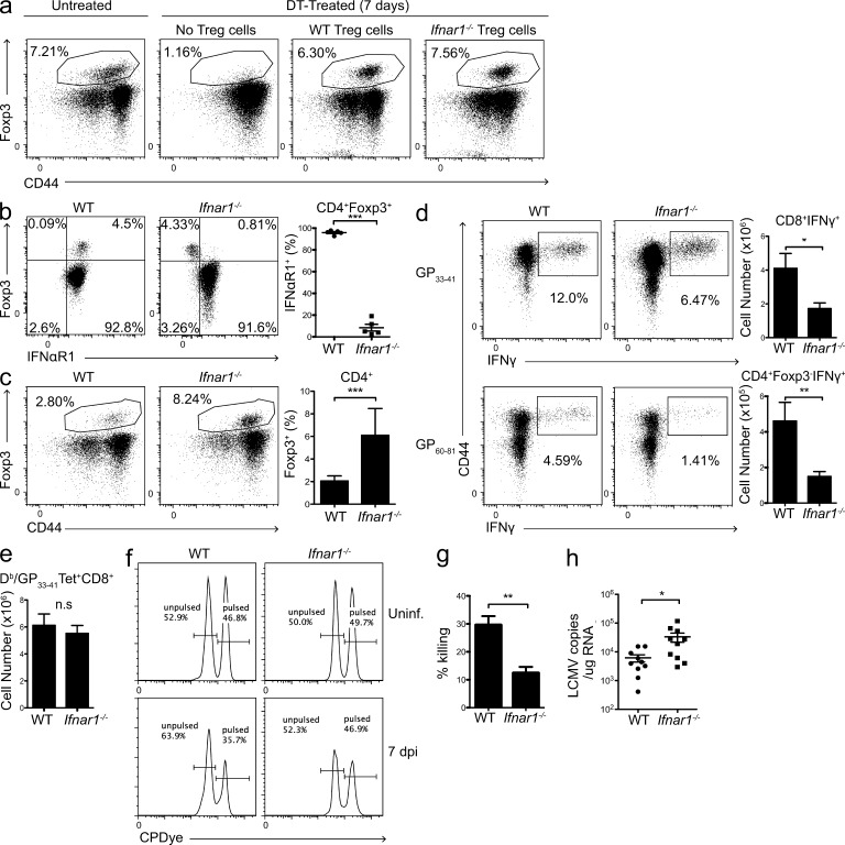
Finally, to determine what effect IFN-mediated inhibition of T reg cells has on the generation of antiviral T cell responses, we modified a previously described T reg cell replacement protocol in which Foxp3DTR mice were treated daily with diphtheria toxin (DT) to deplete mice of endogenous T reg cells, and reconstituted with T reg cells from either WT or Ifnar1−/− donors (Rowe et al., 2011). By 7 d after transfer, both WT and Ifnar1−/− T reg cells were present at similar frequencies in spleen and peripheral lymph nodes that were comparable to those in found in unmanipulated mice (Fig. 7 a and not depicted). Moreover, mice reconstituted with Ifnar1−/− T reg cells showed a selective absence of IFNαR1 expression on T reg cells (Fig. 7 b). However, by 7 dpi, T reg cells in the Ifnar1−/−-replaced mice were present at significantly higher frequency than those in WT-replaced mice (Fig. 7 c). To understand how these changes in T reg cell abundance may impact the antiviral effector T cell response, we examined the abundance and function of LCMV-specific effector T cells in these mice. The absolute number of CD8+ T cells specific for the immunodominant Db/GP33-41 epitope as assessed by tetramer staining was not significantly different between WT- and Ifnar1−/−-replaced mice (Fig. 7 e). However, the proportion and number of CD8+ T cells producing IFN-γ upon restimulation with GP33-41 peptide was significantly reduced in mice reconstituted with Ifnar1−/− T reg cells, and similar results were observed in CD4+Foxp3− T cells stimulated with the GP61-80 peptide (Fig. 7 d). Additionally, mice reconstituted with Ifnar1−/− T reg cells showed a reduced ability to kill GP33-41 peptide-pulsed splenocytes in an in vivo cytotoxicity assay (Fig. 7, f and g) and had significantly higher levels of viral RNA at 7 dpi (Fig. 7 h). Thus, type I IFN-dependent inhibition of T reg cells is essential for the generation of optimal antiviral T cell responses and normal viral clearance.
Figure 7.
Inhibition of T reg cells by type I IFNs is necessary for optimal antiviral immune responses. (a) Representative flow plots showing gated CD4+ T cells from spleens of Foxp3DTR mice left untreated (left) or treated daily for 7 d with 5 µg/kg DT. DT-treated mice were reconstituted with either no T reg cells (center left), WT T reg cells (center right), or Ifnar1−/− T reg cells (right) at the start of DT treatment. Numbers represent frequency of Foxp3+ T reg cells among gated CD4+ T cells. (b) Representative flow cytometry analysis showing IFNαR1 expression on gated CD4+ T cells (left) and summary of IFNαR1 expression on CD4+Foxp3+ T reg cells (right) from peripheral blood of Foxp3DTR mice replaced with WT or Ifnar1−/− T reg cells one day before infection with LCMV. Numbers represent frequency of the indicated quadrants among total CD4+ T cells. n = 5 per group. Data are representative of three independent experiments. (c) Representative flow plots (left) and summary (right) showing frequency of Foxp3+ T reg cells among gated CD4+ T cells in WT- and Ifnar1−/−-replaced Foxp3DTR mice 7 dpi with LCMV-Armstrong. Numbers represent frequency of CD4+Foxp3+ T reg cells among total CD4+ T cells. (d, Left) Representative flow cytometric analysis of IFN-γ production by gated CD8+ T cells (top) and gated CD4+Foxp3− T cells (bottom) by intracellular cytokine staining 7 dpi in WT- and Ifnar1−/−-replaced Foxp3DTR mice after 5-h stimulation of whole splenocytes with GP33-41 (top) or GP61-80 (bottom) peptide. Numbers represent frequency of IFN-γ+ cells among CD8+ (top) and CD4+Foxp3− (bottom) T cells. (d, Right) Absolute number of LCMV peptide-specific IFN-γ+CD8+ (top) and IFN-γ+CD4+Foxp3− T cells (bottom). (e) Absolute number of CD8+ T cells staining positively for Db/GP33-41 tetramer as assessed by flow cytometry. (f) Representative flow cytometric histograms from in vivo cytotoxicity assay showing frequency of CPDyehi-labeled GP33-41 peptide-pulsed splenocytes relative to CPDyelo-labeled unpulsed splenocytes in WT- and Ifnar1−/−-replaced Foxp3DTR mice left uninfected or 7 dpi, 1 h after transfer of splenocytes. (g) Summary of percent killing of peptide-pulsed splenocytes in WT- and Ifnar1−/−-replaced Foxp3DTR mice 7 dpi. (h) LCMV GP RNA expression measured by qPCR in infected spleens and normalized to a standard curve generated using an LCMV-GP plasmid. c–e: n = 10 per group; data are summarized from 3 independent experiments. For all panels, statistical significance was determined using a two-tailed unpaired Student’s t test. *, P < 0.05; **, P < 0.005; ***, P < 0.0001. All data are presented as the mean values ± SEM.
DISCUSSION
Excessive T reg cell activity interferes with efficient pathogen clearance and control in a variety of infectious diseases (Belkaid, 2007). Considering the abundance of T reg cell–activating factors present during infection, the host must use counterregulatory mechanisms to circumvent T reg cell activity and ensure pathogen control. This can be accomplished indirectly in some instances; for example, activation of APCs by TLR or CD40 stimulation protects them from T reg cell–mediated suppression (Serra et al., 2003; Hänig and Lutz, 2008), and pro-inflammatory cytokines such as IL-1β and IL-6 can render effector T cells resistant to T reg cell–mediated suppression (Pasare and Medzhitov, 2003; O’Sullivan et al., 2006). Additionally, effector cells can limit T reg cell activity via IL-2 deprivation (Benson et al., 2012). Type I IFNs are known to regulate several cell types to coordinate effective antiviral immune responses. Here, we define a novel mechanism for the direct repression of T reg cell proliferation during LCMV infection by type I IFNs and demonstrate that this is necessary for production of optimal antiviral T cell responses and efficient viral clearance.
A major challenge T reg cells face during infection is how to maintain self-tolerance while allowing pathogen-specific immune responses to occur. Although excessive T reg cell activity is associated with chronic infection and failure to clear pathogens, too little T reg cell activity during infection can result in autoimmunity and widespread effector T cell activation that impairs pathogen-specific responses (Lund et al., 2008; Fulton et al., 2010; Pace et al., 2012). Interestingly, type I IFNs did not globally inhibit T reg cells during LCMV infection but instead induced a selective decrease in CD44hi T reg cells. Unlike their quiescent CD44lo counterparts, which reside primarily in secondary lymphoid tissues and effectively inhibit T cell priming, CD44hi T reg cells proliferate robustly, abundantly express suppressive effector molecules such as CTLA4, IL-10, and ICOS, and are able to migrate to sites of infection and inflammation via expression of a broad array of tissue-homing receptors (Siegmund et al., 2005; Cretney et al., 2013). Thus, they are poised to inhibit the activity of primed effector T cells. Consistent with this, when T reg cells were made resistant to IFN-mediated inhibition, there was no change in the absolute number of virus-specific CD8+ T cells, suggesting there was no difference in T cell priming or clonal expansion. Rather, LCMV-specific CD8+ T cells were selectively impaired in their effector functions, failing to produce IFN-γ and kill viral peptide-pulsed APCs as efficiently as in mice with WT T reg cells. Several studies have demonstrated that T reg cells can regulate CD8+ and CD4+ T cell effector function independent of their ability to inhibit T cell proliferation via anti-inflammatory cytokines such as TGF-β and IL-10 (Sarween et al., 2004; DiPaolo et al., 2005; Mempel et al., 2006; Sojka and Fowell, 2011). Interestingly, IL-10 and TGF-β production are significantly enhanced in CD44hi T reg cells (Firan et al., 2006; Cretney et al., 2011), and thus our data are consistent with a model in which specific inhibition of CD44hi effector T reg cells allows pathogen-specific effector T cells to carry out their protective functions, whereas the priming of self-reactive T cells in lymphoid tissues remains blocked by CD44lo T reg cells, thereby helping avoid the development of collateral autoimmunity.
That T reg cells and effector T cells both rely on similar signals—like IL-2 and TCR/coreceptor signaling—for their activation and proliferation has made it difficult to understand how these functionally opposed cell types are differentially regulated. However, during LCMV infection, the ability of type I IFNs to enhance the clonal expansion of antigen-specific effector T cells while inhibiting the activation and proliferation of T reg cells is one way in which the activities of effector and T reg cells can be separated. Although LCMV infection results in a co-stimulatory environment that is favorable for both effector T cell and T reg cell proliferation, the parallel expansion of T reg cells is prevented by the direct action of type I IFNs on T reg cells. The precise molecular mechanisms by which IFNs inhibit T reg cell proliferation are not well defined, but our results suggest that IFNs exert direct pro-apoptotic and anti-proliferative effects on T reg cells. There is some evidence for cross talk between IFNαR and TCR/co-receptor pathways, as several signaling components such as Zap70, CD45, and Lck have been shown to interact with IFNαR, and this interaction was required for the anti-proliferative effects of IFN-α in Jurkat T cells (Petricoin et al., 1997). Although IFN-β had no effect on CD3/28-dependent phosphorylation of the downstream PI3K/Akt substrate S6 in T reg cells (unpublished data), it is possible that TCR/co-receptor signaling may modulate the IFNαR signaling pathway in T reg cells. The anti-proliferative effects of type I IFNs in conventional T cells appear to be STAT1-dependent (Bromberg et al., 1996; Tanabe et al., 2005). However, STAT1 expression is reduced in virus-specific CD8+ T cells, and this changes the ratio of different STAT proteins activated by IFNαR signaling, turning this from an anti-proliferative into a pro-proliferative signal required for the optimal expansion of virus-specific cells (Gil et al., 2006, 2012). An inverse mechanism may be at play in T reg cells, where TCR signaling modulates components of the IFNαR signaling pathway to sensitize activated T reg cells to the anti-proliferative effects of type I IFNs. Indeed, we show that CD44hi T reg cells expressed higher levels of IFNαR2, STAT1, and mir-155, and lower levels of Socs1, than CD44lo T reg cells, consistent with their higher responsiveness to IFNs in vitro and in vivo during LCMV infection.
The immunomodulatory effects of type I IFNs are incredibly complex, and several studies have suggested that IFNs may actually promote T reg activity in other contexts, particularly during multiple sclerosis and inflammatory colitis. IFN-β is a commonly prescribed treatment for relapsing-remitting multiple sclerosis, and IFN-β treatment markedly enhanced the function of T reg cells in these patients (de Andrés et al., 2007; Namdar et al., 2010). However, these studies fail to distinguish the direct effect of type I IFNs on T reg cells from their indirect effects on other cell types. In fact, several studies have now demonstrated that the protective functions of IFNs during experimental autoimmune encephalomyelitis depend on IFNαR expression on DCs but not on T cells, and that the enhanced proliferation of T reg cells may be secondary to activation of co-stimulatory molecules like GITRL on DCs (Prinz et al., 2008; Chen et al., 2012; Dann et al., 2012). However, contrary to our results, a recent study demonstrated a direct role for IFN in the maintenance of T reg cells in the mucosa during inflammatory colitis (Lee et al., 2012). This discrepancy may be due to differences in the timing and extent of type I IFN expression in the different models used, as IFNs can exert opposing effects when expressed acutely or chronically. For instance, during LCMV infection, transient IFN production helps promote viral clearance, whereas heightened and prolonged IFN expression promotes viral persistence and immunosuppression (Teijaro et al., 2013; Wilson et al., 2013). Indeed, in the colitis studies mice were given pegylated IFN-α for several weeks, whereas IFNs are only acutely induced for 2–4 d during acute LCMV infection. As excessive type I IFN production is associated with several organ-specific and systemic autoimmune disorders, such as systemic lupus erythematosus, psoriasis, and Sjögren’s syndrome (Baccala et al., 2005), it is essential to determine how IFNs directly and indirectly modulate T reg cell activity in a variety of normal and pathological contexts.
Our data elucidate a novel antiviral mechanism of type I IFNs during acute LCMV infection, and further highlight the degree to which T reg cell activity is sensitive to external cues in the immune environment. Further studies will be required to determine whether prolonged IFN production impacts T reg cell function, and how this may in turn contribute to immune dysfunction in chronic infection and type I IFN-associated autoimmune diseases.
MATERIALS AND METHODS
Mice.
C57BL/6J (B6), CD45.1+ B6 congenic, B6.129P2-Tcrbtm1Mom Tcrdtm1Mom/J (TCR-βδ−/−), and B6.129S2-Cd28tm1Mak/J (Cd28−/−) mice were purchased from The Jackson Laboratory. C57BL/6NTac-IL15tm1Imx N5 (Il15−/−) mice were purchased from Taconic. Foxp3GFP and Foxp3DTR mice were provided by A. Rudensky (Memorial Sloan-Kettering Cancer Center, New York, NY). Ifnar1−/− mice were provided by K. Murali-Krishna (Emory University, Atlanta, GA) and crossed to Cd28−/− and Foxp3GFP mice. All mice were housed and bred at the Benaroya Research Institute (Seattle, WA), and all experiments were performed in accordance within the guidelines of the Institutional Animal Care and Use Committee of the Benaroya Research Institute.
Virus/infections.
LCMV Armstrong 53b was grown in baby hamster kidney cells and titered on Vero cells as previously described (Ahmed et al., 1984). Initial stocks of LCMV Armstrong, BHK cells, and Vero cells were provided by J. Netland and M. Bevan (University of Washington, Seattle, WA). Mice were infected intraperitoneally with 2 × 10⁵ plaque-forming units.
Mixed bone marrow chimeras.
Bone marrow cells were depleted of CD4+ and CD8+ cells using anti-CD4 and anti-CD8 microbeads (Miltenyi Biotec) and injected intravenously into lethally irradiated (1,000 Rad) TCR-βδ−/− mice. Chimeras received 4–8 × 10⁶ cells of a 1:1 mixture of WT (CD45.1+) and Ifnar1−/− (CD45.2+) bone marrow.
Cell isolation.
Cell suspensions were prepared from spleen and peripheral lymph nodes by tissue disruption with glass slides and filtered thru a 40-µM filter. After dissection and removal of Peyer’s patches, intestinal LP lymphocytes were isolated as follows. The intestinal epithelium was stripped, as previously described (Goodman and Lefrancois, 1989), and the remaining intestinal pieces were washed three times in 40 ml of cold RPMI. Intestinal pieces were added to 50 ml of RPMI plus 100 µl 0.5 M MgCl2, 100 µl 0.5 M CaCl2, and 150 U/ml collagenase (Roche). Samples were stirred at 37°C for 1 h, and the released cells were then filtered through nitex. Cells isolated from the LP were pelleted, resuspended in 44% Percoll (GE Healthcare) in RPMI, layered over 67% Percoll, and spun at 2,800 rpm for 20 min. Lymphocytes were isolated from the interface and used for subsequent flow cytometry analyses.
Flow cytometry and cell sorting.
For surface staining, cells were incubated at 4°C for 30 min in staining buffer (HBSS, 2% FBS) with the following directly conjugated antibodies for murine proteins (from BioLegend unless otherwise specified): anti-CD4 (RM4-5), -CD44 (IM7), -CD25 (PC61.5), -CD45.1 (A20), -CD45.2 (104), -CXCR3 (CXCR3-173), -CD62L (MEL-14), –PD-1 (29F.1A12), -ICOS (15F9), -GITR (YGITR-765), -IFNAR1 (MAR1-5A3), –CTLA-4 (UC10-4B9, eBioscience), -CD8 (53–6.7, eBioscience), -IFNAR2 (237526; R&D Systems), IgG1 κ isotype (MOPC-21), and IgG2a isotype (20102; R&D Systems). For intracellular staining, cells were surface stained as described, washed, and permeabilized for 20 min with Fix/Perm buffer (eBioscience) at 4°C. Cells were stained for 30 min at 4°C with the following antibodies: anti–IFN-γ (XMG1; eBioscience), -Foxp3 (FKJ-16s; eBioscience), –Ki-67 (B56; BD), and -STAT1 N terminus (1/Stat1; BD) in PermWash staining medium (eBioscience). For intracellular cytokine staining after restimulation, cell were stimulated with LCMV peptides in 96-well U-bottomed plates (Costar) with 10 µg/ml monensin in 0.2 ml of complete RPMI (RPMI plus 2.05 mM l-glutamine, 10% [vol/vol] fetal calf serum, 50 U/liter penicillin, 50 µg/ml streptomycin, 50 µg/ml gentamycin, 1 mM sodium pyruvate, 1 mM Hepes, and 50 µM β-mercaptoethanol) for 5 h at 37°C, 5% CO2 before staining. GP33-41 (KAVYNFATC) and GP61-80 (GLKGPDIYKGVYQFKSVEFD) peptides were used at 0.1 µg/ml and 10 µg/ml, respectively. For tetramer staining, MHC class I tetramers of H-2Db complexed with LCMV GP33-41 (Fred Hutchinson Cancer Research Center Immune Monitoring Lab) were produced as previously described (Murali-Krishna et al., 1998). Biotinylated complexes were tetramerized using PE-conjugated streptavidin (Molecular Probes). Splenocytes were surface stained as described, washed, and stained for 30 min at 37°C with tetramer in staining buffer. PE-conjugated I-Ab/GP66-77 MHC class II tetramers were a gift from M. Pepper (University of Washington, Seattle, WA). Cells were stained with class II tetramer for 1 h at room temperature, with addition of surface stain markers in the last 20 min, and then washed. Data were acquired on LSRII flow cytometers (BD) and analyzed using FlowJo software (Tree Star). For cell sorting experiments, CD4+ cells were enriched using CD4 Dynabeads (Invitrogen), stained for desired cell surface markers, and isolated using a FACS Aria (BD).
Phospho-STAT staining.
Cells were stimulated for 30 min at 37°C, 5% CO2 in complete RPMI with recombinant murine IFN-β (PBL Biomedical Laboratories) for pSTAT1 staining or recombinant murine IL-2 (PeproTech) for pSTAT5 staining. Cells were harvested and fixed for 20 min in Fix/Perm buffer (BD) at room temperature, washed with Perm/Wash buffer (BD), and fixed in 90% ice cold methanol for 30 min. Cells were washed with Perm/Wash and stained with antibodies against cell surface and intracellular markers, including pSTAT5 (Y694; BD) and pSTAT1 (Y701; BD) in Perm/Wash for 45 min at room temperature. For direct ex vivo pSTAT staining, ∼1/5 of each spleen was ground between glass slides in Fix/Perm buffer, left for 20 min at room temperature, washed, fixed in 90% methanol, and stained as described.
In vitro suppression and T reg cell proliferation assays.
For T reg cell proliferation assays, CD4+CD25+ T reg cells (>90% pure) were isolated from spleens and LNs of WT or Ifnar1−/− mice by magnetic separation using CD4 Dynabeads (Invitrogen) and CD25 microbeads (Miltenyi Biotech). T reg cells were incubated for 9 min at 37°C in 5 µM cell proliferation dye (CPDye) eFluor 670 (eBioscience) in PBS and washed with 100% FBS. APCs were isolated from spleens of WT or Ifnar1−/− mice by depleting splenocytes of T cells using anti-CD4 and anti-CD8 microbeads (Miltenyi Biotech). In each culture well, CPDye-labeled CD4+CD25+ T reg cells were incubated with irradiated (2,500 Rad) APCs at a 1:1 ratio and stimulated with 0.15 µg/ml soluble anti-CD3 (2C11) and 500 U/ml recombinant IL-2 (PeproTech) in the presence or absence of recombinant IFN-β (PBL Biomedical Laboratories) for 72 h at 37°C, 5% CO2. For suppression assays, CD4+Foxp3GFP+ T reg cells were sorted (>95% pure) from spleens and LNs of WT and Ifnar1−/− mice on a FACS Aria (BD). CD4+CD25− Tconv cells (>95% pure) were isolated from spleens and LNs of Ifnar1−/− mice by magnetic separation using CD4 Dynabeads (Invitrogen) and CD25 microbeads (Miltenyi Biotech). CD4+CD25− cells were incubated for 9 min at 37°C in 5 µM CPDye eFluor 670 (eBioscience) in PBS, and washed with 100% FBS. In each culture well, CPDye-labeled CD4+CD25− Tconv cell were incubated with equal numbers of irradiated (2,500 Rad) Ifnar1−/− APCs with or without addition of T reg cells at the indicated ratios, and stimulated with 0.15 µg/ml soluble anti-CD3 (2C11) in the presence or absence of 50 U/ml recombinant IFN-β (PBL Biomedical Laboratories) for 72 h at 37°C, 5% CO2. Data were acquired on LSRII flow cytometers (BD). Division index was calculated from CPDye dilution profiles using FlowJo software (Tree Star). Percent suppression was calculated as: [(Tconv division index without T reg cells)−(Tconv division index with T reg cells)]/(Tconv division index without T reg cells).
T reg cell adoptive transfers and antibody treatments.
CD4+CD25+ T reg cells (>90% pure) were isolated from spleens and LNs of CD45.1+ WT, CD45.2+ Ifnar1−/−, or Ifnar1−/−Cd28−/− mice by magnetic separation using CD4 Dynabeads (Invitrogen) and CD25 microbeads (Miltenyi Biotech). 5–10 × 105 CD4+CD25+ T reg cells were injected intravenously into each recipient CD45.1+CD45.2+ mouse. For transfer of CD44hi T reg cells, Foxp3GFP mice were pretreated with IL-2 complex before T reg cell isolation: 50 µg anti–IL-2 (JES6; BioXCell) was incubated with 1.5 µg recombinant murine IL-2 (carrier free; eBioscience) in PBS overnight at 4°C and injected into donor mice intraperitoneally on days 0, 2, and 4. Mice were sacrificed on day 6 for sorting of CD4+Foxp3GFPCD44hiCD62Llo T reg cells. 5 × 10⁵ sorted T reg cells were transferred per mouse. Mice were infected 1 d after transfer. For IL-2, ICOSL, and B7-1/B7-2 blockade, recipient mice were injected intraperitoneally with a mixture of 100 µg anti–IL-2 (JES6; BioXCell) and 100 µg anti-IL-2 (S4B61; BioXCell), with 100 µg anti-ICOSL (HK5.3; BioXCell), with a mixture of 100 µg anti–B7-1 (16-10A1; BioXCell) and 100 µg anti–B7-1 (GL-1; BioXCell), or with 100 µg rat IgG (Sigma-Aldrich) before T reg cell transfer (day −1) and every 2 d thereafter.
Quantitative PCR.
Fractions of spleens (<5 mg) and sorted cells were stabilized in RNALater. RNA extraction was performed using RNeasy columns (QIAGEN) and cDNA was generated using the Omniscript RT kit (QIAGEN) according to the manufacturer’s instructions. Expression of Ifnb and Socs1 were assessed with Maxima SYBR Green/ROX qPCR Master Mix (Fermentas) and normalized to expression of Gapdh using the following primers: Ifnb, 5′-CTCCACCACAGCCCTCTC-3′ and 5′-CATCTTCTCCGTCATCTCCATAG-3′; Socs1, 5′-CTGCGGCTTCTATTGGGG-3′ and 5′-AAAAGGCAGTCGAAGGTC-3′; and Gapdh, 5′-CCAGTATGACTCCACTCACG-3′ and 5′-GACTCCACGACATACTCAGC-3′. Determination of viral load by qPCR was performed as has been previously described (McCausland and Crotty, 2008). In brief, 1 µg RNA was used in a 20 µl cDNA reaction with SuperScript III Reverse transcription (SSIII; Invitrogen), dNTPs and RT buffer from the Omniscript RT kit, and reverse GP primer at 55°C for 1 h. 5 µl cDNA was used as template for a 25-µl qPCR reaction using Maxima SYBR Green/ROX qPCR Master Mix and GP primers 5′-CATTCACCTGGACTTTGTCAGACTC-3′ and 5′-GCAACTGCTGTGTTCCCGAAAC-3′. Amplification was done for 40 cycles, with each cycle consisting of two steps: 95°C, 15 s; and 60°C, 30 s. Standard curves were generated using linearized pSG5-GP plasmid. For quantitation of miR-155 levels, total RNA was isolated from sorted cells using TRIzol (Invitrogen). 1 µg RNA was used for cDNA synthesis using SSIII reverse transcription kit, followed by real-time PCR using miR-155-specific TaqMan miRNA Assay (Applied Biosystems). Expression was normalized to U6 snRNA (Applied Biosystems). All qPCR analysis was performed in a 7900HT Real Time PCR system (Applied Biosystems).
T reg cell replacement in Foxp3DTR mice.
Donor CD4+CD25+ T reg cells (>90% Foxp3+) were isolated from spleens and LNs of WT or Ifnar1−/− mice as described above. 2–3 × 10⁶ donor T reg cells were transferred intravenously into each Foxp3DTR mouse. Foxp3DTR recipients were treated daily with 5 µg/kg DT (EMD) beginning at the time of transfer. Mice were infected with LCMV 7–8 d after transfer.
In vivo killing assay.
Splenocytes from congenically marked (CD45.1+) mice were incubated for 9 min at 37°C with either 5 µM (CPDyehi) or 1 µM (CPDyelo) CPDye eFluor 670 (eBioscience) in PBS, and washed with 100% FBS. CPDyehi-labeled splenocytes were pulsed for 1 h at 37°C, 5% CO2, with 1 µM GP33-41 peptide in PBS; CPDyelo-labeled splenocytes were unpulsed. Pulsed and unpulsed splenocytes were washed and mixed at a 1:1 ratio, and 107 cells were injected intravenously into each mouse. Mice were sacrificed 1 h after transfer. Percent killing was calculated as: 100−([(%peptide-pulsed splenocytes in infected mice/%unpulsed splenocytes in infected mice)/(%peptide-pulsed in uninfected mice/%unpulsed in uninfected mice)] × 100).
Statistics.
All data are presented as the mean values ± SEM. Statistical significance was determined by one-way ANOVA with Tukey’s post-test, two-tailed unpaired Student’s t test, or two-tailed paired Student’s t test as indicated in figure legends.
Acknowledgments
We wish to thank K. Arumuganathan for assistance in flow cytometry and cell sorting, Dr. Alexander Rudensky for providing Foxp3GFP and Foxp3DTR mice, Drs. Michael Bevan and Jason Netland for initial LCMV stocks and reagents, and Sylvia McCarty for administrative assistance.
This work was funded by grants to D.J.C. Campbell (AR055695, AI067750, AI085130, and HL098067). S. Srivastava is the recipient of a National Cancer Institute training grant from the Department of Immunology at the University of Washington School of Medicine.
The authors declare no competing financial interests.
Footnotes
Abbreviations used:
- CPDye
- cell proliferation dye
- dpi
- days post infection
- DT
- diphtheria toxin
- LCMV
- lymphocytic choriomeningitis virus
- LP
- lamina propria
- SI-LP
- small intestinal LP
References
- Ahmed R., Salmi A., Butler L.D., Chiller J.M., Oldstone M.B. 1984. Selection of genetic variants of lymphocytic choriomeningitis virus in spleens of persistently infected mice. Role in suppression of cytotoxic T lymphocyte response and viral persistence. J. Exp. Med. 160:521–540 10.1084/jem.160.2.521 [DOI] [PMC free article] [PubMed] [Google Scholar]
- Baccala R., Kono D.H., Theofilopoulos A.N. 2005. Interferons as pathogenic effectors in autoimmunity. Immunol. Rev. 204:9–26 10.1111/j.0105-2896.2005.00252.x [DOI] [PubMed] [Google Scholar]
- Belkaid Y. 2007. Regulatory T cells and infection: a dangerous necessity. Nat. Rev. Immunol. 7:875–888 10.1038/nri2189 [DOI] [PubMed] [Google Scholar]
- Belkaid Y., Tarbell K. 2009. Regulatory T cells in the control of host-microorganism interactions*. Annu. Rev. Immunol. 27:551–589 10.1146/annurev.immunol.021908.132723 [DOI] [PubMed] [Google Scholar]
- Benson A., Murray S., Divakar P., Burnaevskiy N., Pifer R., Forman J., Yarovinsky F. 2012. Microbial infection-induced expansion of effector T cells overcomes the suppressive effects of regulatory T cells via an IL-2 deprivation mechanism. J. Immunol. 188:800–810 10.4049/jimmunol.1100769 [DOI] [PMC free article] [PubMed] [Google Scholar]
- Bolacchi F., Sinistro A., Ciaprini C., Demin F., Capozzi M., Carducci F.C., Drapeau C.M., Rocchi G., Bergamini A. 2006. Increased hepatitis C virus (HCV)-specific CD4+CD25+ regulatory T lymphocytes and reduced HCV-specific CD4+ T cell response in HCV-infected patients with normal versus abnormal alanine aminotransferase levels. Clin. Exp. Immunol. 144:188–196 10.1111/j.1365-2249.2006.03048.x [DOI] [PMC free article] [PubMed] [Google Scholar]
- Boyman O., Kovar M., Rubinstein M.P., Surh C.D., Sprent J. 2006. Selective stimulation of T cell subsets with antibody-cytokine immune complexes. Science. 311:1924–1927 10.1126/science.1122927 [DOI] [PubMed] [Google Scholar]
- Bromberg J.F., Horvath C.M., Wen Z., Schreiber R.D., Darnell J.E., Jr 1996. Transcriptionally active Stat1 is required for the antiproliferative effects of both interferon alpha and interferon gamma. Proc. Natl. Acad. Sci. USA. 93:7673–7678 10.1073/pnas.93.15.7673 [DOI] [PMC free article] [PubMed] [Google Scholar]
- Cabrera R., Tu Z., Xu Y., Firpi R.J., Rosen H.R., Liu C., Nelson D.R. 2004. An immunomodulatory role for CD4+CD25+ regulatory T lymphocytes in hepatitis C virus infection. Hepatology. 40:1062–1071 10.1002/hep.20454 [DOI] [PubMed] [Google Scholar]
- Chen M., Chen G., Deng S., Liu X., Hutton G.J., Hong J. 2012. IFN-β induces the proliferation of CD4+CD25+Foxp3+ regulatory T cells through upregulation of GITRL on dendritic cells in the treatment of multiple sclerosis. J. Neuroimmunol. 242:39–46 10.1016/j.jneuroim.2011.10.014 [DOI] [PubMed] [Google Scholar]
- Choi Y.S., Kageyama R., Eto D., Escobar T.C., Johnston R.J., Monticelli L., Lao C., Crotty S. 2011. ICOS receptor instructs T follicular helper cell versus effector cell differentiation via induction of the transcriptional repressor Bcl6. Immunity. 34:932–946 10.1016/j.immuni.2011.03.023 [DOI] [PMC free article] [PubMed] [Google Scholar]
- Clark R.A., Kupper T.S. 2007. IL-15 and dermal fibroblasts induce proliferation of natural regulatory T cells isolated from human skin. Blood. 109:194–202 10.1182/blood-2006-02-002873 [DOI] [PMC free article] [PubMed] [Google Scholar]
- Cretney E., Xin A., Shi W., Minnich M., Masson F., Miasari M., Belz G.T., Smyth G.K., Busslinger M., Nutt S.L., Kallies A. 2011. The transcription factors Blimp-1 and IRF4 jointly control the differentiation and function of effector regulatory T cells. Nat. Immunol. 12:304–311 10.1038/ni.2006 [DOI] [PubMed] [Google Scholar]
- Cretney E., Kallies A., Nutt S.L. 2013. Differentiation and function of Foxp3+ effector regulatory T cells. Trends Immunol. 34:74–80 10.1016/j.it.2012.11.002 [DOI] [PubMed] [Google Scholar]
- Dann A., Poeck H., Croxford A.L., Gaupp S., Kierdorf K., Knust M., Pfeifer D., Maihoefer C., Endres S., Kalinke U., et al. 2012. Cytosolic RIG-I–like helicases act as negative regulators of sterile inflammation in the CNS. Nat. Neurosci. 15:98–106 10.1038/nn.2964 [DOI] [PubMed] [Google Scholar]
- Darrasse-Jèze G., Deroubaix S., Mouquet H., Victora G.D., Eisenreich T., Yao K.H., Masilamani R.F., Dustin M.L., Rudensky A., Liu K., Nussenzweig M.C. 2009. Feedback control of regulatory T cell homeostasis by dendritic cells in vivo. J. Exp. Med. 206:1853–1862 10.1084/jem.20090746 [DOI] [PMC free article] [PubMed] [Google Scholar]
- de Andrés C., Aristimuño C., de Las Heras V., Martínez-Ginés M.L., Bartolomé M., Arroyo R., Navarro J., Giménez-Roldán S., Fernández-Cruz E., Sánchez-Ramón S. 2007. Interferon beta-1a therapy enhances CD4+ regulatory T-cell function: an ex vivo and in vitro longitudinal study in relapsing-remitting multiple sclerosis. J. Neuroimmunol. 182:204–211 10.1016/j.jneuroim.2006.09.012 [DOI] [PubMed] [Google Scholar]
- DiPaolo R.J., Glass D.D., Bijwaard K.E., Shevach E.M. 2005. CD4+CD25+ T cells prevent the development of organ-specific autoimmune disease by inhibiting the differentiation of autoreactive effector T cells. J. Immunol. 175:7135–7142 [DOI] [PubMed] [Google Scholar]
- Firan M., Dhillon S., Estess P., Siegelman M.H. 2006. Suppressor activity and potency among regulatory T cells is discriminated by functionally active CD44. Blood. 107:619–627 10.1182/blood-2005-06-2277 [DOI] [PMC free article] [PubMed] [Google Scholar]
- Fulton R.B., Meyerholz D.K., Varga S.M. 2010. Foxp3+ CD4 regulatory T cells limit pulmonary immunopathology by modulating the CD8 T cell response during respiratory syncytial virus infection. J. Immunol. 185:2382–2392 10.4049/jimmunol.1000423 [DOI] [PMC free article] [PubMed] [Google Scholar]
- Gil M.P., Salomon R., Louten J., Biron C.A. 2006. Modulation of STAT1 protein levels: a mechanism shaping CD8 T-cell responses in vivo. Blood. 107:987–993 10.1182/blood-2005-07-2834 [DOI] [PMC free article] [PubMed] [Google Scholar]
- Gil M.P., Ploquin M.J., Watford W.T., Lee S.H., Kim K., Wang X., Kanno Y., O’Shea J.J., Biron C.A. 2012. Regulating type 1 IFN effects in CD8 T cells during viral infections: changing STAT4 and STAT1 expression for function. Blood. 120:3718–3728 10.1182/blood-2012-05-428672 [DOI] [PMC free article] [PubMed] [Google Scholar]
- Golding A., Rosen A., Petri M., Akhter E., Andrade F. 2010. Interferon-alpha regulates the dynamic balance between human activated regulatory and effector T cells: implications for antiviral and autoimmune responses. Immunology. 131:107–117 [DOI] [PMC free article] [PubMed] [Google Scholar]
- Goodman T., Lefrancois L. 1989. Intraepithelial lymphocytes. Anatomical site, not T cell receptor form, dictates phenotype and function. J. Exp. Med. 170:1569–1581 10.1084/jem.170.5.1569 [DOI] [PMC free article] [PubMed] [Google Scholar]
- Hänig J., Lutz M.B. 2008. Suppression of mature dendritic cell function by regulatory T cells in vivo is abrogated by CD40 licensing. J. Immunol. 180:1405–1413 [DOI] [PubMed] [Google Scholar]
- Havenar-Daughton C., Kolumam G.A., Murali-Krishna K. 2006. Cutting Edge: The direct action of type I IFN on CD4 T cells is critical for sustaining clonal expansion in response to a viral but not a bacterial infection. J. Immunol. 176:3315–3319 [DOI] [PubMed] [Google Scholar]
- Herman A.E., Freeman G.J., Mathis D., Benoist C. 2004. CD4+CD25+ T regulatory cells dependent on ICOS promote regulation of effector cells in the prediabetic lesion. J. Exp. Med. 199:1479–1489 10.1084/jem.20040179 [DOI] [PMC free article] [PubMed] [Google Scholar]
- Hisaeda H., Maekawa Y., Iwakawa D., Okada H., Himeno K., Kishihara K., Tsukumo S., Yasutomo K. 2004. Escape of malaria parasites from host immunity requires CD4+ CD25+ regulatory T cells. Nat. Med. 10:29–30 10.1038/nm975 [DOI] [PubMed] [Google Scholar]
- Homann D., Teyton L., Oldstone M.B. 2001. Differential regulation of antiviral T-cell immunity results in stable CD8+ but declining CD4+ T-cell memory. Nat. Med. 7:913–919 10.1038/90950 [DOI] [PubMed] [Google Scholar]
- Huehn J., Siegmund K., Lehmann J.C., Siewert C., Haubold U., Feuerer M., Debes G.F., Lauber J., Frey O., Przybylski G.K., et al. 2004. Developmental stage, phenotype, and migration distinguish naive- and effector/memory-like CD4+ regulatory T cells. J. Exp. Med. 199:303–313 10.1084/jem.20031562 [DOI] [PMC free article] [PubMed] [Google Scholar]
- Iwai H., Abe M., Hirose S., Tsushima F., Tezuka K., Akiba H., Yagita H., Okumura K., Kohsaka H., Miyasaka N., Azuma M. 2003. Involvement of inducible costimulator-B7 homologous protein costimulatory pathway in murine lupus nephritis. J. Immunol. 171:2848–2854 [DOI] [PubMed] [Google Scholar]
- Kolumam G.A., Thomas S., Thompson L.J., Sprent J., Murali-Krishna K. 2005. Type I interferons act directly on CD8 T cells to allow clonal expansion and memory formation in response to viral infection. J. Exp. Med. 202:637–650 10.1084/jem.20050821 [DOI] [PMC free article] [PubMed] [Google Scholar]
- Lee S.E., Li X., Kim J.C., Lee J., González-Navajas J.M., Hong S.H., Park I.K., Rhee J.H., Raz E. 2012. Type I interferons maintain Foxp3 expression and T-regulatory cell functions under inflammatory conditions in mice. Gastroenterology. 143:145–154 10.1053/j.gastro.2012.03.042 [DOI] [PMC free article] [PubMed] [Google Scholar]
- Long M., Adler A.J. 2006. Cutting edge: Paracrine, but not autocrine, IL-2 signaling is sustained during early antiviral CD4 T cell response. J. Immunol. 177:4257–4261 [DOI] [PMC free article] [PubMed] [Google Scholar]
- Luft T., Pang K.C., Thomas E., Hertzog P., Hart D.N., Trapani J., Cebon J. 1998. Type I IFNs enhance the terminal differentiation of dendritic cells. J. Immunol. 161:1947–1953 [PubMed] [Google Scholar]
- Lund J.M., Hsing L., Pham T.T., Rudensky A.Y. 2008. Coordination of early protective immunity to viral infection by regulatory T cells. Science. 320:1220–1224 10.1126/science.1155209 [DOI] [PMC free article] [PubMed] [Google Scholar]
- McCausland M.M., Crotty S. 2008. Quantitative PCR technique for detecting lymphocytic choriomeningitis virus in vivo. J. Virol. Methods. 147:167–176 10.1016/j.jviromet.2007.08.025 [DOI] [PMC free article] [PubMed] [Google Scholar]
- Mempel T.R., Pittet M.J., Khazaie K., Weninger W., Weissleder R., von Boehmer H., von Andrian U.H. 2006. Regulatory T cells reversibly suppress cytotoxic T cell function independent of effector differentiation. Immunity. 25:129–141 10.1016/j.immuni.2006.04.015 [DOI] [PubMed] [Google Scholar]
- Min B., Thornton A., Caucheteux S.M., Younes S.A., Oh K., Hu-Li J., Paul W.E. 2007. Gut flora antigens are not important in the maintenance of regulatory T cell heterogeneity and homeostasis. Eur. J. Immunol. 37:1916–1923 10.1002/eji.200737236 [DOI] [PubMed] [Google Scholar]
- Mozzillo N., Ascierto P. 2012. Reduction of circulating regulatory T cells by intravenous high-dose interferon alfa-2b treatment in melanoma patients. Clin. Exp. Metastasis. 29:801–805 10.1007/s10585-012-9504-2 [DOI] [PubMed] [Google Scholar]
- Müller U., Steinhoff U., Reis L.F., Hemmi S., Pavlovic J., Zinkernagel R.M., Aguet M. 1994. Functional role of type I and type II interferons in antiviral defense. Science. 264:1918–1921 10.1126/science.8009221 [DOI] [PubMed] [Google Scholar]
- Murali-Krishna K., Altman J.D., Suresh M., Sourdive D.J., Zajac A.J., Miller J.D., Slansky J., Ahmed R. 1998. Counting antigen-specific CD8 T cells: a reevaluation of bystander activation during viral infection. Immunity. 8:177–187 10.1016/S1074-7613(00)80470-7 [DOI] [PubMed] [Google Scholar]
- Namdar A., Nikbin B., Ghabaee M., Bayati A., Izad M. 2010. Effect of IFN-β therapy on the frequency and function of CD4+CD25+ regulatory T cells and Foxp3 gene expression in relapsing–remitting multiple sclerosis (RRMS): a preliminary study. J. Neuroimmunol. 218:120–124 10.1016/j.jneuroim.2009.10.013 [DOI] [PubMed] [Google Scholar]
- Nguyen K.B., Salazar-Mather T.P., Dalod M.Y., Van Deusen J.B., Wei X.Q., Liew F.Y., Caligiuri M.A., Durbin J.E., Biron C.A. 2002. Coordinated and distinct roles for IFN-αβ, IL-12, and IL-15 regulation of NK cell responses to viral infection. J. Immunol. 169:4279–4287 [DOI] [PubMed] [Google Scholar]
- O’Sullivan B.J., Thomas H.E., Pai S., Santamaria P., Iwakura Y., Steptoe R.J., Kay T.W., Thomas R. 2006. IL-1β breaks tolerance through expansion of CD25+ effector T cells. J. Immunol. 176:7278–7287 [DOI] [PubMed] [Google Scholar]
- Pace L., Vitale S., Dettori B., Palombi C., La Sorsa V., Belardelli F., Proietti E., Doria G. 2010. APC activation by IFN-α decreases regulatory T cell and enhances Th cell functions. J. Immunol. 184:5969–5979 10.4049/jimmunol.0900526 [DOI] [PubMed] [Google Scholar]
- Pace L., Tempez A., Arnold-Schrauf C., Lemaitre F., Bousso P., Fetler L., Sparwasser T., Amigorena S. 2012. Regulatory T cells increase the avidity of primary CD8+ T cell responses and promote memory. Science. 338:532–536 10.1126/science.1227049 [DOI] [PubMed] [Google Scholar]
- Pasare C., Medzhitov R. 2003. Toll pathway-dependent blockade of CD4+CD25+ T cell-mediated suppression by dendritic cells. Science. 299:1033–1036 10.1126/science.1078231 [DOI] [PubMed] [Google Scholar]
- Petricoin E.F., III, Ito S., Williams B.L., Audet S., Stancato L.F., Gamero A., Clouse K., Grimley P., Weiss A., Beeler J., et al. 1997. Antiproliferative action of interferon-α requires components of T-cell-receptor signalling. Nature. 390:629–632 10.1038/37648 [DOI] [PubMed] [Google Scholar]
- Prinz M., Schmidt H., Mildner A., Knobeloch K.P., Hanisch U.K., Raasch J., Merkler D., Detje C., Gutcher I., Mages J., et al. 2008. Distinct and nonredundant in vivo functions of IFNAR on myeloid cells limit autoimmunity in the central nervous system. Immunity. 28:675–686 10.1016/j.immuni.2008.03.011 [DOI] [PubMed] [Google Scholar]
- Riley C.H., Jensen M.K., Brimnes M.K., Hasselbalch H.C., Bjerrum O.W., Straten P.T., Svane I.M. 2011. Increase in circulating CD4+CD25+Foxp3+ T cells in patients with Philadelphia-negative chronic myeloproliferative neoplasms during treatment with IFN-α. Blood. 118:2170–2173 10.1182/blood-2011-03-340992 [DOI] [PubMed] [Google Scholar]
- Rowe J.H., Ertelt J.M., Aguilera M.N., Farrar M.A., Way S.S. 2011. Foxp3+ regulatory T cell expansion required for sustaining pregnancy compromises host defense against prenatal bacterial pathogens. Cell Host Microbe. 10:54–64 10.1016/j.chom.2011.06.005 [DOI] [PMC free article] [PubMed] [Google Scholar]
- Sakaguchi S., Yamaguchi T., Nomura T., Ono M. 2008. Regulatory T cells and immune tolerance. Cell. 133:775–787 10.1016/j.cell.2008.05.009 [DOI] [PubMed] [Google Scholar]
- Sarween N., Chodos A., Raykundalia C., Khan M., Abbas A.K., Walker L.S. 2004. CD4+CD25+ cells controlling a pathogenic CD4 response inhibit cytokine differentiation, CXCR-3 expression, and tissue invasion. J. Immunol. 173:2942–2951 [DOI] [PubMed] [Google Scholar]
- Serra P., Amrani A., Yamanouchi J., Han B., Thiessen S., Utsugi T., Verdaguer J., Santamaria P. 2003. CD40 ligation releases immature dendritic cells from the control of regulatory CD4+CD25+ T cells. Immunity. 19:877–889 10.1016/S1074-7613(03)00327-3 [DOI] [PubMed] [Google Scholar]
- Shafiani S., Dinh C., Ertelt J.M., Moguche A.O., Siddiqui I., Smigiel K.S., Sharma P., Campbell D.J., Way S.S., Urdahl K.B. 2013. Pathogen-specific Treg cells expand early during mycobacterium tuberculosis infection but are later eliminated in response to Interleukin-12. Immunity. 38:1261–1270 10.1016/j.immuni.2013.06.003 [DOI] [PMC free article] [PubMed] [Google Scholar]
- Siegmund K., Feuerer M., Siewert C., Ghani S., Haubold U., Dankof A., Krenn V., Schön M.P., Scheffold A., Lowe J.B., et al. 2005. Migration matters: regulatory T-cell compartmentalization determines suppressive activity in vivo. Blood. 106:3097–3104 10.1182/blood-2005-05-1864 [DOI] [PMC free article] [PubMed] [Google Scholar]
- Simpson T.R., Quezada S.A., Allison J.P. 2010. Regulation of CD4 T cell activation and effector function by inducible costimulator (ICOS). Curr. Opin. Immunol. 22:326–332 10.1016/j.coi.2010.01.001 [DOI] [PubMed] [Google Scholar]
- Sojka D.K., Fowell D.J. 2011. Regulatory T cells inhibit acute IFN-γ synthesis without blocking T-helper cell type 1 (Th1) differentiation via a compartmentalized requirement for IL-10. Proc. Natl. Acad. Sci. USA. 108:18336–18341 10.1073/pnas.1110566108 [DOI] [PMC free article] [PubMed] [Google Scholar]
- Tanabe Y., Nishibori T., Su L., Arduini R.M., Baker D.P., David M. 2005. Cutting edge: role of STAT1, STAT3, and STAT5 in IFN-αβ responses in T lymphocytes. J. Immunol. 174:609–613 [DOI] [PubMed] [Google Scholar]
- Tanaka S., Maeda S., Hashimoto M., Fujimori C., Ito Y., Teradaira S., Hirota K., Yoshitomi H., Katakai T., Shimizu A., et al. 2010. Graded attenuation of TCR signaling elicits distinct autoimmune diseases by altering thymic T cell selection and regulatory T cell function. J. Immunol. 185:2295–2305 10.4049/jimmunol.1000848 [DOI] [PubMed] [Google Scholar]
- Tang Q., Henriksen K.J., Boden E.K., Tooley A.J., Ye J., Subudhi S.K., Zheng X.X., Strom T.B., Bluestone J.A. 2003. Cutting edge: CD28 controls peripheral homeostasis of CD4+CD25+ regulatory T cells. J. Immunol. 171:3348–3352 [DOI] [PubMed] [Google Scholar]
- Teijaro J.R., Ng C., Lee A.M., Sullivan B.M., Sheehan K.C., Welch M., Schreiber R.D., de la Torre J.C., Oldstone M.B. 2013. Persistent LCMV infection is controlled by blockade of type I interferon signaling. Science. 340:207–211 10.1126/science.1235214 [DOI] [PMC free article] [PubMed] [Google Scholar]
- Theofilopoulos A.N., Baccala R., Beutler B., Kono D.H. 2005. Type I interferons (α/β) in immunity and autoimmunity. Annu. Rev. Immunol. 23:307–335 10.1146/annurev.immunol.23.021704.115843 [DOI] [PubMed] [Google Scholar]
- Trinchieri G. 2010. Type I interferon: friend or foe? J. Exp. Med. 207:2053–2063 10.1084/jem.20101664 [DOI] [PMC free article] [PubMed] [Google Scholar]
- Williams M.A., Tyznik A.J., Bevan M.J. 2006. Interleukin-2 signals during priming are required for secondary expansion of CD8+ memory T cells. Nature. 441:890–893 10.1038/nature04790 [DOI] [PMC free article] [PubMed] [Google Scholar]
- Wilson E.B., Yamada D.H., Elsaesser H., Herskovitz J., Deng J., Cheng G., Aronow B.J., Karp C.L., Brooks D.G. 2013. Blockade of chronic type I interferon signaling to control persistent LCMV infection. Science. 340:202–207 10.1126/science.1235208 [DOI] [PMC free article] [PubMed] [Google Scholar]
- Xia J., Liu W., Hu B., Tian Z., Yang Y. 2010. IL-15 promotes regulatory T cell function and protects against diabetes development in NK-depleted NOD mice. Clin. Immunol. 134:130–139 10.1016/j.clim.2009.09.011 [DOI] [PubMed] [Google Scholar]
- Zhang X., Sun S., Hwang I., Tough D.F., Sprent J. 1998. Potent and selective stimulation of memory-phenotype CD8+ T cells in vivo by IL-15. Immunity. 8:591–599 10.1016/S1074-7613(00)80564-6 [DOI] [PubMed] [Google Scholar]
- Zhao J., Zhao J., Fett C., Trandem K., Fleming E., Perlman S. 2011. IFN-γ– and IL-10–expressing virus epitope-specific Foxp3+ T reg cells in the central nervous system during encephalomyelitis. J. Exp. Med. 208:1571–1577 10.1084/jem.20110236 [DOI] [PMC free article] [PubMed] [Google Scholar]


