Abstract
The strain-specific modifier Ssm1 is responsible for the strain-dependent methylation of particular E. coli gpt-containing transgenic sequences. Here, we identify Ssm1 as the KRAB-zinc finger (ZF) gene 2610305D13Rik located on distal chromosome 4. Ssm1b is a member of a gene family with an unusual array of three ZFs. Ssm1 family members in C57BL/6 (B6) and DBA/2 (D2) mice have various amino acid changes in their ZF domain and in the linker between the KRAB and ZF domains. Ssm1b is expressed up to E8.5; its target transgene gains partial methylation by this stage as well. At E9.5, Ssm1b mRNA is no longer expressed but by then its target has become completely methylated. By contrast, in D2 embryos the transgene is essentially unmethylated. Methylation during B6 embryonic development depends on Dnmt3b but not Mecp2. In differentiating B6 embryonic stem cells methylation spreads from gpt to a co-integrated neo gene that has a similarly high CpG content as gpt, but neo alone is not methylated. In adult B6 mice, Ssm1b is expressed in ovaries, but in other organs only other members of the Ssm1 family are expressed. Interestingly, the transgene becomes methylated when crossed into some, but not other, wild mice that were kept outbred in the laboratory. Thus, polymorphisms for the methylation patterns seen among laboratory inbred strains are also found in a free-living population. This may imply that mice that do not have the Ssm1b gene may use another member of the Ssm1 family to control the potentially harmful expression of certain endogenous or exogenous genes.
Keywords: Ssm1, KRAB-zinc finger genes, DNA methylation, Gene inactivation, ES cells, Mouse development
INTRODUCTION
The role of epigenetic modification in the control of gene expression has been abundantly demonstrated, including the need for silencing of most of the genome while allowing tissue-specific expression of a subset of sequences. DNA methylation is involved in gene silencing. The known essential de novo DNA methyltransferases do not have DNA sequence specificity, so other factors are presumed to direct the methylases to specific targets. In the case of the maintenance methyltransferase Dnmt1 hemimethylated DNA may suffice. However, for de novo methylation, a targeting mechanism must exist. The strain-specific modifier Ssm1 is a candidate for a novel targeting factor as it causes specific gene silencing via DNA methylation and chromatin compaction (Padjen et al., 2005).
An extensive analysis has been carried out with a target of Ssm1, the HRD transgene (supplementary material Fig. S1), which is a complex construct designed to study V(D)J recombination (Engler et al., 1991). When HRD is carried in certain inbred strains of mice (Mus musculus), such as C57BL/6 (B6), it is highly methylated at CpG nucleotides. HRD is, however, unmethylated in other strains, such as DBA/2 (D2) (Engler et al., 1991; Weng et al., 1995; Padjen et al., 2005). Unmethylated HRD transgenes are transcribed and undergo V(D)J recombination (Engler and Storb, 1999). When an unmethylated HRD is crossed into B6 or any one of six other methylating strains that we examined (Engler et al., 1991) it becomes methylated within one generation, leading to the conclusion that Ssm1b is dominant. Both HRD-methylating and non-methylating strains are spread throughout the phylogeny of inbred laboratory mice (Tsang et al., 2005), suggesting either that it represents an ancient polymorphism found in the wild ancestors of laboratory mice or that a mutation occurred very early in the history of mouse domestication which then assorted itself among inbred strains. As we show below, the former hypothesis appears to be correct. Since the B6 phenotype is dominant [(B6×D2)F1 mice methylate HRD], in this paper we use the designation Ssm1b when a B6 allele is homo- or heterozygous in the embryonic stem cells (ESCs)/mice under investigation and Ssm1d when the ESCs/mice are homozygous D2. As discussed in detail below, we do not know whether Ssm1b and Ssm1d are allelic variants or come from different loci because the region of distal chromosome 4 where Ssm1b resides is not available for other strains besides B6. We postulate that Ssm1d mice express a related Ssm1 gene that is responsible for the suppression of related targets.
Within the original HRD transgene, we have identified a discrete segment, derived from the gpt gene of E. coli, that is the major determinant for Ssm1-mediated methylation (Engler et al., 1998). Methylation spreads into the surrounding chromosome in a strain-dependent fashion, and the methylation status is independent of the transgene integration site and transgene copy number (Engler et al., 1998), suggesting that the level of Ssm1 modifier is not limiting within this range.
A detailed analysis of transgenic embryos has shown that methylation occurs around the time of implantation, coincident with global methylation changes of endogenous loci (Weng et al., 1995). Analysis of post-implantation embryos revealed that strain-specific methylation is initiated prior to embryonic day (E) 6.5 in Ssm1b mice (Weng et al., 1995). A strain-independent pattern of partial methylation occurs in the trophectoderm (Weng et al., 1995). To address earlier stages, ESCs were derived from E3.5 blastocysts of Ssm1b and Ssm1d mice carrying HRD transgenes (Weng et al., 1995). Some methylation of the HRD transgene was found in undifferentiated ESCs of both mouse strains. Upon differentiation, HRD became more methylated in Ssm1b but less methylated in Ssm1d cells.
The HRD transgene is in active chromatin in Ssm1d adults, but heterochromatic in adult Ssm1b mice (Padjen et al., 2005). In undifferentiated ESCs of both strains, the transgene is in a chromatin state intermediate between active and inactive. This intermediate state is still observed in both B6 and D2 ESCs 1 week after removal of leukemia inhibitory factor (LIF) and feeder fibroblasts, except that in B6 the HRD transgene becomes associated with the methylated DNA-binding protein Mecp2. After differentiation in culture, in B6 ESCs HRD is heterochromatic, whereas in D2 the HRD transgene assumes an active chromatin state (Padjen et al., 2005). HRD transgenic RNA is expressed in D2 in all stages, but in B6 only in undifferentiated ESCs and during the first 3.5 days after differentiation in culture. HRD RNA in B6 is already reduced at 7 days of differentiation, and is not expressed in E10 embryos. The first increase in HRD DNA methylation in B6 precedes the loss of expression of the HRD transgene. However, complete methylation coincides with chromatin compaction, at which time HRD transcription ceases.
The Ssm1/HRD system is unique in that a genetically defined modifier directs methylation to a defined target sequence. Understanding the molecular details of this process should help determine how methylation and chromatin patterns are established, and perhaps to what extent silencing is controlled as opposed to occurring by default (lack of activating signals).
Here we describe the identification of the Ssm1 gene and the other Ssm1 family members, their expression during development and in adult mice, the gain of HRD methylation during development, the role of Dnmt3b and Mecp2, the methylation of HRD in wild mice, and finally a model concerning the function of Ssm1 and the Ssm1b versus Ssm1d strain dichotomy.
RESULTS
Mapping and identification of the Ssm1 gene
Originally, Ssm1 was localized to a ∼10 cM region on distal chromosome 4 using a BXD recombinant inbred panel (see figure 5 in Engler et al., 1991; Taylor, 1989). Map position was refined by a 500 mouse backcross (Engler and Storb, 2000) and further with a 2000 mouse intercross.
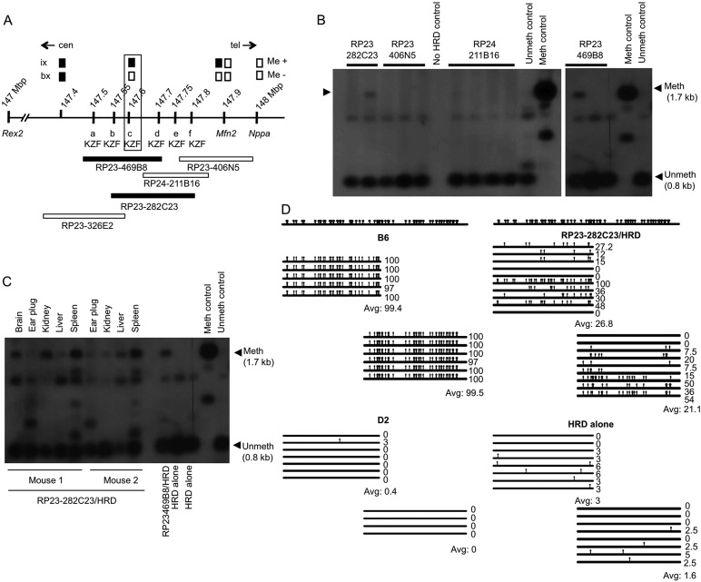
One of the backcross (bx) offspring defines the centromeric border of the Ssm1 candidate region (Fig. 1A) (Engler and Storb, 2000). Its genome is D2 at Nppa (148.0 Mb on chromosome 4; NCBI Annotation Release 103) and D2 at several more centromeric loci until a single nucleotide polymorphism (SNP) at 147.4 Mb. This mouse does not methylate HRD (Me–), thus placing Ssm1 telomeric of 147.4 Mb. All other analyses were consistent with this location (not shown).
Fig. 1.
Mapping and identification of the Ssm1 gene. (A) Map of distal chromosome 4. Me+ and Me– indicate methylation present or absent of HRD in recombinants (ix, intercross; bx, backcross). Boxes indicate genotype: black, B6 genotype; white, D2 genotype. a, b, c, d, e, f are six candidate KZF genes. Position (Mb) on chromosome 4 is indicated (NCBI Annotation, Release 103); at 147.6 Mb Ssm1b is outlined. Beneath are five overlapping BACs. Centromeric/telomeric (cen/tel) orientation is indicated. (B) Southern blot showing HRD (see map in supplementary material Fig. S1) methylation in BAC RP23-282C23 or BAC RP23-469B8 transgenic mice. Meth, methylated; unmeth, unmethylated band. Different lanes for the same BAC represent littermates. (C) Southern blot showing HRD methylation in two mice carrying independent integrations of BAC RP23-282C23. (D) Bisulfite analysis: increased HRD/gpt methylation in tail DNA from mouse 1 of C. HRD alone, littermates without BAC. Each horizontal line represents the sequence from an individual bacterial colony. Vertical bars, meCpGs. The percentage of CpGs methylated is indicated. Above is shown all the CpGs in this sequence. First part of gpt analyzed, 530 nt; second part of gpt analyzed, 540 nt; overlap between the two parts of gpt, 162 nt.
In order to define the telomeric end of the interval containing Ssm1, a 2000 mouse intercross (4000 meioses) was analyzed. The informative recombinant from the intercross (ix) is shown at the top of Fig. 1A. The recombinant chromosome is B6 from 147.4 to 147.9 Mb, but D2 at Mfn2 (147.9) and Nppa (148.0) as well as at distal loci. When crossed with a D2 tester HRD transgenic mouse, offspring inheriting the recombinant chromosome methylated the HRD transgene (Me+), showing that Ssm1 is centromeric of Nppa. Again, all other mapping data were consistent with this assignment (not shown). Thus, Ssm1 resides in a 0.5 Mb interval between 147.4 and 147.9 Mb on mouse chromosome 4 (Fig. 1A).
About a dozen genes are found in this interval, ranging from well characterized to only predicted. Of these, six genes were plausible candidates; all are C2-H2 zinc finger (ZF) genes containing KRAB domains N-terminal of the ZFs (Fig. 1A). One attractive model for Ssm1 action is that the ZFs bind the DNA target and the KRAB domain recruits repressive factors.
To functionally assess the KRAB-ZF (KZF) genes, BACs from a B6 library carrying at least one of the Ssm1b candidates were introduced into Ssm1d fertilized eggs carrying the HRD target. A BAC containing Ssm1b should cause methylation of HRD in the normally non-methylating strain. Five overlapping BACs contain all of the candidate genes (Fig. 1A). Ssm1d C3H/HeJ females were mated with Ssm1d D2 males carrying hemizygous HRD. Batches of (C3H×D2)F1 eggs were injected with one of the five B6 BACs. Three of the BACs had no effect on HRD transgene methylation but mice with either of two overlapping BACs (RP23-469B8 and RP23-282C23) showed a dramatic increase in methylation of HRD (Fig. 1B-D). These BACs share the complete KZF ‘c’ gene (147.6 Mb; boxed). HRD methylation was initially analyzed by Southern blots of tail DNA (Fig. 1B) and in various organs (Fig. 1C) and was further confirmed by bisulfite analysis (Fig. 1D).
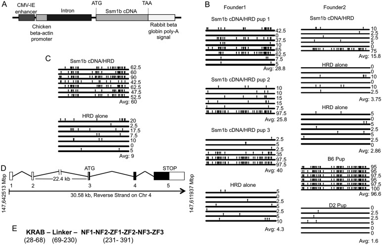
Although it was possible that Ssm1 was an miRNA or other regulatory RNA (see below), we first investigated the candidate KZF gene at 147.6 Mb that caused HRD methylation. A cDNA transgene was made in the vector pCXN2 (Fig. 2A) (Niwa et al., 1991), which is known to be functional in undifferentiated and differentiating ESCs (Alexopoulou et al., 2008). Two 147.6 cDNA transgenic lines (founders 1 and 2) showed methylation of HRD by Southern blots and bisulfite sequencing (Fig. 2B). Since some copies of HRD are unmethylated, we assume that only a subset of cells in the early embryo express the KZF transgenic cDNA; mosaic integration of transgenes has been observed frequently (Wilkie et al., 1986; Chandler et al., 2007). Some transgenic lines did not methylate HRD but we have no evidence to suggest that these lines expressed the 147.6 cDNA transgenes at the appropriate stage of development.
Fig. 2.
HRD gains methylation upon introduction of Ssm1b cDNA into mice. (A) The Ssm1b cDNA construct that was introduced into a non-methylating mouse strain (DBA/C3H). (B) Bisulfite analysis: gain of HRD methylation in newborn pups from two independent founder mice with the Ssm1b cDNA transgene. B6 and D2 pups provide controls for complete and low methylation, respectively. (C) Bisulfite analysis showing gain and spread of methylation in the next generation of pups [(founder 1×DBA)F1] carrying the Ssm1b cDNA transgene. (D) Ssm1b gene structure showing exons (1-5) and introns on chromosome 4. Black box, translated sequence; white box, untranslated sequence. (E) Ssm1b protein domains (amino acid boundaries are numbered). NF1-NF3 are non-functional ZFs, whereas ZF1-ZF3 are functional ZFs.
Further generations from founder 1 that carried the 147.6 cDNA were also analyzed and they showed almost complete HRD methylation (Fig. 2C). Thus, the presence of the 147.6 KZF gene leads to methylation of HRD and we therefore conclude that the KZF gene at position 147.6 Mb is Ssm1. The HRD-methylating Ssm1 gene will henceforth be referred to as Ssm1b.
To confirm that the 147.6 cDNA causes HRD methylation also in ESCs, the same 147.6 cDNA transgene in the pCXN2 vector was furnished with a FLAG tag just before the STOP codon and introduced into D2 ESC lines (supplementary material Fig. S3A). Two independent D2 ESC lines carrying the 147.6-FLAG cDNA transgene showed expression of Ssm1b-FLAG (supplementary material Fig. S3B) and increased HRD methylation (by bisulfite analysis) already in undifferentiated ESCs (supplementary material Fig. S3C), with a further rise in HRD DNA methylation upon differentiation for 7 days after removal of LIF and feeder cells (supplementary material Fig. S3C). Similar to the bisulfite analyses of the Ssm1b transgenic mice (Fig. 2B), the HRD methylation levels varied considerably between sequences of HRD DNA clones (supplementary material Fig. S3C).
Further, we conclude that Ssm1b function is based on the KZF protein; the 147.6 cDNA contains no known miRNA and has no similarity with known miRNAs (miRBase, http://www.mirbase.org).
NCBI designated the Ssm1b gene as 2610305D13Rik and the mRNA as NM_145078. A difference found in the Ssm1b mRNA that we amplified in comparison to the published sequence NM_145078 is the addition of 29 nt at the end of exon 1 (supplementary material Fig. S2). These 29 nt correspond to a sequence in the Ssm1b gene and appear to be due to differential RNA splicing (lack of excision of one short intron). The Ssm1b gene has five exons, with one very large intron (∼22 kb) between exon 2 and exon 3 (Fig. 2D). In addition, there is an initiator motif (TCATTCT) and a downstream promoter element (GGTCA) (supplementary material Fig. S2), which together comprise a core promoter (Burke and Kadonaga, 1996, 1997; Yang et al., 2007). That this is the promoter region for Ssm1b has yet to be verified experimentally. Ssm1b encodes a KZF protein of 407 amino acids with one KRAB-A box (Urrutia, 2003; Vissing et al., 1995), a 218 amino acid linker, and three functional C2-H2 ZFs (Fig. 2E; supplementary material Fig. S2). A variant appears to be expressed in Ssm1d mice (as discussed below). Ssm1b and Ssm1d might be allelic variants (although a contig of distal chromosome 4 does not seem to exist for any of the Ssm1d strains) with related targets and repressive functions (see below).
Expression of Ssm1 in ESCs
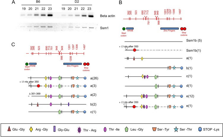
Using primers 18F and 2515R (supplementary material Table S1) a 2.5 kb product was amplified and sequenced from the cDNAs obtained from B6 and D2 ESCs (Fig. 3A). Interestingly, the B6 ESCs expressed not only Ssm1b mRNA, but also several related mRNAs that have SNPs resulting in altered amino acids in the ZF and linker regions, but not in the KRAB domain (Fig. 3B). Since none of the other mRNAs maps to the Ssm1 candidate region in distal chromosome 4 (Fig. 1A), they are not involved in the methylation of HRD. One of these other mRNAs has identical ZFs to Ssm1b (c, Fig. 3B) but has changes in the linker. Thus, the linker might be involved in the target specificity as well.
Fig. 3.
Expression of Ssm1 in B6 and D2 ESCs. (A) RT-PCR of beta actin and Ssm1 in B6 and D2 undifferentiated ESCs. The number of PCR cycles is indicated. (B) Ssm1 mRNAs in B6 ESCs. Each horizontal line represents the mRNA sequence of each member of the Ssm1 family amplified. Numbers in parentheses indicate the number of bacterial clones for each sequence. The region shown is coding sequence of Ssm1 from nt 276 to nt 1499 (supplementary material Fig. S2). Lines labeled Ssm1b indicate sequences matching the genomic sequence of Ssm1b and hence considered the Ssm1b cDNA. Lines marked a, b, c, d and e represent the other Ssm1 family sequences. Alternative splice forms of the same sequence with either addition (+13 or +39) or deletion (Δ) of nucleotides are labeled with the same letter as the original sequence. These changes lie within the translated RNA. In some splice forms the addition of 13 nt leads to a STOP codon 153 nt further 3′. Vertical marks indicate silent SNPs differing from the Ssm1b sequence. SNPs causing amino acid changes are marked with a symbol with the actual amino acid changes depicted beneath. Red top line displays the Ssm1 region with vertical marks for all SNPs with nucleotide positions indicated. The positions of the KRAB (nt 357-477, supplementary material Fig. S2) and ZF (nt 966-1448) domains and the start (nt 276-278) and STOP (nt 1497-1499) codons are also depicted. (C) Expression of Ssm1 in D2 ESCs. There is no expression of Ssm1b in the D2 ESCs.
Another surprising observation is that none of the Ssm1b family related genes was identified in the mouse genome using the BLAST tool from NCBI (http://blast.ncbi.nlm.nih.gov/Blast.cgi). The chromosome that contains the Ssm1b gene still has unsequenced gaps and it is possible that some or all of the Ssm1-like genes reside in these gaps.
Several Ssm1 family mRNAs are also expressed in D2 ESCs (Fig. 3C). D2 ESCs do not, however, express Ssm1b mRNA. Ssm1b mRNA and all of the Ssm1 family mRNAs share unusual ZFs: two C-C/H-H fingers (ZF1, ZF2) are followed by an inactive Y-C/H-H and a third functional C-C/H-H finger (ZF3) (Fig. 2E; supplementary material Fig. S2). The other five KZF genes mapping near Ssm1b on distal chromosome 4 (Fig. 1A) do not have the unusual ZFs; all contain larger numbers of functional ZFs. The levels of Ssm1b and Ssm1 family mRNAs are one-third the levels in ESCs after 4 days differentiation of ESCs, and halved again by 7 days (supplementary material Fig. S4A).
Clearly, the Ssm1b sequence is not seen in Ssm1d ESCs among 34 sequenced Ssm1 cDNAs, whereas in B6 six of the 23 sequences are Ssm1b (Fig. 3B,C). Thus, Ssm1d cells either do not express Ssm1b or, if they do, then Ssm1b mRNA would be, at the most, one-ninth the level in Ssm1b. However, transfected Ssm1b in Ssm1d ESCs causes higher methylation of HRD DNA (supplementary material Fig. S3). These findings suggest that it is indeed the protein sequence of Ssm1b that causes the methylation, rather than higher expression of Ssm1b.
Expression of Ssm1 in early mouse development
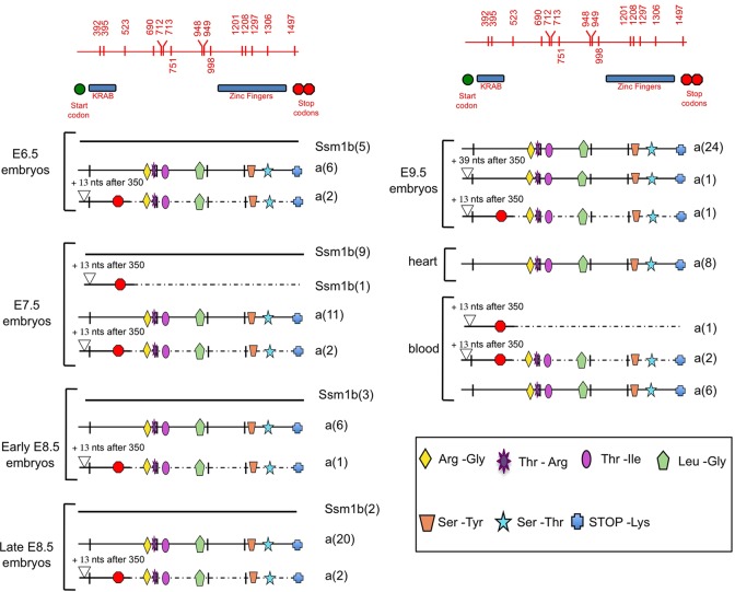
Ssm1b is expressed at the blastocyst stage as shown above (ESCs are derived from the inner cell mass of blastocysts). To analyze the expression of Ssm1b at further stages of mouse embryonic development, mRNA was collected from E6.5, E7.5, E8.5 and E9.5 B6 embryos, and Ssm1b cDNA was amplified using primers 18F and 2515R (supplementary material Table S1). Ssm1b mRNA was expressed in E6.5, E7.5 and E8.5 (early and late) embryos in addition to the other Ssm1-like mRNAs. But in the E9.5 embryos only other Ssm1 family mRNAs were found (Fig. 4). Presumably, the Ssm1b protein binds to its genomic targets before E9.5.
Fig. 4.
Expression of Ssm1 in early mouse development. B6 females were crossed to B6 males and embryos from various stages of development were analyzed for the expression of Ssm1b (see Fig. 3B legend for details). Ssm1b was expressed up to late E8.5 together with the other Ssm1 family sequences. At E9.5 and in heart and blood, Ssm1b expression ceased while the other Ssm1 family sequences continued to be expressed.
Ssm1 expression was also analyzed in various organs in an adult B6 mouse (Fig. 4). Certain organs, such as the heart, showed high expression levels of Ssm1 family mRNA but none (except for the ovaries, not shown) expressed Ssm1b. Thus, Ssm1b expression seems to be tightly regulated and only active up to E8.5.
Analysis of HRD methylation during development
HRD methylation in extra-embryonic tissue (EET) had been analyzed by Southern blot of E6.5 to E12.5 post-implantation embryos (Weng et al., 1995). In contrast to the embryo proper, there was no differential methylation between D2 and (B6×D2)F1 EET (supplementary material Table S2); instead, both showed intermediate bands suggesting that Ssm1b might not be expressed in EET.
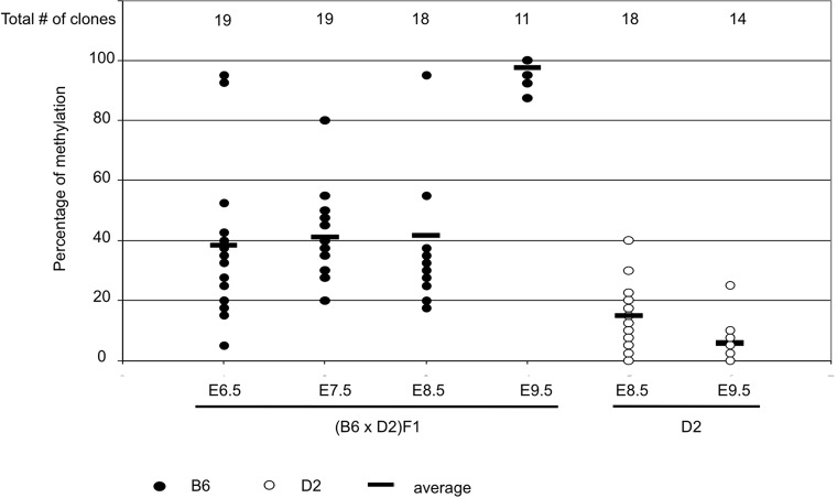
In the current study, embryos at different stages of development (E6.5-9.5) from (B6×D2)F1 crosses were analyzed for HRD (gpt) methylation. At E6.5, HRD was not completely methylated but was significantly more methylated than in later stage D2 embryos (Fig. 5; supplementary material Fig. S5). In E7.5 and E8.5 (B6×D2)F1 embryos, HRD methylation was about the same on average, although at E8.5 two sequences were almost completely methylated (supplementary material Fig. S5). In contrast to the low level of methylation in D2 at E9.5, B6 showed essentially complete methylation of HRD by E9.5 (P<0.0002). This steep increase of the spreading of methylation between E8.5 and E9.5 is unlikely to be due to Ssm1b protein since its mRNA is not seen at E9.5 (Fig. 4). Instead, it might be related to the switch from Dnmt3b to Dnmt3a that occurs around this stage (Watanabe et al., 2002). This would mean that spreading of DNA methylation requires de novo methyltransferase activity.
Fig. 5.
HRD-gpt methylation during early mouse development. Gain in HRD-gpt methylation (bisulfite analysis) from E6.5 to E9.5 (B6×D2)F1 mouse embryos (see supplementary material Fig. S4). Circles represent percentages of CpGs methylated per gpt sequence from individual bacterial colonies. E8.5 and E9.5 embryos from D2 mice are shown as controls. To compare two sets of methylated sequences, a Mann-Whitney two-sample test (a non-parametric rank test) was performed on the percentages of methylated sites per clone (Mann and Whitney, 1947).
Association of Ssm1b with Dnmt3b
Because the earliest sign of HRD inactivation in Ssm1b mice is the increased DNA methylation in (B6×D2)F1 ESCs compared with D2 ESCs (Weng et al., 1995; Padjen et al., 2005), Ssm1b might operate by de novo DNA methylation. The Ssm1b gene product lacks a methyltransferase domain and hence it is likely that it works in concert with one of the de novo methyltransferases, Dnmt3a and/or Dnmt3b (Okano et al., 1999). To determine the role of Dnmt3b in Ssm1b function, the HRD transgene was crossed into a Dnmt3b knockout background. Multiple crosses were required to obtain E10.5 embryos that have a homozygous deletion of Dnmt3b, possess one Ssm1b allele and contain the HRD transgene that was unmethylated in the Ssm1d parent (supplementary material Fig. S6).
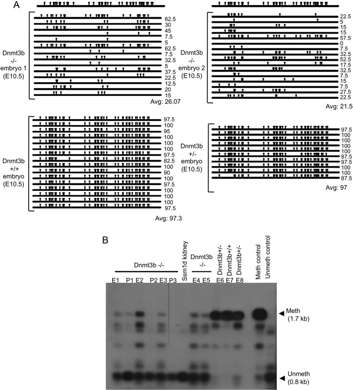
In the Dnmt3b−/− embryos in an Ssm1b background and their placentas, the HRD transgene was mostly unmethylated (by Southern blot analysis), whereas in littermates that were either Dnmt3b+/+ or Dnmt3b+/− the HRD transgene was completely methylated (Fig. 6B). This result was confirmed by bisulfite analysis, where all the clones from the Dnmt3b−/− embryos show greatly decreased levels of methylation (Fig. 6A), suggesting that Ssm1b activity is linked to de novo DNA methylation. The few HRD transgene copies with higher methylation might have been caused by Dnmt3a activity (Borgel et al., 2010). Thus, HRD methylation in Ssm1b mice depends mainly on de novo DNA methylation by Dnmt3b. Dnmt3b mutations are frequent in the human ICF syndrome (Owen and Bowie, 1972). Since 30-40% of patients have no Dnmt3b mutations, a co-factor such as Ssm1b might be mutated.
Fig. 6.
Role of Dnmt3b in HRD methylation. (A) Bisulfite analysis (see Fig. 1 legend). (B) Southern blot (see Fig. 1 legend). There is low HRD methylation in B6 E10.5 embryos lacking Dnmt3b compared with Dnmt3b+ littermates. E1-E8: E10.5 embryos; P1-P3: E10.5 placentas.
Role of the methylated DNA-binding protein Mecp2 in Ssm1b function
Since it is possible that Ssm1 plays a role in targeting of the methyl-CpG (meCpG) DNA-binding domain (MBD) protein Mecp2 to HRD (gpt), HRD was crossed into either of two types of Mecp2-deficient mice, one with a truncation of Mecp2 within exon 4 (Shahbazian et al., 2002) and the other with complete inactivation of Mecp2 (Guy et al., 2001). On the Ssm1b background, the mice lacking Mecp2 activity still methylate HRD (supplementary material Fig. S8). This suggests that the readout of the Ssm1b-associated DNA methylation does not require Mecp2. It has not been determined whether Ssm1 interacts with other MBD proteins.
HRD methylation and Ssm1b expression in wild-derived mice
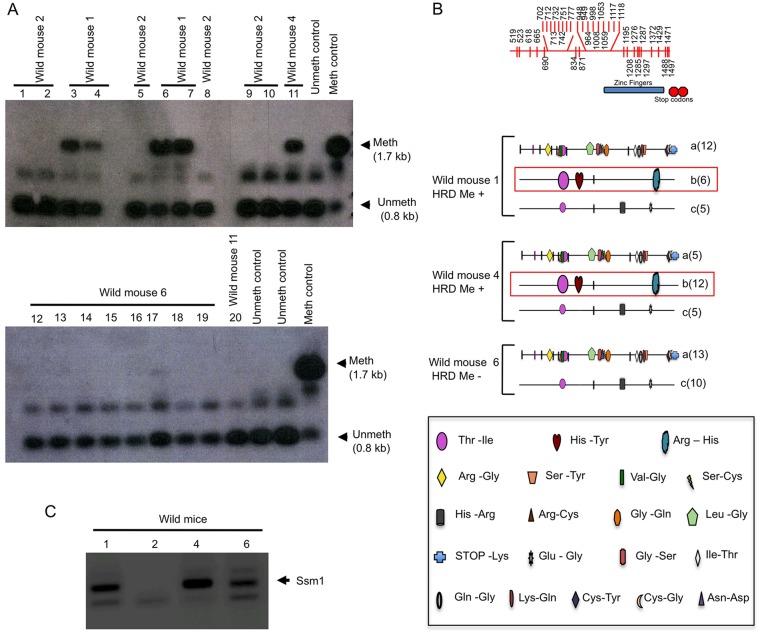
To determine whether Ssm1b might be an artifact of laboratory mice, we analyzed HRD methylation in mice recently descended from wild ancestors. D2 mice with an unmethylated HRD transgene were crossed with outbred wild-derived mice (Miller et al., 2002) and methylation of HRD was determined in the offspring. We found that they are a mix of HRD-methylating and non-methylating mice (Fig. 7A). Offspring of some wild mice showed complete methylation of HRD, whereas others showed no HRD methylation. Thus, polymorphisms for the methylation patterns seen among laboratory inbred strains are also found in a free-living population.
Fig. 7.
An Ssm1b-like gene in HRD/gpt-methylating wild-derived mice. (A) Southern blot of liver DNA from offspring of wild female mice mated to D2-HRD males. Methylation of HRD is shown by offspring 3, 4, 6 and 7 from wild mouse 1 and offspring 11 from wild mouse 4; no methylation of HRD is apparent in offspring 1, 2, 5, 9 and 10 from wild mouse 2, offspring 12-19 from wild mouse 6, and offspring 20 from wild mouse 11. (B) Tail DNA from wild-derived mothers was analyzed for the presence of part of the Ssm1 gene by PCR amplification with primers 495F and 2515R. The red box indicates sequence very similar to Ssm1b that is seen in females with HRD-methylating offspring (HRD Me+) but is absent in the female whose offspring did not methylate HRD (HRD Me–). (C) PCR amplification of Ssm1 from tail DNA of wild-derived mice 1, 2, 4 and 6 using primers 495F and 2515R. Various primers complementary to this region were tested but were unsuccessful in amplifying Ssm1 in wild-derived mouse 2.
To determine whether there were any Ssm1b or Ssm1-like genes in wild mice we obtained tail DNA from the mothers of both the HRD-methylating and non-methylating wild mice and amplified the Ssm1 region using primers 495F and 2515R (supplementary material Table S1), which amplify the last exon of the Ssm1 gene family. HRD-methylating wild mice #1 and #4 have a gene that is almost identical to Ssm1b (Fig. 7B, ‘b’), but with one amino acid change (R→H) at a non-conserved position in the last functional ZF (Thomas and Schneider, 2011) and T→I and H→Y in the linker between the KRAB and ZF domains (Fig. 7B). Non-methylating mouse #6 does not have an identical gene to Ssm1b but a similar one that is also present in the methylating mice. The R→H change in the ZF of Me+ mice #1 and #4 might interfere with zinc coordination by the His-His domain; therefore, it is uncertain whether this gene product is responsible for HRD methylation. This uncertainty is supported by the vastly different ratios of ‘b’ to ‘a+c’ between the two Me+ mice. These ratios are low in B6 mRNA (Fig. 3B) and genomic DNA (not shown). Possibly, the primers that amplify the Ssm1 family genes in laboratory mice do not match the related gene in wild mice. In fact, for Me– wild mouse #2 the 495F and 2515R primers did not amplify any Ssm1-like gene, nor did any other B6 primer pairs in the Ssm1b gene (Fig. 7C). These primers were originally designed to amplify the linker, the ZFs and the 3′UTR of the Ssm1b gene (supplementary material Fig. S2). The forward primers, including 495F, are located in the linker; thus, even silent SNPs might have been incompatible. Therefore, whether the gene responsible for HRD methylation in wild mice is Ssm1b remains to be ascertained.
HRD is repressed when it is transfected into established B6 ESCs and the repression spreads to adjacent sequences
ESCs derived from HRD transgenic (B6×D2)F1 mice show partial HRD methylation that is greater than the methylation level in D2 ESCs (Padjen et al., 2005). This might indicate that Ssm1b begins to function at the blastocyst stage or that Ssm1b needs to interact with its target at an earlier pre-blastocyst stage. To distinguish between these possibilities an HRD-neo transgene (supplementary material Fig. S7C) was transfected into established B6 ESCs.
Similar to the findings with ESCs derived from HRD transgenic (B6×D2)F1 mice, the gpt region of the transfected HRD transgene shows partial methylation in the undifferentiated ESCs, i.e. ESCs grown on fetal fibroblasts and LIF, but gains complete methylation upon differentiation, as seen in all three clones with independent transfection of HRD-neo (supplementary material Fig. S7B). Thus, the presence of Ssm1b at the ESC stage is sufficient for the later methylation of gpt.
The neo part of the transfected HRD-neo transgene is unmethylated in the undifferentiated B6 ESCs but becomes methylated upon differentiation (supplementary material Fig. S7A). By contrast, the control transgene neo alone (without gpt) stayed unmethylated even after differentiation (supplementary material Fig. S7A). One explanation for the unexpected partial methylation of the neo gene in the B6-neo-10 cell line at D28 (supplementary material Fig. S7A) could be its integration into a methylated region of the B6 genome. Thus, the complete methylation of HRD and its spreading to adjacent DNA sequences occurs during ESC differentiation and does not require the presence of the HRD target before the blastocyst/ESC stage, suggesting that Ssm1b begins to act in undifferentiated and early differentiating ESCs. Also, this analysis shows that HRD (and presumably its gpt portion) and not neo (which has a similarly high CpG/CpX ratio to gpt: 9.4% in neo versus 8.4% in gpt) is a specific target for DNA methylation by Ssm1b and it is the spreading of methylation from HRD to the adjacent neo gene that leads to the complete methylation of neo.
DISCUSSION
The Ssm1b gene is located on mouse distal chromosome 4 and is a member of a novel family of KZF genes. As our study shows, Ssm1b is responsible for the specific methylation of the HRD transgene in mice. The Ssm1b gene by itself does not encode a methyltransferase, since it lacks the conserved ‘transferase’ sequence present in all DNA methyltransferases from bacteria to man (Bestor, 2000), but Ssm1b causes HRD methylation via the de novo methyltransferase Dnmt3b. Since Dnmt3b does not have any sequence specificity beyond CpG dinucleotides (Okano et al., 1998; Yoder et al., 1997), the study of HRD methylation by Ssm1 might provide insights into the mechanisms that determine how specific loci are targeted for methylation and heterochromatin formation early in development.
Ssm1b belongs to a family of related KZFs genes with only three functional C2-H2 ZFs interspersed with three non-functional ones (Fig. 2E; supplementary material Fig. S2). We broadly label these unusual KZF genes as the Ssm1 family. The Ssm1 family is thus unique in terms of the organization of its ZFs. It has two non-functional ZFs, followed by two functional ones, a non-functional ZF, and finally a third functional ZF (Fig. 2E). In addition, the Ssm1 genes possess a fairly long and unique linker region between the KRAB and ZF domains. The role of the linker in KZF proteins is not known but it has been found that the location of KZFs in the nucleus varies depending on the linker sequence (Fleischer et al., 2006). Thus, the unique nature of the ZFs and linker is likely to be responsible for the targeting of Ssm1b protein to specific regions of the genome.
Polymorphisms in Ssm1 genes are not an artifact of domestication as we also observed them in wild-derived mice, leading to the conclusion that this is indeed a conserved, and presumably important, gene family. This raises the question of what maintains such polymorphisms in nature. A possible answer is that certain environmental conditions favor one set of genes and that other conditions favor the alternative set. What such hypothetical conditions might be is of course unknown. However, mice, as the most widespread mammalian species on earth other than humans (Berry and Bronson, 1992), are ideally suited to address this question, but trying to answer it would require a broad census of multiple wild populations.
With regards to the expression and function of Ssm1 family genes during development, the Ssm1b protein seems to be functional in undifferentiated ESCs. There is already more methylation of HRD/gpt in undifferentiated B6 ESCs than in undifferentiated D2 ESCs (Padjen et al., 2005). When HRD is transfected into established B6 ESCs (supplementary material Fig. S7B) gpt becomes methylated to the same percentage as in undifferentiated ESCs from HRD transgenic B6 mice (Padjen et al., 2005), suggesting that, indeed, Ssm1b most likely acts in blastocysts, if not earlier in development.
The early expression of Ssm1b in blastocysts is supported by a transcriptome analysis comparing ESCs grown in fetal calf serum (as our ESCs) and cells grown without serum in 2i medium (Marks et al., 2012). The latter are more undifferentiated and presumably more like cells of the inner cell mass. One of the genes expressed in the Marks et al. study was Ssm1b (2610305D13Rik) on chromosome 4. Three ESC lines grown in 2i medium had an average rpkm (reads per kb transcript per million mapped sequence reads) of 194.1, whereas the ESC lines grown in serum medium had an average rpkm of only 61.3. Thus, if these numbers are accepted as linear relationships, Ssm1b expression increases 3.2-fold in ESCs grown in 2i medium compared with the cells grown in serum.
If cells grown in 2i medium are indeed more undifferentiated, the finding that Ssm1b expression is higher in these cells is in line with our observation that Ssm1b expression is reduced upon differentiation: Ssm1b expression drops by almost half upon differentiating the ESCs for 4 days (supplementary material Fig. S4B), as compared with ‘undifferentiated’ ESCs grown with LIF on feeder cells (Fig. 3B). Thus, our data and the data from Marks et al. confirm that Ssm1b is expressed in the inner cell mass cells during mouse development. Identifying a possibly earlier stage of Ssm1b expression and function during pre-implantation embryonic development is an important goal that we are addressing in a separate study.
At the undifferentiated stage, both Ssm1b and Ssm1d ESCs show a biphasic pattern of active and inactive chromatin (Padjen et al., 2005). This agrees with recent reports that multiple genes in ESCs (Giadrossi et al., 2007; Bernstein et al., 2007), as well as stem cells in general (Azuara et al., 2006), have a mixed chromatin pattern. These findings were suggested to indicate that genes in stem cells are poised for a choice of either activation or inactivation. Thus, HRD behaves like an endogenous gene. However, we cannot rule out the possibility that the ‘biphasic’ pattern might be the result of heterogeneity in the cell population. An unusual observation about the Ssm1 system is that, once differentiation is fully underway, different mouse strains treat HRD chromatin in opposite ways. Divergence between the strains appears to be initiated ∼7 days after differentiation of ESCs in culture: at this stage, the only sign of the initiation of HRD inactivation in Ssm1b cells is a significant increase in Mecp2 binding to the gpt core of HRD and a slight reduction of HRD mRNA (Padjen et al., 2005). In the corresponding Ssm1b E7.5 embryos, Ssm1b is still expressed, but expression ceases after E8.5 and HRD has become completely methylated at E9.5. Therefore, Ssm1b must have initiated methylation at a stage earlier than E9.5, and this methylation then spread across the transgene during further development.
The active demethylation of meCpGs has recently been attributed to the TET proteins that convert 5-methylcytosine (5mC) to 5-hydroxymethylcytosine (5hmC), and further to 5-formylcytosine (5fC) and 5-carboxycytosine (5caC), leading to replacement of meCpG by cytosine and thus DNA demethylation (reviewed by Bhutani et al., 2011). It appears that tissue-specific genes become actively demethylated in preparation for their activation. By contrast, the 5meC form of HRD/gpt induced during early embryonic development through Ssm1b does not undergo these dynamic changes; instead, HRD DNA remains methylated in fetal and adult life. This finding lends support to our preliminary ChIP-Seq analysis of endogenous Ssm1b target genes (not shown), which has revealed repetitive DNA sequences, such as SINEs, as the potential targets of Ssm1b. If unique tissue-specific genes were the targets, in an adult tissue such as liver in which over 50% of the cells are hepatocytes that express the metallothionein (MT) gene, which is, like the HRD gene, driven by the MT promoter, a significant proportion of the gpt sequences should be unmethylated. Although the bisulfite assays that we performed would not distinguish 5hmC from 5mC, it is unlikely that HRD would remain 100% methylated if it underwent the first step of demethylation. In fact, in mice that were treated with cadmium to induce MT expression, the HRD transgene was only expressed in Ssm1d mice, in which its DNA was not methylated (Weng et al., 1995).
So far, preliminary ChIP-Seq data (unpublished observations) seem to indicate that the endogenous targets of Ssm1b are repetitive DNA sequences. The repression of another group of repetitive sequences, intracisternal A particles (IAPs), was found to be due to a different KZF gene (Rowe et al., 2010). We propose that Ssm1 proteins are required for the repression early in development of repetitive DNA sequences, the expression of which would swamp the function of unique genes (Li et al., 1992). Many repetitive elements are suppressed by DNA methylation (Walsh et al., 1998). It is thus likely that the multiple variant mRNAs of the Ssm1 family that we have found (and perhaps extended family members that were not RT-PCR amplified using primers specific for Ssm1b) are specific for different, but related, repetitive sequences. The Ssm1 family proteins have many amino acid polymorphisms in the linker and the ZF region that could be responsible for differential target specificities. For instance, it appears that gpt is a target only for Ssm1b. Presumably, the Ssm1b protein binds to a specific motif in gpt via its functional ZFs, whereas other Ssm1 family proteins do not bind gpt owing to amino acid changes in their ZFs that prevent their recognition of gpt as a target. Although gpt is not a target of Ssm1-like proteins, presumably various interrelated repetitive sequences will be targets. The Ssm1 gene family might be just one of several families of multi-loci genes that are responsible for the essential repression of different repetitive genomic elements. One reason why a repressor such as Ssm1 was not discovered earlier might be that the target recognition sequences are short and, because of the redundancy of the system, were not found. We do not know whether, in analogy with Zfp57 (Takikawa et al., 2013), Ssm1b targets imprinted genes.
The finding of gpt as a discrete target for Ssm1b, together with the finding that neo with a similarly high density of CpGs is not a target, makes it unlikely that Ssm1 recognizes all foreign, potentially parasitic DNA sequences. There are no obvious repeats in gpt as potential methylation targets (Reinhart et al., 2002). Thus, it is possible that Ssm1b recognizes a specific sequence within gpt that it binds to and initiates methylation, and endogenous targets might share some common features with gpt.
All Ssm1-like genes that we have found encode the identical KRAB protein domain. KRAB is a repressive domain that interacts with KRAB-associated protein (KAP1; also known as Trim28). Ssm1b may bind to its targets and recruit methyltransferases either directly or indirectly by interaction through the KRAB domain with proteins that are, or bind to, de novo methyltransferases. Spreading of methylation beyond the target sequences might be due to propagation of chromatin structure (Groner et al., 2010). Our previous experiments showed that DNA methylation of HRD precedes certain DNA chromatin modifications (Padjen et al., 2005). This raises the question of whether Ssm1b protein induces DNA methylation by interacting directly with Dnmt3b and/or other de novo DNA methyltransferases and steering them to potential endogenous targets, or whether it interacts with MBD proteins or other suppressive factors to direct them to partly methylated or unmethylated DNA. As we have shown, Ssm1-associated DNA methylation does not require Mecp2, although it is still possible that Ssm1b interacts with other MBD proteins. It is also possible that Ssm1b recruits a chromatin-modifying factor that we have not yet examined.
Thus, Ssm1b is a novel gene that points to a new family of KZF genes initiating specific DNA methylation and chromatin modification. The Ssm1 family is likely to be involved in the repression of repetitive DNA sequences in the epigenetic control of early development, and discovering the details of its function provides an exciting challenge.
MATERIALS AND METHODS
Transgenic mice
Experiments with mice were performed in compliance with USDA animal welfare and PHS humane animal care guidelines.
Backcrosses and intercrosses to map Ssm1
The 500 mouse backcross was described by Engler and Storb (Engler and Storb, 2000). For intercrosses, (D2×B6)F1 mice were mated with each other and 2000 progeny were analyzed for HRD methylation.
ESCs
ESCs from HRD transgenic mice of both (B6×D2)F1 and D2 strains were isolated by A. Weng (Weng et al., 1995). B6 ESCs were obtained from the University of Chicago Transgenic Mouse Facility, D2 ESCs from Teruhiko Wakayama (Wakayama et al., 2001) and mouse embryonic fibroblasts (DR4) from Stanford Transgenic Research Center. The HRD-neo construct consists of the EcoRI-HindIII HRD transgene (Engler and Storb, 1987) linked to pko-neo (Sambrook et al., 1989). The neo gene alone was obtained from the pMC1 Neo-PolyA vector (Agilent Technologies).
BACs and cDNA transgenes
Bacterial artificial chromosomes (BACs) were obtained from Children's Hospital Oakland Research Institute (CHORI). Ssm1b cDNA transgenes were prepared from Ssm1b mRNA from undifferentiated B6 ESCs (Fig. 3) and cloned into the cDNA expression vector pCXN2 (Niwa et al., 1991). All BAC and Ssm1b cDNA transgenic mice were made at the University of Chicago Transgenic Mouse Facility. For ESCs, the FLAG tag was introduced immediately before the STOP codon of the Ssm1b cDNA (supplementary material Fig. S3A), cloned into pCXN2 and transfected into the D2 ESCs (Tompers and Labosky, 2004).
RT-PCR for Ssm1 transcripts
RNA was isolated using RNA STAT60 (CS-110, AMSBIO) or the RNAqueous-4PCR kit (AM1914, Ambion). cDNA was made using the Superscript III first-strand synthesis system (18080-051, Invitrogen). Ssm1b cDNA was amplified using primers 18F and 2515R (supplementary material Table S1), gel-purified using the GeneJet gel extraction kit (K0691, Fermentas) and cloned into a PCR cloning vector (240205, Stratagene). Plasmid DNA was isolated using the GeneJet plasmid mini prep kit (K0502, Fermentas).
PCR for analyzing the Ssm1 gene in wild mice
DNA was isolated from tails of outbred descendants of wild mice trapped in Idaho (Miller et al., 2002). The Ssm1 region was amplified using primers 495F and 2515R (supplementary material Table S1). Female wild mice were mated with HRD transgenic male D2 mice. Liver DNA of the offspring was analyzed for HRD methylation by Southern blot (Padjen et al., 2005).
Bisulfite sequencing
Bisulfite sequencing of gpt and neo was carried out as described previously (Padjen et al., 2005).
Dnmt3b-deficient mice
Dnmt3b+/− mice on the B6 background were obtained from Novartis (Ueda et al., 2006). The breeding scheme is detailed in supplementary material Fig. S6.
Mecp2-deficient mice
Two lines of Mecp2-deficient mice were obtained from Jackson Laboratory: B6.129P2(C)-Mecp2tm1.Bird/J (Guy et al., 2001) and B6.129S-Mecp2itm1Hzo/J (Shahbazian et al., 2002). HRD transgenic male offspring lacking Mecp2 provided the necessary experimental combination (Ssm1b/d, HRD, Mecp2−/−).
Supplementary Material
Acknowledgements
We thank L. Degenstein of the University of Chicago Transgenic Mouse Facility for production of transgenic mice and valuable discussions, D. Nicolae for statistical analysis, I. Swanson for Ssm1 PCR assays of wild mice, L. Godley for Dnmt3b primer design, R. Chaillet and L. Doglio for thoughtful comments, W. Buikema of the University of Chicago DNA Sequencing Facility for DNA sequencing, T. Wakayama for D2 ESCs and J. Miyazaki for the pCXN2 vector.
Footnotes
Competing interests
The authors declare no competing financial interests.
Author contributions
S.R. supervised the production and analysis of the BAC and Ssm1b cDNA transgenic mice and the Dnmt3b−/− embryos, co-designed with U.S. and carried out most of the experiments reported in this paper, and wrote the paper together with U.S. P.E. designed and produced the HRD transgene, made HRD transgenic mice and discovered the strain-specific methylation, designed and carried out the mapping of Ssm1b, created variants of HRD and discovered that gpt is the Ssm1b target. G.B. performed all the mouse breedings and assisted in all the molecular biology experiments. L.M. carried out the experiments in supplementary material Fig. S4A, checked the BAC sequences conferring the Ssm1 effect for the presence of miRNAs, and cloned and sequenced Ssm1 cDNA that was then used to make transgenic mice. A.P. crossed HRD transgenic DBA males with female wild-derived mice. S.A. established the colony of wild-derived mice and kept them outbred. T.M. provided ideas and discussion throughout this study and edited the paper. U.S. directed the study and wrote the paper together with S.R.
Funding
The studies were supported by National Institutes of Health (NIH) grants [RO3HD05827 and AI047380] and a Pilot Project grant from the University of Chicago Cancer Research Center to U.S.; support for the S.A. laboratory was by an NIH Nathan Shock Center grant [P30 AG13319]. S.R. was supported by a postdoctoral fellowship from the Cancer Research Institute. Deposited in PMC for release after 12 months.
Supplementary material
Supplementary material available online at http://dev.biologists.org/lookup/suppl/doi:10.1242/dev.105726/-/DC1
References
- Alexopoulou A. N., Couchman J. R., Whiteford J. R. (2008). The CMV early enhancer/chicken beta actin (CAG) promoter can be used to drive transgene expression during the differentiation of murine embryonic stem cells into vascular progenitors. BMC Cell Biol. 9, 2 10.1186/1471-2121-9-2 [DOI] [PMC free article] [PubMed] [Google Scholar]
- Azuara V., Perry P., Sauer S., Spivakov M., Jørgensen H. F., John R. M., Gouti M., Casanova M., Warnes G., Merkenschlager M., et al. (2006). Chromatin signatures of pluripotent cell lines. Nat. Cell Biol. 8, 532-538 10.1038/ncb1403 [DOI] [PubMed] [Google Scholar]
- Bernstein B. E., Meissner A., Lander E. S. (2007). The mammalian epigenome. Cell 128, 669-681 10.1016/j.cell.2007.01.033 [DOI] [PubMed] [Google Scholar]
- Berry R. J., Bronson F. H. (1992). Life history and bioeconomy of the house mouse. Biol. Rev. Camb. Philos. Soc. 67, 519-550 10.1111/j.1469-185X.1992.tb01192.x [DOI] [PubMed] [Google Scholar]
- Bestor T. H. (2000). The DNA methyltransferases of mammals. Hum. Mol. Genet. 9, 2395-2402 10.1093/hmg/9.16.2395 [DOI] [PubMed] [Google Scholar]
- Bhutani N., Burns D. M., Blau H. M. (2011). DNA demethylation dynamics. Cell 146, 866-872 10.1016/j.cell.2011.08.042 [DOI] [PMC free article] [PubMed] [Google Scholar]
- Borgel J., Guibert S., Li Y., Chiba H., Schübeler D., Sasaki H., Forné T., Weber M. (2010). Targets and dynamics of promoter DNA methylation during early mouse development. Nat. Genet. 42, 1093-1100 10.1038/ng.708 [DOI] [PubMed] [Google Scholar]
- Burke T. W., Kadonaga J. T. (1996). Drosophila TFIID binds to a conserved downstream basal promoter element that is present in many TATA-box-deficient promoters. Genes Dev. 10, 711-724 10.1101/gad.10.6.711 [DOI] [PubMed] [Google Scholar]
- Burke T. W., Kadonaga J. T. (1997). The downstream core promoter element, DPE, is conserved from Drosophila to humans and is recognized by TAFII60 of Drosophila. Genes Dev. 11, 3020-3031 10.1101/gad.11.22.3020 [DOI] [PMC free article] [PubMed] [Google Scholar]
- Chandler K. J., Chandler R. L., Broeckelmann E. M., Hou Y., Southard-Smith E. M., Mortlock D. P. (2007). Relevance of BAC transgene copy number in mice: transgene copy number variation across multiple transgenic lines and correlations with transgene integrity and expression. Mamm. Genome 18, 693-708 10.1007/s00335-007-9056-y [DOI] [PMC free article] [PubMed] [Google Scholar]
- Engler P., Storb U. (1987). High-frequency deletional rearrangement of immunoglobulin kappa gene segments introduced into a pre-B-cell line. Proc. Natl. Acad. Sci. U.S.A. 84, 4949-4953 10.1073/pnas.84.14.4949 [DOI] [PMC free article] [PubMed] [Google Scholar]
- Engler P., Storb U. (1999). Hypomethylation is necessary but not sufficient for V(D)J recombination within a transgenic substrate. Mol. Immunol. 36, 1169-1173 10.1016/S0161-5890(99)00124-8 [DOI] [PubMed] [Google Scholar]
- Engler P., Storb U. (2000). A linkage map of distal mouse chromosome 4 in the vicinity of Ssm1, a strain-specific modifier of methylation. Mamm. Genome 11, 694-695 10.1007/s003350010124 [DOI] [PubMed] [Google Scholar]
- Engler P., Haasch D., Pinkert C. A., Doglio L., Glymour M., Brinster R., Storb U. (1991). A strain-specific modifier on mouse chromosome 4 controls the methylation of independent transgene loci. Cell 65, 939-947 10.1016/0092-8674(91)90546-B [DOI] [PubMed] [Google Scholar]
- Engler P., Doglio L. T., Bozek G., Storb U. (1998). A cis-acting element that directs the activity of the murine methylation modifier locus Ssm1. Proc. Natl. Acad. Sci. U.S.A. 95, 10763-10768 10.1073/pnas.95.18.10763 [DOI] [PMC free article] [PubMed] [Google Scholar]
- Fleischer S., Wiemann S., Will H., Hofmann T. G. (2006). PML-associated repressor of transcription (PAROT), a novel KRAB-zinc finger repressor, is regulated through association with PML nuclear bodies. Exp. Cell Res. 312, 901-912 10.1016/j.yexcr.2005.12.005 [DOI] [PubMed] [Google Scholar]
- Giadrossi S., Dvorkina M., Fisher A. G. (2007). Chromatin organization and differentiation in embryonic stem cell models. Curr. Opin. Genet. Dev. 17, 132-138 10.1016/j.gde.2007.02.012 [DOI] [PubMed] [Google Scholar]
- Groner A. C., Meylan S., Ciuffi A., Zangger N., Ambrosini G., Dénervaud N., Bucher P., Trono D. (2010). KRAB-zinc finger proteins and KAP1 can mediate long-range transcriptional repression through heterochromatin spreading. PLoS Genet. 6, e1000869 10.1371/journal.pgen.1000869 [DOI] [PMC free article] [PubMed] [Google Scholar]
- Guy J., Hendrich B., Holmes M., Martin J. E., Bird A. (2001). A mouse Mecp2-null mutation causes neurological symptoms that mimic Rett syndrome. Nat. Genet. 27, 322-326 10.1038/85899 [DOI] [PubMed] [Google Scholar]
- Li E., Bestor T. H., Jaenisch R. (1992). Targeted mutation of the DNA methyltransferase gene results in embryonic lethality. Cell 69, 915-926 10.1016/0092-8674(92)90611-F [DOI] [PubMed] [Google Scholar]
- Mann H. B., Whitney D. R. (1947). On a test of whether one of two random variables is stochastically larger than the other. Ann. Math. Statist. 18, 1-164 10.1214/aoms/1177730491 [DOI] [Google Scholar]
- Marks H., Kalkan T., Menafra R., Denissov S., Jones K., Hofemeister H., Nichols J., Kranz A., Stewart A. F., Smith A., et al. (2012). The transcriptional and epigenomic foundations of ground state pluripotency. Cell 149, 590-604 10.1016/j.cell.2012.03.026 [DOI] [PMC free article] [PubMed] [Google Scholar]
- Miller R. A., Harper J. M., Dysko R. C., Durkee S. J., Austad S. N. (2002). Longer life spans and delayed maturation in wild-derived mice. Exp. Biol. Med. 227, 500-508 [DOI] [PubMed] [Google Scholar]
- Niwa H., Yamamura K., Miyazaki J. (1991). Efficient selection for high-expression transfectants with a novel eukaryotic vector. Gene 108, 193-199 10.1016/0378-1119(91)90434-D [DOI] [PubMed] [Google Scholar]
- Okano M., Xie S., Li E. (1998). Cloning and characterization of a family of novel mammalian DNA (cytosine-5) methyltransferases. Nat. Genet. 19, 219-220 10.1038/890 [DOI] [PubMed] [Google Scholar]
- Okano M., Bell D. W., Haber D. A., Li E. (1999). DNA methyltransferases Dnmt3a and Dnmt3b are essential for de novo methylation and mammalian development. Cell 99, 247-257 10.1016/S0092-8674(00)81656-6 [DOI] [PubMed] [Google Scholar]
- Owen C. A., Jr, Bowie E. J. (1972). Intravascular coagulation-fibrinolysis (ICF) syndrome. Ann. Clin. Lab. Sci. 2, 26-30 [PubMed] [Google Scholar]
- Padjen K., Ratnam S., Storb U. (2005). DNA methylation precedes chromatin modifications under the influence of the strain-specific modifier Ssm1. Mol. Cell. Biol. 25, 4782-4791 10.1128/MCB.25.11.4782-4791.2005 [DOI] [PMC free article] [PubMed] [Google Scholar]
- Reinhart B., Eljanne M., Chaillet J. R. (2002). Shared role for differentially methylated domains of imprinted genes. Mol. Cell. Biol. 22, 2089-2098 10.1128/MCB.22.7.2089-2098.2002 [DOI] [PMC free article] [PubMed] [Google Scholar]
- Rowe H. M., Jakobsson J., Mesnard D., Rougemont J., Reynard S., Aktas T., Maillard P. V., Layard-Liesching H., Verp S., Marquis J., et al. (2010). KAP1 controls endogenous retroviruses in embryonic stem cells. Nature 463, 237-240 10.1038/nature08674 [DOI] [PubMed] [Google Scholar]
- Sambrook J., Fritsch E. F., Maniatis T. (1989). Molecular Cloning: A Laboratory Manual, 2nd edn Cold Spring Harbor: Cold Spring Harbor Laboratory Press [Google Scholar]
- Shahbazian M. D., Young J. I., Yuva-Paylor L. A., Spencer C. M., Antalffy B. A., Noebels J. L., Armstrong D. L., Paylor R., Zoghbi H. Y. (2002). Mice with truncated MeCP2 recapitulate many Rett syndrome features and display hyperacetylation of histone H3. Neuron 35, 243-254 10.1016/S0896-6273(02)00768-7 [DOI] [PubMed] [Google Scholar]
- Takikawa S., Wang X., Ray C., Vakulenko M., Bell F. T., Li X. (2013). Human and mouse ZFP57 proteins are functionally interchangeable in maintaining genomic imprinting at multiple imprinted regions in mouse ES cells. Epigenetics 8, 1268-1279 10.4161/epi.26544 [DOI] [PMC free article] [PubMed] [Google Scholar]
- Taylor B. A. (1989). Recombinant inbred strains. In Genetic Variation in the Laboratory Mouse (ed. Lyon M. L., Searle A. G.), pp. 773-796 Oxford: Oxford University Press [Google Scholar]
- Thomas J. H., Schneider S. (2011). Coevolution of retroelements and tandem zinc finger genes. Genome Res. 21, 1800-1812 10.1101/gr.121749.111 [DOI] [PMC free article] [PubMed] [Google Scholar]
- Tompers D. M., Labosky P. A. (2004). Electroporation of murine embryonic stem cells: a step-by-step guide. Stem Cells 22, 243-249 10.1634/stemcells.22-3-243 [DOI] [PubMed] [Google Scholar]
- Tsang S., Sun Z., Luke B., Stewart C., Lum N., Gregory M., Wu X., Subleski M., Jenkins N. A., Copeland N. G., et al. (2005). A comprehensive SNP-based genetic analysis of inbred mouse strains. Mamm. Genome 16, 476-480 10.1007/s00335-005-0001-7 [DOI] [PubMed] [Google Scholar]
- Ueda Y., Okano M., Williams C., Chen T., Georgopoulos K., Li E. (2006). Roles for Dnmt3b in mammalian development: a mouse model for the ICF syndrome. Development 133, 1183-1192 10.1242/dev.02293 [DOI] [PubMed] [Google Scholar]
- Urrutia R. (2003). KRAB-containing zinc-finger repressor proteins. Genome Biol. 4, 231 10.1186/gb-2003-4-10-231 [DOI] [PMC free article] [PubMed] [Google Scholar]
- Vissing H., Meyer W. K.-H., Aagaard L., Tommerup N., Thiesen H.-J. (1995). Repression of transcriptional activity by heterologous KRAB domains present in zinc finger proteins. FEBS Lett. 369, 153-157 10.1016/0014-5793(95)00728-R [DOI] [PubMed] [Google Scholar]
- Wakayama T., Tabar V., Rodriguez I., Perry A. C. F., Studer L., Mombaerts P. (2001). Differentiation of embryonic stem cell lines generated from adult somatic cells by nuclear transfer. Science 292, 740-743 10.1126/science.1059399 [DOI] [PubMed] [Google Scholar]
- Walsh C. P., Chaillet J. R., Bestor T. H. (1998). Transcription of IAP endogenous retroviruses is constrained by cytosine methylation. Nat. Genet. 20, 116-117 10.1038/2413 [DOI] [PubMed] [Google Scholar]
- Watanabe D., Suetake I., Tada T., Tajima S. (2002). Stage- and cell-specific expression of Dnmt3a and Dnmt3b during embryogenesis. Mech. Dev. 118, 187-190 10.1016/S0925-4773(02)00242-3 [DOI] [PubMed] [Google Scholar]
- Weng A., Magnuson T., Storb U. (1995). Strain-specific transgene methylation occurs early in mouse development and can be recapitulated in embryonic stem cells. Development 121, 2853-2859 [DOI] [PubMed] [Google Scholar]
- Wilkie T. M., Brinster R. L., Palmiter R. D. (1986). Germline and somatic mosaicism in transgenic mice. Dev. Biol. 118, 9-18 10.1016/0012-1606(86)90068-0 [DOI] [PubMed] [Google Scholar]
- Yang C., Bolotin E., Jiang T., Sladek F. M., Martinez E. (2007). Prevalence of the Initiator over the TATA box in human and yeast genes and identification of DNA motifs enriched in human TATA-less core promoters. Gene 389, 52-65 10.1016/j.gene.2006.09.029 [DOI] [PMC free article] [PubMed] [Google Scholar]
- Yoder J. A., Soman N. S., Verdine G. L., Bestor T. H. (1997). DNA (cytosine-5)-methyltransferases in mouse cells and tissues: studies with a mechanism-based probe. J. Mol. Biol. 270, 385-395 10.1006/jmbi.1997.1125 [DOI] [PubMed] [Google Scholar]
Associated Data
This section collects any data citations, data availability statements, or supplementary materials included in this article.


