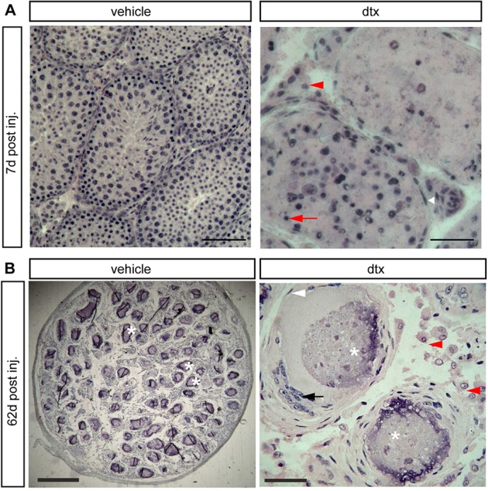
Fig. 5.

Testicular histology following SC ablation at pnd18. (A) Testicular histology of mice 7 d after ablation at pnd18. Testes retained tubular architecture with representative PTMCs (white arrowhead) surrounding tubules and LCs in the interstitium (red arrowhead). Seminiferous tubules exhibited altered spermatogenesis and apoptotic GCs were clearly visible (red arrow). Scale bars: 100 µm (left), 50 µm (right). (B) In adult mice (pnd80) injected at pnd18, the tubular structure of the testis remained intact, although the tubules were marked by calcium salt deposits in the lumen (asterisks). PTMCs were present around the tubules (white arrowhead), forming multilayers in places. The lumen of some of the tubules contained unidentified cells (black arrow). LCs (B, red arrowheads) were present between the tubules. Scale bars: 400 µm (left), 50 µm (right).
