Abstract
Programmed cell death-1 (PD-1) is a crucial negative regulator of CD8 T cell development and function, yet the mechanisms that control its expression are not fully understood. Through a non-biased DNase I hypersensitivity assay, four novel regulatory regions within the Pdcd1 locus were identified. Two of these elements flank the locus, bind the transcriptional insulator protein CTCF, and interacted with each other, creating a potential regulatory compartmentalization of the locus. In response to T cell activation signaling, NFATc1 bound to two of the novel regions that function as independent regulatory elements. STAT binding sites were identified in these elements as well. In splenic CD8 T cells, TCR-induced PD-1 expression was augmented by interleukin 6 and 12, inducers of STAT3 and STAT4 activity, respectively. IL-6 or IL-12 on its own did not induce PD-1. Importantly, STAT3/4 and distinct chromatin modifications were associated with the novel regulatory regions following cytokine stimulation. The NFATc1/STAT regulatory regions were found to interact with the promoter region of the Pdcd1 gene providing a mechanism for their action. Together these data add multiple novel distal regulatory regions and pathways to the control of PD-1 expression and provide a molecular mechanism by which proinflammatory cytokines, such as IL-6 or IL-12 can augment PD-1 expression.
Keywords: T Cells, Cell Surface Molecules, Transcription Factors, Gene Regulation, PD-1
Introduction
Programmed Death 1 (PD-1), encoded by Pdcd1, is a transmembrane protein that is highly expressed on the surface of immune cells during chronic immune activation and in a variety of cancers (1–4). Following engagement with its ligands, PD-L1/L2, signaling through PD-1 leads to an exhaustive phenotype wherein T cells lose their effector functions and ability to proliferate (5). In both in vitro and in vivo settings, blockade of PD-1 — PD-L1/L2 interactions results in reinvigoration of CD8 T cell effector functions and reduced viral loads in experimental systems (6–9). Recently, PD-1/PD-L1 blockade has been shown to be an efficacious treatment for some late stage cancers (10–12). Despite its clear importance in immune function, the mechanisms by which PD-1 is regulated are still poorly understood.
The transient upregulation of PD-1 during acute viral infection has been attributed to the action of nuclear factor of activated T cells c1 (NFATc1 or NFAT2) binding to a conserved region located upstream of the Pdcd1 promoter termed Conserved Region C (CR-C) (13). cFos was identified as a factor that binds to CR-B, a promoter proximal element that was necessary for maximal induction by NFATc1 (14). Additionally, an interferon-stimulated regulatory element (ISRE), located in CR-C, was reported to enhance and prolong PD-1 transcription upon T cell and macrophage activation (15, 16). In contrast to these factors, T-bet has been shown to negatively regulate PD-1 in CD8 T cells during LCMV infection (17). Other reports have also suggested a role for B lymphocyte-induced maturation protein-1 (Blimp-1) in modulating PD-1 expression, although no direct role for that factor has been reported (18).
DNA methylation, a transcriptionally repressive epigenetic modification, was found to be dynamically modulated in antigen-specific CD8 T cells and inversely correlated with PD-1 expression during effector (on) and memory (off) phases following an acute viral infection with LCMV (19). During chronic LCMV infection, exhausted CD8 T cells, which express high levels of PD-1, became and remained hypomethylated at the CR-B and CR-C regions of Pdcd1, suggesting that the presence of antigen may drive expression and the control of DNA methylation (19). However, analysis of Pdcd1 DNA methylation in antigen-specific CD8 T cells of HIV infected individuals showed that despite viral control through HAART or the patients’ natural immune response (elite controllers) the Pdcd1 locus remains demethylated (20). These observations suggest that early immune events may establish epigenetic modifications of the locus that are maintained irrespective of antigen levels.
Multiple cytokines have been shown to regulate PD-1, including several in the common γ-chain family (IL-2, IL-7, IL-15, and IL-21) and Type I IFNs (IFN-α and IFN-β) (15, 16, 21). IL-6, which acts through STAT3, has been shown to predict antiviral responses in individuals coinfected with HIV and HCV where high levels of IL-6 in the serum correlate with non-responding individuals (22, 23). STAT3 is critical for differentiation and function of CD4 T cell subsets including TH17, TH2, T follicular helper (TFH), and T regulatory cells (Treg), as well as memory formation of CD4 and CD8 T cells (24–28). In addition to IL-6, the cytokines IL-10 and IL-21 signal through the JAK family of proteins culminating in STAT3 activation (29). IL-10 has been shown to directly inhibit CD4 responses and blockade of IL-10 signaling leads to clearance of chronic LCMV infection, suggesting that STAT3 plays a role in viral persistence (30, 31). The above reports suggest that multiple cytokines can regulate PD-1. However, with the exception of IFN-α inducing responses from an ISRE located in CR-C, no direct effect of cytokine induced factors regulating Pdcd1 gene expression has been shown (15, 16).
All current known regulators of Pdcd1 are located in or adjacent to the previously described CR-B and CR-C regulatory regions that reside within the first 1.2 kb upstream of the transcription start site (TSS) (13–15, 17, 19, 20). However, in many genes it is common that distal regulatory elements can be found more than 10 kb away from the TSS (32–34). To determine if Pdcd1 is regulated by distal elements, a nonbiased approach was employed across the murine Pdcd1 locus. The results identified four novel distal regulatory regions. Two of these elements flanked the locus and bound the insulator protein CCCTC-binding factor (CTCF). The third element, located upstream of CR-C, bound NFATc1 and STAT3 or STAT4 in response to TCR and IL-6 or IL-12 signaling, respectively. The final region, located close to the downstream CTCF site also bound NFATc1 and STAT3 or STAT4. The chromatin structure of each regulatory region is altered in response to T cell activation and cytokine stimulation, with unique patterns arising in response to each stimulus. Each of the novel NFAT/STAT elements was found to interact with the Pdcd1 promoter region in CD8 T cells, demonstrating that they are likely to contribute directly to Pdcd1 transcription. Together, these results introduce new pathways that integrate multiple mechanisms to stimulate and regulate PD-1 in response to different environmental cues.
Materials and Methods
Cells and Culture
The murine T lymphoma cell line EL4 was obtained from the American Type Culture Collection. Primary splenic CD8 T cells were isolated from C57BL/6 mice by magnetic bead separation (Miltenyi Biotech Inc., Auburn, CA) according to manufacturer’s recommendations. All cells were cultured in RPMI 1640 supplemented with 5% fetal bovine serum (Sigma-Aldrich Co, St. Louis, MO), 5% bovine calf serum (Thermo Fisher Scientific Inc., Waltham, MA), 10mM HEPES, 1mM sodium pyruvate, 4.5 g/L glucose, and 100 U/ml penicillin/streptomycin. Cells were treated with 50 ng/ml phorbol 12-myristate 13-acetate (PMA) (Sigma-Aldrich), 2 μM ionomycin (Io) (Sigma-Aldrich), 1 μg/ml cyclosporin A (CsA) (Sigma-Aldrich), 500U IFN-α (eBioscience Inc., San Diego, CA), 20 ng/mL IL-2 (Miltenyi) 10 ng/mL IL-4 (Miltenyi), 20 ng/mL IL-6 (Miltenyi), 20 ng/mL IL-12 (Miltenyi), 20 ng/mL IFN-γ (Miltenyi), or CD3/CD28 activation beads (Life Technologies Co, Grand Island, NY) according to manufacturer’s protocol where indicated. All mouse experiments were conducted in accordance with approved Emory University institutional animal care and use protocols.
PCR based DNase I Hypersensitivity Assays
The protocol for PCR-based DNase I hypersensitivity screening was previously described (13). Briefly, nuclei were isolated and treated with increasing concentrations of DNase I (Worthington Biochemical Co, Lakewood, NJ). Concentration ranges for DNase I were determined empirically for each lot and cell type by titrating DNase I for its ability to digest CR-C as the positive control but not regions previously found to be resistant to DNase I (e.g., +6.3 region). Following purification of the digested DNA, PCR was performed across the Pdcd1 locus using a set of 59 primer pairs (Supplemental Table 1). For conventional PCR assays, PCR amplicons were designed to span approximately 1kb of the Pdcd1 locus. After visualization of the PCR products by agarose gel, band intensities were quantified using ImageJ (35). DNase I hypersensitivity was determined by using band intensities across increasing DNase I concentrations and calculating a slope from a best-fit line to represent the loss of signal. Slopes were normalized to the known DNase I hypersensitive CR-C region. For a higher resolution DNase I hypersensitivity assay, real-time PCR amplicons were designed to be 400–600 bp. Following amplification, values were normalized to a known DNase I insensitive region located within the Ciita gene, which is not expressed in these cells (13, 36). DNase I hypersensitivity was again determined by calculating a slope using the normalized threshold values across increasing DNase I concentrations. All DNase I assay primers are listed in Supplemental Table 1. DNase I hypersensitivity assays were performed at least three times from independent cultures or preparations of splenic CD8 T cells from mice.
Gene Reporter Assays
DNA sequences corresponding to the DNase I hypersensitive sites were cloned into the firefly luciferase reporter vectors pGL3-Promoter or pGL3-Basic (Promega Co, Madison, WI) using NheI (New England Biolabs Inc., Ipswitch, MA). Plasmids were transiently transfected into EL4 cells by nucleofection using kit L protocols as described by Lonza Inc. (Allendale, NJ). At 16 hours post transfection, cells were treated with PMA/Io, IL-6, and/or IL-12 where indicated and incubated for 24 hours. For experiments using CsA, cells were treated for 2 hours with CsA before addition of PMA/Io. Lysates were collected 24 hours after PMA and ionomycin addition. The Dual-Luciferase Reporter Assay System (Promega) was used to quantify luciferase activity. Firefly luciferase was normalized to the co-transfected, constitutively expressing Renilla luciferase gene (pRLTK). The average fold change in normalized luciferase activity was calculated from three independent transfections and is shown with standard deviation.
RNA isolation and RT-PCR analysis
The RNeasy kit (Qiagen Sciences Inc., Germantown, MD) was used to isolate total RNA. Reverse transcription assays were carried out using SuperScript II reverse transcriptase (Life Technologies) and 1 μg of RNA. Real-time PCR analyses on three independent RNA preparations were conducted to quantify Pdcd1 mRNA. Pdcd1 levels were normalized to 18s ribosomal RNA.
Chromatin Immunoprecipitation Assays
Chromatin Immunoprecipitation (ChIP) assays were previously described (37, 38). Cells were cross-linked in 1% formaldehyde for 15 minutes. Chromatin was sonicated to an average size of 400–600 bp and then 2 μg was immunoprecipitated with antibodies raised against NFATc1 (Thermo Scientific), p-STAT1 (Cell Signaling Technologies Inc., Danvers, MA), p-STAT3 (Cell Signaling) p-STAT4 (Cell Signaling Technologies), p-STAT5 (Cell Signaling), p-STAT6 (Cell Signaling), RNA Polymerase II (EMD Millipore Co., Billerica, MA), p300 (Santa Cruz Biotechnology Inc., Santa Cruz, CA), H3K27ac (EMD Millipore), H3K4me1 (EMD Millipore), H3K4me3 (EMD Millipore), or CTCF (Rockland Immunochemicals Inc., Gilbertsville, PA). A negative control IgG (EMD Millipore) was also used in parallel. Following immunoprecipitation (IP) and washing, DNA from each IP was quantified by real-time PCR using a standard curve from sonicated murine genomic DNA. Each ChIP was performed from three independently isolated cell preparations and plotted as the average percent pulldown of input chromatin with standard deviation.
Chromatin Confirmation Capture (3C)
3C assays were performed from modified procedures that have previously been described (39, 40). Briefly, 107 EL4 or 3 × 10⁶ CD8 T cells were suspended in RPMI supplemented with 5% fetal bovine serum and 5% bovine calf serum. Formaldehyde was added to a final concentration of 1% and cells were incubated for 10 min at room temperature. Following incubation, glycine was added to a final concentration of 125 mM to quench the cross-linking reaction. Cross-linked nuclei were isolated and digested overnight with PstI (New England Biolabs). After digestion, PstI was heat inactivated and the reaction was diluted 1:40 in ligation buffer and ligated overnight with T4 DNA ligase (New England Biolabs). Following purification, quantitative PCR using primer sets (Supplemental Table 1) located adjacent to PstI sites was used to identify 3C ligation products. To generate material for a 3C standard curve, the RP23-4L19 bacterial artificial chromosome (Children’s Hospital Oakland Research Institute, Oakland, CA), which contains the entire Pdcd1 locus, was PstI digested and religated to generate all possible PstI-PstI end-joined fragments. This DNA was then used with the same 3C primer sets to provide a qPCR standard curve. 3C data is presented as the average cross-linking frequency with the standard deviation from three biological replicates.
Results
The Pdcd1 locus contains multiple DNase I hypersensitive sites
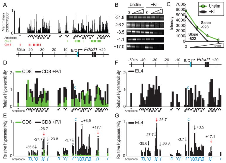
The Pdcd1 locus in the mouse is homologous to other mammals from ~−10kb upstream the transcription start site (TSS) through and downstream of the gene (Figure 1A). Surprisingly, whereas the mouse sequences further upstream (−10 to −50kb) are similar among rodents, these sequences diverge in the human where PDCD1 is located on chromosome 2. There is no homology between mouse and humans from −10 to −31kb. From −31 to −50kb, the murine sequence is homologous to a region ~123–134kb upstream of the CHD1 gene on human chromosome 5. This might suggest that common elements regulating Pdcd1 gene expression would be contained within the homologous regions of mouse chromosome 1 and human chromosome 2 that encode the Pdcd1/PDCD1 loci. Chromatin sensitive to digestion by DNase I is indicative of potential regulatory regions (41). Using this conceptual approach, we previously identified and showed that the CR-B and CR-C regulatory regions of Pdcd1 were differentially sensitive to DNase I digestion in unstimulated and PMA/Io stimulated CD8 T cells and EL4 cells (13). To identify additional regulatory elements, a PCR-based strategy employing 59 amplicons, was used to scan ~70 kb of the Pdcd1 locus for DNase I hypersensitive regions (Figure 1A). Amplicons of 0.9–1.3 kb in length covered all the unique, non-repetitive DNA sequences within the locus. Splenic CD8 T cells were isolated from C57BL/6 mice and immediately processed or treated with PMA/Io for 24h a time point in which PD-1 expression is induced to a high level ex vivo (13). Representative amplicons displaying hypersensitivity in response to PMA/Io (−3.5 and −1.1 (CR-C)), constitutive hypersensitivity (−26.2 and +17.0), and background levels of hypersensitivity (−31.8) to DNase I are shown in Figure 1B. A representative sample for each amplicon from the entire set is provided in Supplemental Figure 1A. To quantitate these relative sensitivities, amplicon intensity was measured using ImageJ and a slope was generated to identify regions that lost signal with increasing DNase I concentrations (Figures 1B and C). Using this strategy more than 70 kb of the Pdcd1 locus was scanned (Figure 1D). It should be noted that the regions not scanned contained repetitive element sequences (42). Stimulation of splenic CD8 T cells with PMA/Io led to an increase in DNase I sensitivity throughout the locus (Figure 1D). Regions showing either a differential sensitivity between control and stimulated CD8 T cells greater than a 2 fold change or a greater relative hypersensitivity than CR-C were chosen for further characterization as these could indicate areas essential for Pdcd1 expression and regulation.
Figure 1. The Pdcd1 locus contains multiple inducible DNase I hypersensitive sites.
(A) Schematic of the PD-1 locus showing relative positions to the TSS, conservation of sequences among mammals defined by MULTIZ alignment, and the human chain alignment of chromosome 2 (green) and 5 (red) (58, 70). Amplicons (Black boxes) used for conventional PCR-based DNase I hypersensitivity analysis are shown as is the previously defined CR-B/C regulatory region (B/C, blue). Using increasing amounts of DNase I, each of the 59 amplicons were used to assess the DNase I hypersensitivity of splenic CD8 T cells (control or stimulated with PMA and ionomycin (+P/I) ex vivo for 24h). Each PCR amplicon is between 0.9–1.3 kb in length. (B) Select examples of the 59 amplicons that were evaluated by conventional PCR are shown and their position from the TSS is indicated. (C) Each of the 59 amplicons was quantitated using ImageJ software. The bands from the +17.0 PCR shown in B were used as an example with slopes determined by linear regression. (D) Relative DNase I hypersensitivity of CD8 T cells for unstimulated (green) and PMA/Io cultured cells (black). DNase I sensitivity was calculated by taking the negative value of the slope following ImageJ quantitation and normalizing to the previously known hypersensitive region CR-C. Amplicon locations are shown below. The amplicon representing CR-C is shown with a blue C. (E) A higher resolution DNase I hypersensitivity map was constructed using real-time PCR on PMA/Io stimulated CD8 T cells. Asterisks above bars indicate regions that were chosen for further analyses that showed a statistically significant (p <0.05) increase in DNase I hypersensitivity over control samples. Black and red arrows denote regions chosen for further study and the location of these regions with respect to the TSS is indicated. The DNase I sensitivity for each of the regions indicated by the arrows was statistically significant (p <0.05). Amplicons are displayed as blue boxes along the bottom. (F) Relative hypersensitivity of the Pdcd1 locus in the murine EL4 T cell line using the same methodology as in CD8 T cells from D. (G). Quantitative real-time PCR analysis of DNase I hypersensitivity in EL4 cells using the methodology from E. The data from these experiments were averaged from three independent cell preparations.
To obtain a higher resolution profile of the potential regulatory elements, primers were designed to produce amplicons between 400–600 bp across each of the regions of interest determined from Figure 1D. Quantitative real-time PCR was performed on DNase I digested CD8 T cell DNA (Figure 1E). Analysis from these studies showed two peaks of DNase I sensitivity in control, unstimulated cells located at −26.7 kb and +17.0 kb from the TSS (Figure 1E, green bars, red arrows). The sensitivity of these regions to DNase I increased following PMA/Io stimulation, further supporting the possibility that these regions may be vital for PD-1 expression.
The murine EL4 T lymphoma cell line has been previously used to characterize constitutive PD-1 expression and regulation (13). DNase I hypersensitivity was also assessed in EL4 cells by the same PCR-based DNase I hypersensitivity assay and ImageJ amplicon analysis (Figure 1F and Supplemental Figure 1B), as well as quantitated using real-time PCR (Figure 1G). Comparison of CD8 T cells stimulated with PMA/Io and EL4 cells showed similar DNase I hypersensitivity profiles. Five of these regions were hypersensitive to DNase I in both EL4 and CD8 T cells and contained sequence homology across multiple species (Black arrows in Figures 1E and 1G). These regions were therefore chosen for further study. Thus, this DNase I hypersensitivity screen identified seven potential regulatory regions across the 70 kb encompassing the Pdcd1 locus.
Two DNase I Hypersensitive sites act as calcineurin-dependent regulatory elements
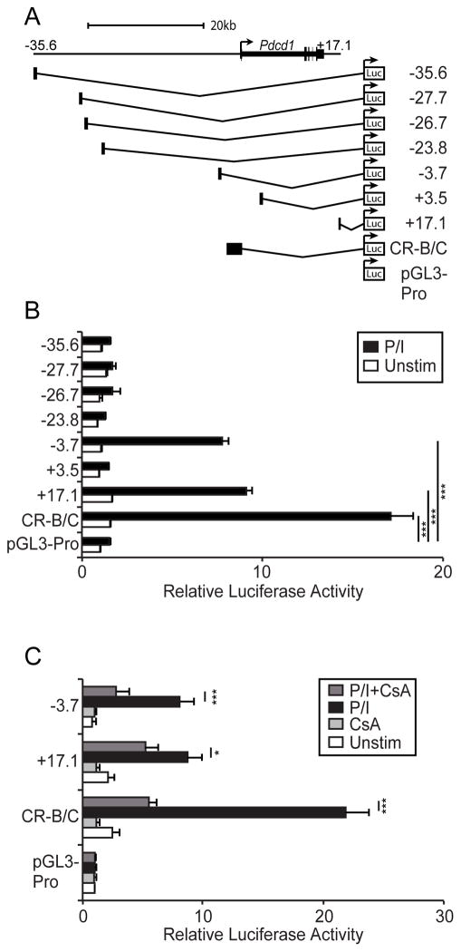
To determine if any of the chosen hypersensitive sites have transcriptional regulatory activity, 300–600 bp sequences representing the selected DNase I hypersensitive regions were cloned into the pGL3-Promoter luciferase reporter vector (Figure 2A). The constructs were transiently transfected into EL4 cells, which constitutively express PD-1, and luciferase activity was determined. As with the previously described CR-B/C construct, two regions, −3.7 and +17.1, showed substantial increases in relative luciferase activity upon PMA/Io stimulation (Figure 2B). This suggests that these regions may play a role in regulating PD-1 during T cell activation. Regions −35.6 and −27.7, which did not display statistically different DNase I hypersensitivity and were chosen as negative controls, displayed no transcription stimulating activity. The two remaining regions, located at −23.8 and +3.5 did not display induced activation and were not characterized further. Previously, we showed that calcineurin-NFAT pathway is critical to PMA/Io induced PD-1 expression through CR-C (13).
Figure 2. Two novel regulatory regions respond to T cell activation.
(A) DNase I hypersensitive sites selected from Figures 1E and 1G were cloned into the pGL3-Promoter firefly luciferase reporter vector. (B) Luciferase assays from EL4 cells transiently transfected with the construct indicated. Following transfection cells were cultured for 16h then stimulated with PMA/Io (P/I) for 24h. Firefly luciferase activity was quantitated, normalized to the cotransfected Renilla luciferase plasmid, and plotted relative to the empty pGL3-promoter (pGL3-Pro) vector. Data shown are the average of three independent experiments with standard deviation. Two-way ANOVA statistical tests comparing each stimulated sample to the empty vector were performed with *** representing P<0.001. (C) Constructs responding to PMA/Io were tested for their ability to respond to cyclosporine A (CsA) in transfection assays performed as above except that some cultures were treated for 2 hours with CsA prior to PMA/Io stimulation. The averages of three independent luciferase transfections were plotted. Two-way ANOVA comparing PMA/Io stimulated samples −/+ CsA were performed. ***P<0.001, *P<0.05.
To determine if the calcineurin pathway was also involved with the activity observed for the −3.7 and +17.1 regulatory regions, luciferase assays were carried out in the presence/absence of cyclosporine A (CsA). CsA blocks calcineurin-dependent dephosphorylation of NFAT and subsequent translocation into the nucleus (43, 44). Here, CsA inhibited/reduced the PMA/Io dependent activation of both regulatory regions, as well as the previously defined CR-B/C region, suggesting a role for an NFAT family member in the activity of these regions (Figure 2C).
IL-6 and IL-12 leads to increased expression and a change in the chromatin structure of Pdcd1
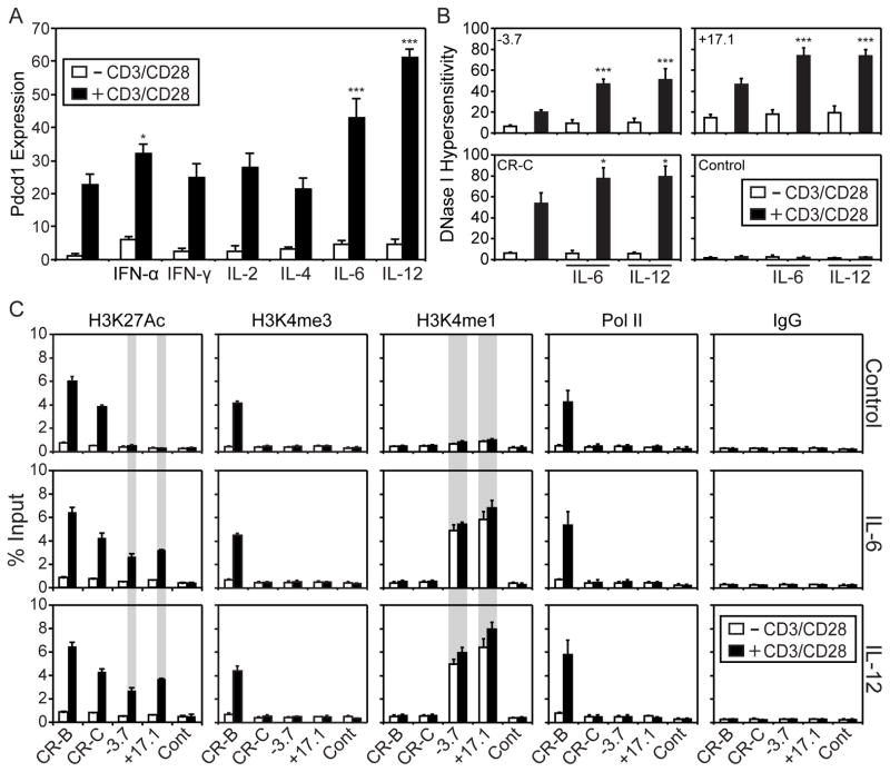
In silico analysis using the JASPAR database identified several putative STAT binding sites within −3.7 and +17.1, suggesting that STATs could play a role in the regulation of PD-1 (45). Analysis of ChIP-seq data on CD4 T cell subsets identified CR-C, −3.7, and +17.1, as being bound by multiple STAT family members (46, 47). Therefore, to test which if any STATs could stimulate PD-1 expression, CD8 T cells were isolated from C57BL/6 mice and stimulated ex vivo with or without CD3/CD28 beads in the presence or absence of IFN-α (STAT1/2), IFN-γ (STAT1), IL-2 (STAT5), IL-4 (STAT6), IL-6 (STAT3), or IL-12 (STAT4) (Figure 3A). On their own, none of these cytokines induced Pdcd1 gene expression. However, when coupled with CD3/CD28 bead stimulation, only IFN-α, IL-6 and IL-12 enhanced Pdcd1 gene expression (Figure 3A). While IFN-α was shown previously to enhance PD-1 expression in both macrophages and T cells through binding of the ISGF3 complex to an ISRE located in CR-C (15, 16), IL-6 and IL-12 enhancement of Pdcd1 suggests a potential role for STAT3 and STAT4 in augmenting PD-1 expression.
Figure 3. IL-6 and IL-12 stimulation induces distinct chromatin modifications at Pdcd1 regulatory regions.
(A) Pdcd1 mRNA expression from splenic CD8 T cells that were cultured ex vivo with CD3/CD28 bead stimulation in the presence or absence of IFN-α, IFN-γ, IL-2, IL-4, IL-6, or IL-12 for 24h was measured by real-time RT-PCR. Data is presented as the relative Pdcd1 expression normalized to 18s rRNA from three biological replicates with standard deviation. The student’s t-test was used to determine significance of activated vs. activated and cytokine treated CD8 T cells. (B) Primary splenic CD8 T cells were cultured for 24h in the presence or absence of CD3/CD28 beads, IL-6, and/or IL-12 as indicated and subsequently analyzed for DNase I hypersensitivity at −3.7, +17.1, CR-C and a control (+18.2) region. The significance of activated vs. cytokine treated, activated CD8 T cells was determined by student’s t-test. (C) Quantitative ChIP assays using antibodies against H3K27ac, H3K4me1, H3K4me3, Pol II, and a control IgG were performed from splenic CD8 T cells cultured for 24h with and without CD3/CD28 beads, IL-6, and/or IL-12 as indicated. Data were presented as the average percent chromatin input from three independent experiments. Error bars represent standard deviation. Student’s t test comparisons between control and IL-6 or IL-12 treated samples showing a significance of p<0.001 are indicated by gray shading across the ChIP samples. In the other panels p values are represented as follows: *, P<0.05; ***, P<0.001
An increase in Pdcd1 expression suggests the chromatin architecture of the locus could be changed by IL-6 and IL-12 treatment. To determine if this is the case, DNase I hypersensitivity assays were conducted on primary splenic CD8s stimulated with CD3/CD28 beads and/or IL-6 and IL-12. Both IL-6 and IL-12 stimulation of activated CD8 T cells led to an increase in the DNase I hypersensitivity of CR-C, −3.7, and +17.1, but not of a control region (Figure 3B). IL-6 and IL-12 treatment had no effect on their own.
Enhancer regions are marked by H3K4me1 and when combined with H3K27ac, denote active enhancers (48, 49). In addition, H3K4me3 marks the promoters of active genes. Chromatin immunoprecipitation assays (ChIP) were used to determine the presence of each of these marks in splenic CD8 T cells cultured with CD3/CD28 beads, IL-6, and/or IL-12. CR-B and CR-C were marked with H3K27ac in response to T cell activation, irrespective of cytokine treatment (Figure 3C). CR-B was also marked by high levels of H3K4me3 in response to T cell activation, correlating with active gene expression. The −3.7 and +17.1 regions acquired H3K27ac in response to T cell activation and cytokine stimulation, but not with either alone. In contrast, H3K4me1 was only present at −3.7 and +17.1 in response to cytokine stimulation, irrespective of whether the T cells were activated. Some active enhancers have also been shown to bind RNA polymerase II (PolII) (50). Here, PolII binding was only observed at CR-B (a region that is close to the TSS) in response to CD3/CD28 stimulation and not at the other regulatory regions or a control upstream region. A control IgG ChIP at each of the sites showed no reactivity and demonstrates specificity for the assay. In some cases, active enhancers have been reported to express short transcripts known as eRNAs (51). The presence of such transcripts was assessed in CD8 T cells stimulated with CD3/CD28 beads with and without IL-6 or IL-12. No significant levels of transcripts were observed irrespective of the treatment (data not shown).
NFATc1, STAT3, STAT4, and p300 bind to the −3.7 and +17.1 regions
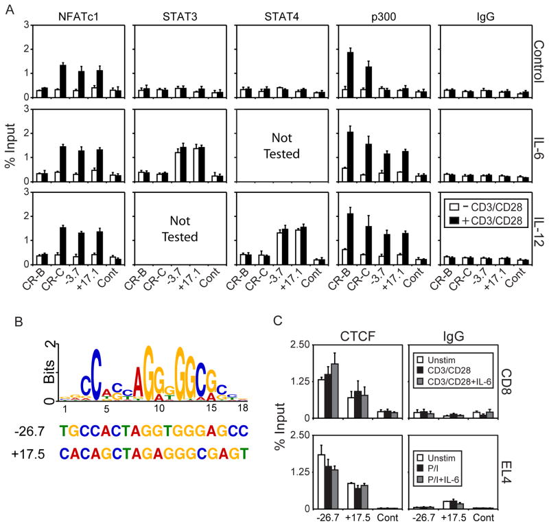
To demonstrate that STAT3, STAT4, and NFAT bind to the regulatory regions, ChIP was performed using antibodies to NFATc1, STAT3, and STAT4. For these experiments, splenic CD8 T cells were cultured for 24 hours with or without CD3/CD28 beads and in the presence/absence of IL-6 or IL-12. NFATc1 was bound to CR-C, −3.7, and +17.1 in CD3/CD28 bead activated cells, irrespective of cytokine stimulation (Figure 4A). STAT3 and STAT4 bound only to −3.7 and +17.1 in cells stimulated with IL-6 or IL-12, respectively. STAT binding was not dependent on T cell activation. The transcription factor p300, a histone acetyltransferase that can catalyze H3K27 acetylation (52, 53), is often found at active enhancers. Using ChIP, p300 was found at CR-B and CR-C in response to CD3/CD28 stimulation (Figure 4A). The −3.7 and +17.1 elements were also bound by p300, but only in response to both CD3/CD28 and cytokine treatment. A control sequence or control IgG antibody showed only background levels of binding. Thus, NFATc1 binds in response to T cell activation, while STAT binding is induced through cytokine stimulation. p300 binding correlates with H3K27ac and is differentially recruited to the regulatory regions through either T cell activation at CR-B and CR-C, or through T cell activation combined with cytokine signaling at −3.7 and +17.1.
Figure 4. NFATc1, STAT3, STAT4, p300, and CTCF bind to the Pdcd1 locus.
Quantitative ChIP for NFATc1, STAT3, STAT4, p300 and CTCF binding to the regions indicated was performed. All ChIP data is presented as average percent input from three independent experiments with standard deviation. An IgG control antiserum was used as a specificity control for antibody binding. (A) ChIP for NFATc1, STAT3, STAT4, p300, or a control IgG from splenic CD8 T cells stimulated for 24h with CD3/CD28 beads, IL-6, and/or IL-12 as indicated. (B) Consensus CTCF binding sequence in logo format aligned with the DNA sequences from −3.7 and +17.1 is shown. (C) ChIP for CTCF or a control IgG from CD8 T cells stimulated with CD3/CD28 beads, or beads and IL-6 and EL4 cells stimulated with PMA/Io or PMA/Io (P/I) and IL-6.
CTCF binding sites flank the Pdcd1 gene
CTCF, the only known mammalian insulator protein, has previously been shown to be essential for formation of regulatory loops that segregate commonly regulated genes from other nearby genes (39, 40). ChIP-seq experiments from whole thymus identified two regions within the Pdcd1 locus (54), −26.7 and +17.5, which were identified in our initial screen as being sensitive to DNase I in the absence of T cell activation (Figure 1). To generate a consensus CTCF logo, sites bound by CTCF in mouse whole thymus mapped by the ENCODE Consortium corresponding to the central 100 bp of each peak were analyzed using the MEME-ChIP software package (55). The CTCF site depicted in Figure 4B was the top scoring motif (p-value = 4.73e-243). Conventional qPCR-based ChIP using CTCF antibodies confirmed that CTCF was bound to both these sites in primary CD8 T cells, as well as in EL4 cells (Figure 4C). Moreover, CTCF binding levels did not change in response to PMA/Io, CD3/CD28 beads, and/or IL-6 treatment. It should be noted that CTCF binding to the +17.5 fragment is downstream of the DNA sequences that contain NFAT and STAT binding sites and was not included in the +17.1 reporter constructs used above. These data suggest that these CTCF sties may form the outer boundaries for Pdcd1 regulatory regions.
STAT and NFAT binding sites are critical for regulatory function of the −3.7 and +17.1 regulatory regions
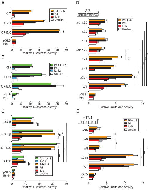
To assess whether the newly identified regulatory regions could respond to IL-6 or IL-12, luciferase reporter gene transfections were carried out in EL4 cells followed by IL-6, IL-12 and/or PMA/Io stimulation. The +17.1 region showed a significant increase in luciferase activity after addition of either IL-6 or IL-12 to PMA/Io treated EL4 cells (Figures 5A and 5B). In contrast, the −3.7 and CR-C regions did not display an increase in activity following cytokine treatment. Neither IL-6 nor IL-12 activated expression of the reporter vector without PMA/Io induction, suggesting that TCR signaling is required to initiate Pdcd1 expression.
Figure 5. NFATc1 and STAT3 binding sites are necessary for regulatory activity of the −3.7 and +17.1 regulatory regions.
(A and B) EL4 cells were transfected with the indicated construct as in Figure 2 and stimulated simultaneously with PMA/Io (P/I) and/or the cytokine indicated. At 24h post stimulation, luciferase activity was determined and normalized to the cotransfected Renilla plasmid. (C) The −3.7 and +17.1 regions were cloned into the previously described Pdcd1 promoter–CR-B–pGL3-Basic–luciferase reporter expression vector (13). (D and E) NFAT and STAT binding sites identified in silico (indicated as N or S, respectively), were deleted in the pGL3-Promoter based constructs. The resulting constructs were subsequently transfected into EL4 cells and stimulated with PMA/Io and/or IL-6 as indicated. Δ indicates which sequence was deleted. ΔCon represents a random deletion and serves as a negative control. Data from three independent experiments were averaged and plotted as relative luciferase to pGL3-Promoter (pGL3-Pro) or to pGL3-Basic with standard deviation. Two-way ANOVA tests were used to determine the significance between samples. In A and B, the statistical tests compared PMA/Io stimulation to PMA/Io and IL-6; or PMA/Io stimulation to PMA/Io and IL-12. In C, PMA/Io stimulated cultures were compared to unstimulated, PMA/Io +IL-6, and PMA/Io +IL-12 cultures. PMA/Io stimulated cultures of −3.7, +17.1, and CR-B/C constructs were also compared to the PMA/Io stimulated CR-B construct. Statistical differences in D and E were assessed between the wild-type construct and the deletion constructs stimulated with PMA/Io. Additional analysis between the single and double deletion constructs were also assessed as indicated. *, P<0.05; **, P<0.01; and ***, P<0.001.
To confirm that the −3.7 and +17.1 regions function to promote Pdcd1 expression, the two regions were cloned into the previously described CR-B reporter construct, which uses the Pdcd1 promoter and CR-B instead of a heterologous promoter (13). In agreement with the above experiments, the −3.7/B and +17.1/B region containing vectors displayed a significant increase in luciferase activity in response to PMA/Io (Figure 5C). The +17.1/B vector also showed an additional increase in luciferase activity when costimulated with IL-6 or IL-12. These results confirm that the −3.7 and +17.1 regions are able to enhance the activity of the Pdcd1 promoter.
To assess the role of both the NFAT and STAT sites in driving the activity of these regions, the putative binding sites were identified using JASPAR (45) and then deleted in their respective pGL3-Promoter reporter constructs. The constructs were subsequently transfected into EL4 cells and stimulated with PMA/Io and/or IL-6. The −3.7 region contains two predicted STAT (S1 (chr1:95952803-95952809), S2 (chr1:95952814-95952820)) and two NFAT (N1 (chr1:95952661-95952667), N2 (chr1:95952686-95952691)) binding sites. Loss of either predicted NFAT site (ΔN1 or ΔN2) or STAT binding site (ΔS1 or ΔS2) led to a decrease in luciferase activity (Figure 5D). Deletion of both NFAT sites in concert (ΔN1ΔN2) resulted in less luciferase activity than deletion of either NFAT site alone. Unlike the NFAT sites, elimination of both STAT sites together (ΔS1ΔS2) showed no further loss of luciferase activity. Because the loss of luciferase activity was in response to PMA/Io, this suggests minimally that these sites are integral to the activity of this region in EL4 cells. A random control mutation (ΔCon (chr1:95952934-95952941)) showed a slight increase in luciferase activity, suggesting that not all mutations within the DNase I hypersensitive region negatively affect the reporter gene expression/activity.
Luciferase reporter vectors carrying the deletions of the STAT (ΔS (chr1:95932036-95932042)) or NFAT (ΔN (chr1:95932003-95932009)) binding sites in the +17.1 region showed a significant loss of luciferase activity compared to wild type, with the NFAT mutation showing a more dramatic loss of expression (Figure 5E). Importantly, there was no change when a control deletion (ΔCon (chr1:95932096-95932101)) is introduced into another part of the region. A construct containing the double STAT and NFAT mutation (ΔNS) showed similar levels of luciferase to that of the single NFAT mutation (ΔN).
As shown above, deletion of the STAT binding sites in the −3.7 or +17.1 construct resulted in a decrease of luciferase activity in PMA/Io only treated cells (Figures 5D and 5E), suggesting that these sites were active in the absence of added cytokine. To determine if this was the case, ChIP of EL4 cells following PMA/Io stimulation was performed using antibodies to STAT1, STAT3, STAT4, STAT5, and STAT6. The results showed inducible binding of NFATc1 and STAT1 at −3.7 and +17.1 upon PMA/Io treatment, whereas NFATc1 but not STAT1 bound CR-C following PMA/Io stimulation (Supplemental Figure 2). There was no detectable binding of STAT3, STAT4, STAT5, or STAT6 compared to control IgG at any region queried. These data suggest that following PMA/Io stimulation of EL4 cells, STAT1 binding is activated and is able to augment expression of the reporter constructs described above. Thus, mutation of the STAT sites in −3.7 and +17.1 alter the PMA/Io stimulated activity.
Dynamic interactions between the −3.7 region and two insulator regions creates a unique chromatin architecture during Pdcd1 activation
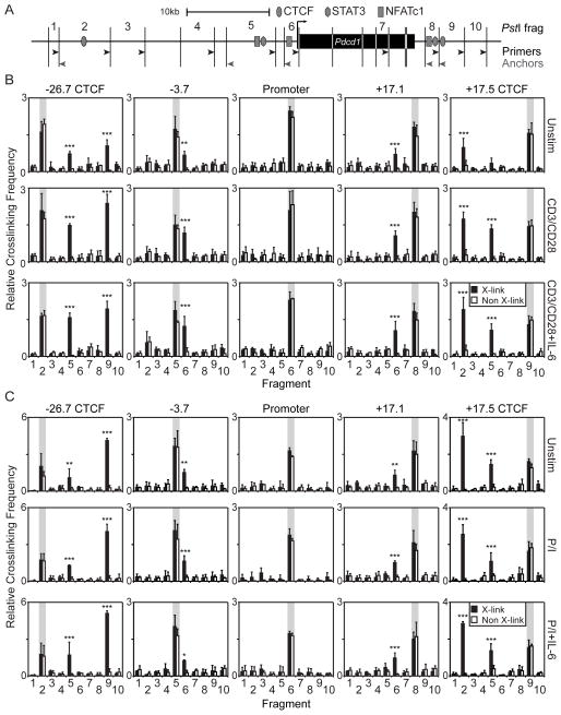
Distal enhancers can carry out their effects by interacting with promoters through long-range chromatin loops. (56). Detection of these interactions can be accomplished using chromatin confirmation capture (3C) assays, which define spatial relationships formed by DNA bound factors (57). To determine the organization of the Pdcd1 locus and if that chromatin organization changes during Pdcd1 expression, 3C assays were performed on both CD8 T and EL4 cells. A map of the Pdcd1 locus and the positions of the 3C fragments is shown in Figure 6A. In this 3C assay system, the restriction enzyme PstI was chosen to provide the greatest resolution between regulatory elements. Cells were left untreated, activated (CD3/CD28 beads for CD8 T cells or PMA/Io for EL4), or activated and treated with IL-6 prior to preparation of 3C libraries. Five 3C anchor fragments, representing the flanking CTCF sites (Figure 6A, fragments 2 and 9), the +3.7 and −17.1 regulatory elements (fragments 5 and 8, respectively), and the Pdcd1 promoter (fragment 6) were chosen to identify interactions between regulatory regions.
Figure 6. Multiple long-range interactions occur across the Pdcd1 locus.
(A) Schematic of the Pdcd1 locus with CTCF, NFATc1, and STAT3 binding sites indicated. Vertical lines denote PstI sites. Arrowheads denote locations of anchors (gray) or fragment test primers (black) used in the 3C assays. (B) 3C assays of splenic CD8 T cells unstimulated or activated with CD3/CD28 beads for 24h in the presence or absence of IL-6. Anchor primers are indicated at the top of each column and fragment test primers are indicated in the abscissa. The region used as the anchor is shaded in gray. Crosslinked and non-crosslinked samples are indicated by black and white bars, respectively. (C) 3C assays were performed on EL4 cells untreated, activated with PMA/Io, or PMA/Io and IL-6 for 24h. Relative crosslinking frequency is defined as the average of the real-time PCR based values for each 3C amplicon divided by a non-specific control amplicon. Data is presented as the average relative crosslinking efficiency from three experiments plus standard deviation. Two-way ANOVA comparing crosslinked and non-crosslinked samples was performed to determine significant changes in crosslinking frequency. ***, P<0.001; **, P<.01; and *, P<0.05.
In CD8 T cells, an interaction between the flanking CTCF sites bound to PstI fragments 2 (−26.7 region) and 9 (+17.5 region) was observed irrespective of CD3/CD28 stimulation or IL-6 treatment (Figure 6B). Using 3C anchor primers associated with the flanking CTCF sites, interactions between the CTCF sites (fragments 2 and 9) and the −3.7 region (fragment 5) were observed in unstimulated cells. Upon activation, the interaction frequency of CTCF sites and the −3.7 region increased (p<0.001), but addition of IL-6 had no effect on the interactions (Figure 6B). A similar set of interactions between the CTCF sites and the −3.7 region were constitutively observed in EL4 cells (Figure 6C). However, as these cells are already expressing PD-1, it was not surprising that no increase in CTCF–CTCF or CTCF – −3.7 interactions was observed in response to PMA/Io stimulation or IL-6 treatment (Figure 6C). When the promoter element was used as an anchor, interactions were not observed between the −3.7 or the +17.1 regions. However, when 3C anchors were placed on the opposite sides of the restriction fragments, reproducible interactions between −3.7 and +17.1 regions with the Pdcd1 promoter fragment were readily detected. In CD8 T cells, a significant increase in the −3.7 – Promoter interaction upon T cell activation (p<0.01) that was unaffected by IL-6 was observed (Figure 6B). This interaction was constitutive and unchanged with respect to treatment in EL4 cells. Interactions between −17.1 and the promoter region were constitutive in both CD8 T and EL4 cells. Because the change in anchor primers results in different DNA ligations being detected in the 3C assay, this indicates that there is a distinct spatial orientation of the regulatory elements and DNA fragment ends that could be readily available for 3C ligation. This possibility could provide an explanation for the inability to detect interactions between the 3C −3.7 anchor and the CTCF sites. No interactions were observed with randomly chosen fragments (1, 3, 4, 5, or 10) across the locus, demonstrating specificity of the assay. Experiments assessing the accessibility of the locus in crosslinked cells were performed and demonstrated that PstI can access all restriction sites used with relatively equal efficiency (Supplemental Figure 3). Taken together, these results describe a unique chromatin architecture for the Pdcd1 locus that is enhanced when Pdcd1 is activated in primary CD8 T cells.
Discussion
The cis-acting elements that are known to control Pdcd1 expression were limited to promoter proximal regulatory regions identified from sequence conservation and DNase I hypersensitivity assays a number of years ago (13). Using an unbiased DNase I hypersensitivity assay approach to discover potential regulatory regions (41), a number of complex, distal regulatory elements located both upstream and downstream of the TSS were discovered. Two of the DNase I hypersensitive elements (−3.7kb and +17.1 kb) displayed regulatory activity when transfected into the PD-1 constitutive expressing T cell line EL4. Two additional elements were constitutively hypersensitive and bound the transcriptional insulator factor CTCF. The downstream elements showed a high degree of homology with human sequences, including consensus binding sequences and binding by ChIP-seq for STATs, NFATc1, and CTCF (54, 58), suggesting that the conservation is important for the regulation of this locus. The −3.7 and −26.7 elements are well conserved in rat but not in humans. However, in humans, ChIP-seq data shows STAT and NFATc1 binding and H3K4me1 to an upstream region located approximately 4.8 kb upstream of the TSS, suggesting a conservation of regulatory mechanisms. Chip-seq experiments also suggest that CTCF binds approximately 7.5 kb upstream of the TSS of the human PDCD1 gene (54). Thus, the arrangement and functionality of elements appears to be conserved between species. Together with CR-B, CR-C, and the T-bet binding site, the new data presented here raises the total number of cis-elements that could potentially regulate Pdcd1 to seven (13, 17). As discussed below, the discovery of a role for STAT factors to directly regulate PD-1 expression broadens the overall mechanisms by which this gene can respond to cytokine stimulation and local immune environments.
Previously, NFATc1 was found to modulate PD-1 expression through CR-C (13). Here, through sensitivity to cyclosporine A and ChIP assays, NFATc1 was found to be a necessary, but not sufficient, mediator of the transcriptional activity of the −3.7 and +17.1 regulatory elements. While these elements may coordinate TCR-mediated induction of Pdcd1 with CR-C, their full activity is unlikely to be realized until STATs are bound, as active enhancer modifications (p300, H3K27ac, and H3K4me1) did not appear unless both NFATc1 and the STATs were bound. The presence of multiple cis-elements bound by the same key regulatory factors is found in other systems (59–61), and we propose that the multiple elements identified here could provide synergy and/or greater activation potential than a single element. Interactions between each of the new NFATc1 elements and the Pdcd1 promoter region fragment (which contained CR-C) were observed, supporting the concept that all three of these NFATc1 elements participate in activating transcription of Pdcd1.
An in silico search for transcription factor-binding sites at the −3.7 and +17.1 regulatory regions revealed a potential role for multiple STAT family proteins. ChIP-seq experiments on differentiated CD4 T cells suggested that STAT3 and STAT4 could be binding to these sites in CD8 T cells as well (46, 47). Indeed, IL-6 and IL-12 stimulation of activated CD8 T cells led to STAT3 or STAT4 binding, respectively, at the −3.7 and +17.1 regulatory regions. Importantly, in ex vivo stimulated CD8 T cells, IL-6 or IL-12 stimulation led to an increase in Pdcd1 gene expression over activated cells that received no cytokine. No effect of cytokine treatment alone was observed on Pdcd1 expression, suggesting that NFATc1-mediated initiation of Pdcd1 transcription is required for STAT3 and STAT4 to effect PD-1 levels. Thus, both −3.7 and 17.1 are complex regulatory elements that bind both NFATc1 and STAT3 or STAT4. Dissection of the −3.7 and +17.1 elements by site mutagenesis and reporter assays showed that both the NFAT and STAT elements were functional. Upon cytokine stimulation, the +17.1 reporter construct showed an increase in luciferase activity, although there was no increase from the −3.7 construct. The failure of −3.7 to respond to IL-6 and IL-12 stimulation may be due to the fact that upon PMA stimulation, EL4 cells produce IFN-γ, IL-2, and IL-4 (62, 63). Thus, it is possible that these cytokines, which activate STAT1, STAT5, and STAT6, respectively, could already be contributing to the expression of the −3.7-region reporter construct and that further stimulation by IL-6 or IL-12 could not be detected. Indeed, ChIP assays showed STAT1 binding to the −3.7 region in PMA/Io activated EL4 cells. Interestingly, +17.1 was also bound by STAT1 in these cells, however IL-6 stimulation augmented the reporter activity. The differences between the regulatory elements could reflect the fact that −3.7 has two sites compared to a single site in +17.1 or the relative binding capacity of these elements in vivo for STAT proteins and, whether under the endogenous situation, the elements are designed to work in concert. The finding of STAT1 binding in EL4 cells would also suggest a potential role for interferons in regulating Pdcd1 activity through these elements under specific conditions. Although a role for IFN-γ in the system used here was not observed, a role for type I interferon’s, which utilize STAT1 was previously described to use an ISRE located in CR-C (15, 16).
Although Pdcd1 expression requires T cell activation, there is a change in the chromatin architecture at −3.7 and +17.1 regions in response to cytokine treatment alone as H3K4me1 was observed at these regions following just cytokine stimulation. This suggests that the −3.7 and +17.1 regions become “poised” enhancers in response to cytokine stimulation. The induced binding of p300 and concomitant accumulation of H3K27ac in the presence of TCR and cytokine signaling imply that p300 is part of the Pdcd1 induction mechanism and likely is involved in activating these enhancers. These data suggest that the elements function differently with CR-C responding to only TCR/NFATc1 signaling and the distal elements (−3.7 and +17.1) functioning as the cytokine response elements. This may reflect a built in protective feature for making sure that PD-1 is not expressed on cells that are exposed to cytokines but have not received an antigen-specific activation signal.
Analysis of published ChIP-seq data on CD4 T cell subsets stimulated with various cytokines to induce STAT3, STAT4, STAT5, and STAT6 identified seven sites within the Pdcd1 locus that bound multiple STATs (46, 47, 54). All of the peaks were enriched for STAT3 and STAT4, with four of these peaks represented by CR-C, −3.7, +17.1, and +17.5. The three remaining peaks were located at −2.7 kb, the promoter region, and 1.2 kb downstream of the TSS. As shown here, quantitative ChIP in CD8 T cells stimulated with IL-6 and IL-12 for each of these sites confirmed binding of STAT3 and STAT4 to −3.7 and +17.1 (Figure 4). Additional ChIP assays showed binding to the −2.7 site (data not shown); however, no STAT binding was observed at CR-C or any of the other peaks (data not shown). This suggests that there is some background to the ChIP-seq datasets or that binding in those assays represents CD4 T cell subset-specific pattern. If the latter is the case, then a greater role for STAT factors in controlling PD-1 expression in other T cell subtypes is likely.
CTCF can function to block the activity of enhancers in cis and/or function as a chromatin boundary factor to prevent the spreading of heterochromatin (64). CTCF has previously been shown to control the chromatin architecture and expression of genes in the major histocompatibility complex class II and interferon-γ loci, as well as V(D)J recombination (39, 40, 65, 66). Regulation of gene expression by CTCF is thought to be due to the formation of long-range interactions that bring enhancers and promoters together (36, 39, 67). Here, two CTCF sites flank the Pdcd1 gene and likely serve to isolate it from its neighboring genes. As such, the CTCF-CTCF interactions have the potential to create a regulatory domain that would insulate Pdcd1 from external enhancers and/or focus the regulatory potential of the elements defined here on the Pdcd1 promoter.
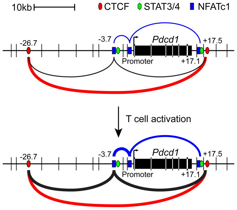
The 3C experiments produced a complex set of data suggesting that four different interactions are present in PD-1 expressing CD8 T cells. The first set reflects the constitutive CTCF–CTCF site interactions mentioned above (Figure 7, red lines). A second set of interactions occurs between either CTCF site and the −3.7 region (Figure 7, black lines). In CD8 T cells, these interactions increase upon NFAT stimulation. Curiously, reciprocal interactions between these sites were not observed when the anchors were placed on the opposing sides of the restriction fragments. This suggests that there are spatial constraints established by the elements that firmly establish the orientation of the free DNA ends, restricting the ability of the ends to be ligated, and ultimately detected in the 3C assay. The third and fourth set of interactions occurs between the −3.7 region and the promoter and the +17.1 region and the promoter (blue lines). As above, these interactions were orientation specific, suggesting that the interactions between the elements, promoter, and CTCF sites define a critical structure that might be required to control Pdcd1 expression.
Figure 7. Schematic of long-range cis-element interactions at the Pdcd1 locus that occur following CD8 T cell activation.
CTCF, NFATc1, and STAT3/4 binding sites are shown. PstI restriction enzyme sites are represented by vertical black lines. CTCF—CTCF (red lines), −3.7—CTCF (black lines), −3.7—Promoter (blue lines) and +17.1—Promoter (blue lines) interactions are shown with the relative strength represented by line thickness.
Taken together, our findings have expanded the role of NFATc1 and added STAT3, STAT4, and CTCF to the list of factors contributing to PD-1 expression. Intriguingly, STAT3 or STAT4 could explain how PD-1 is expressed at high levels in chronic infections, a condition in which NFATc1 translocation to the nucleus is impaired (68). The use of STAT family members to regulate PD-1 allows for environmental cues to regulate PD-1 expression. This suggests that regulation of PD-1 on CD8 T cells could be accomplished by targeting other cell types that produce cytokines that signal CD8 T cells. Previously, it has been shown that IL-2 inhibits IL-17 expression through STAT5 binding to the IL-17 locus and this repression could be reversed by the addition of IL-6, which induces STAT3 binding at the same sites (69). Thus, a similar system may occur where cytokine stimulation leads to different STAT members binding to the same sites to control PD-1 expression. Such control would be envisioned to reflect cell type accessibility to the different sites, as well as cell type expression of the various cytokine receptors.
Supplementary Material
Acknowledgments
We would like to thank all the members of the lab for their comments and assistance supporting this work, including Dr. C. Schaer, B. Barwick, and A. Bally.
Abbreviations
- 3C
Chromatin Confirmation Capture
- Blinp-1
B lymphocyte-induced maturation protein-1
- ChIP
Chromatin Immunoprecipitation
- CR
Conserved Region
- CsA
Cyclosporine A
- CTCF
CCCTC-binding factor
- eRNA
enhancer RNA
- H3
Histone H3
- Io
Ionomycin
- ISGF3
Interferon-Stimulated Gene Factor 3
- ISRE
Interferon-Stimulated Regulatory Element
- K4me1
Lysine 4 monomethylation
- K4me3
Lysine 4 trimethylation
- K27ac
Lysine 27 acetylation
- PolII
RNA Polymerase II
- PD-1
Programmed Death-1
- TSS
Transcription Start Site
Footnotes
This work was supported by grants from The NIH to JMB and RA (P01 AI080192)
Disclosures
Dr. Ahmed has patents and receives licensing fees related to the PD-1 pathway.
References
- 1.Day CL, Kaufmann DE, Kiepiela P, Brown JA, Moodley ES, Reddy S, Mackey EW, Miller JD, Leslie AJ, DePierres C, Mncube Z, Duraiswamy J, Zhu B, Eichbaum Q, Altfeld M, Wherry EJ, Coovadia HM, Goulder PJR, Klenerman P, Ahmed R, Freeman GJ, Walker BD. PD-1 expression on HIV-specific T cells is associated with T-cell exhaustion and disease progression. Nature. 2006;443:350–354. doi: 10.1038/nature05115. [DOI] [PubMed] [Google Scholar]
- 2.Fourcade J, Sun Z, Benallaoua M, Guillaume P, Luescher IF, Sander C, Kirkwood JM, Kuchroo V, Zarour HM. Upregulation of Tim-3 and PD-1 expression is associated with tumor antigen‚Äìspecific CD8+ T cell dysfunction in melanoma patients. The Journal of Experimental Medicine. 2010;207:2175–2186. doi: 10.1084/jem.20100637. [DOI] [PMC free article] [PubMed] [Google Scholar]
- 3.Wherry EJ, Ha SJ, Kaech SM, Haining WN, Sarkar S, Kalia V, Subramaniam S, Blattman JN, Barber DL, Ahmed R. Molecular Signature of CD8+ T Cell Exhaustion during Chronic Viral Infection. Immunity. 2007;27:670–684. doi: 10.1016/j.immuni.2007.09.006. [DOI] [PubMed] [Google Scholar]
- 4.Akbay EA, Koyama S, Carretero J, Altabef A, Tchaicha JH, Christensen CL, Mikse OR, Cherniack AD, Beauchamp EM, Pugh TJ, Wilkerson MD, Fecci PE, Butaney M, Reibel JB, Soucheray M, Cohoon TJ, Janne PA, Meyerson M, Hayes DN, Shapiro GI, Shimamura T, Sholl LM, Rodig SJ, Freeman GJ, Hammerman PS, Dranoff G, Wong KK. Activation of the PD-1 pathway contributes to immune escape in EGFR-driven lung tumors. Cancer discovery. 2013 doi: 10.1158/2159-8290.CD-13-0310. [DOI] [PMC free article] [PubMed] [Google Scholar]
- 5.Zajac AJ, Blattman JN, Murali-Krishna K, Sourdive DJD, Suresh M, Altman JD, Ahmed R. Viral Immune Evasion Due to Persistence of Activated T Cells Without Effector Function. The Journal of Experimental Medicine. 1998;188:2205–2213. doi: 10.1084/jem.188.12.2205. [DOI] [PMC free article] [PubMed] [Google Scholar]
- 6.Barber DL, Wherry EJ, Masopust D, Zhu B, Allison JP, Sharpe AH, Freeman GJ, Ahmed R. Restoring function in exhausted CD8 T cells during chronic viral infection. Nature. 2006;439:682–687. doi: 10.1038/nature04444. [DOI] [PubMed] [Google Scholar]
- 7.Nakamoto N, Cho H, Shaked A, Olthoff K, Valiga ME, Kaminski M, Gostick E, Price DA, Freeman GJ, Wherry EJ, Chang KM. Synergistic Reversal of Intrahepatic HCV-Specific CD8 T Cell Exhaustion by Combined PD-1/CTLA-4 Blockade. PLoS pathogens. 2009;5:e1000313. doi: 10.1371/journal.ppat.1000313. [DOI] [PMC free article] [PubMed] [Google Scholar]
- 8.Dyavar Shetty R, Velu V, Titanji K, Bosinger SE, Freeman GJ, Silvestri G, Amara RR. PD-1 blockade during chronic SIV infection reduces hyperimmune activation and microbial translocation in rhesus macaques. The Journal of Clinical Investigation. 2012;122:1712–1716. doi: 10.1172/JCI60612. [DOI] [PMC free article] [PubMed] [Google Scholar]
- 9.Porichis F, Kwon DS, Zupkosky J, Tighe DP, McMullen A, Brockman MA, Pavlik DF, Rodriguez-Garcia M, Pereyra F, Freeman GJ, Kavanagh DG, Kaufmann DE. Responsiveness of HIV-specific CD4 T cells to PD-1 blockade. Blood. 2011;118:965–974. doi: 10.1182/blood-2010-12-328070. [DOI] [PMC free article] [PubMed] [Google Scholar]
- 10.Brahmer JR, Drake CG, Wollner I, Powderly JD, Picus J, Sharfman WH, Stankevich E, Pons A, Salay TM, McMiller TL, Gilson MM, Wang C, Selby M, Taube JM, Anders R, Chen L, Korman AJ, Pardoll DM, Lowy I, Topalian SL. Phase I Study of Single-Agent Anti-Programmed Death-1 (MDX-1106) in Refractory Solid Tumors: Safety, Clinical Activity, Pharmacodynamics, and Immunologic Correlates. Journal of Clinical Oncology. 2010;28:3167–3175. doi: 10.1200/JCO.2009.26.7609. [DOI] [PMC free article] [PubMed] [Google Scholar]
- 11.Brahmer JR, Tykodi SS, Chow LQM, Hwu WJ, Topalian SL, Hwu P, Drake CG, Camacho LH, Kauh J, Odunsi K, Pitot HC, Hamid O, Bhatia S, Martins R, Eaton K, Chen S, Salay TM, Alaparthy S, Grosso JF, Korman AJ, Parker SM, Agrawal S, Goldberg SM, Pardoll DM, Gupta A, Wigginton JM. Safety and Activity of Anti-PD-L1 Antibody in Patients with Advanced Cancer. New England Journal of Medicine. 2012;366:2455–2465. doi: 10.1056/NEJMoa1200694. [DOI] [PMC free article] [PubMed] [Google Scholar]
- 12.Topalian SL, Drake CG, Pardoll DM. Targeting the PD-1/B7-H1(PD-L1) pathway to activate anti-tumor immunity. Current Opinion in Immunology. 2012;24:207–212. doi: 10.1016/j.coi.2011.12.009. [DOI] [PMC free article] [PubMed] [Google Scholar]
- 13.Oestreich KJ, Yoon H, Ahmed R, Boss JM. NFATc1 Regulates PD-1 Expression upon T Cell Activation. The Journal of Immunology. 2008;181:4832–4839. doi: 10.4049/jimmunol.181.7.4832. [DOI] [PMC free article] [PubMed] [Google Scholar]
- 14.Xiao G, Deng A, Liu H, Ge G, Liu X. Activator protein 1 suppresses antitumor T-cell function via the induction of programmed death 1. Proceedings of the National Academy of Sciences. 2012;109:15419–15424. doi: 10.1073/pnas.1206370109. [DOI] [PMC free article] [PubMed] [Google Scholar]
- 15.Terawaki S, Chikuma S, Shibayama S, Hayashi T, Yoshida T, Okazaki T, Honjo T. IFN-α Directly Promotes Programmed Cell Death-1 Transcription and Limits the Duration of T Cell-Mediated Immunity. The Journal of Immunology. 2011;186:2772–2779. doi: 10.4049/jimmunol.1003208. [DOI] [PubMed] [Google Scholar]
- 16.Cho HY, Lee SW, Seo SK, Choi IW, Choi I, Lee SW. Interferon-sensitive response element (ISRE) is mainly responsible for IFN-alpha-induced upregulation of programmed death-1 (PD-1) in macrophages. Biochimica et biophysica acta. 2008;1779:811–819. doi: 10.1016/j.bbagrm.2008.08.003. [DOI] [PubMed] [Google Scholar]
- 17.Kao C, Oestreich KJ, Paley MA, Crawford A, Angelosanto JM, Ali MAA, Intlekofer AM, Boss JM, Reiner SL, Weinmann AS, Wherry EJ. Transcription factor T-bet represses expression of the inhibitory receptor PD-1 and sustains virus-specific CD8+ T cell responses during chronic infection. Nat Immunol. 2011;12:663–671. doi: 10.1038/ni.2046. [DOI] [PMC free article] [PubMed] [Google Scholar]
- 18.Shin H, Blackburn SD, Intlekofer AM, Kao C, Angelosanto JM, Reiner SL, Wherry EJ. A Role for the Transcriptional Repressor Blimp-1 in CD8+ T Cell Exhaustion during Chronic Viral Infection. Immunity. 2009;31:309–320. doi: 10.1016/j.immuni.2009.06.019. [DOI] [PMC free article] [PubMed] [Google Scholar]
- 19.Youngblood B, Oestreich KJ, Ha SJ, Duraiswamy J, Akondy RS, West EE, Wei Z, Lu P, Austin JW, Riley JL, Boss JM, Ahmed R. Chronic Virus Infection Enforces Demethylation of the Locus that Encodes PD-1 in Antigen-Specific CD8+ T Cells. Immunity. 2011;35:400–412. doi: 10.1016/j.immuni.2011.06.015. [DOI] [PMC free article] [PubMed] [Google Scholar]
- 20.Youngblood B, Noto A, Porichis F, Akondy RS, Ndhlovu ZM, Austin JW, Bordi R, Procopio FA, Miura T, Allen TM, Sidney J, Sette A, Walker BD, Ahmed R, Boss JM, Sékaly RP, Kaufmann DE. Cutting Edge: Prolonged Exposure to HIV Reinforces a Poised Epigenetic Program for PD-1 Expression in Virus-Specific CD8 T Cells. The Journal of Immunology. 2013;191:540–544. doi: 10.4049/jimmunol.1203161. [DOI] [PMC free article] [PubMed] [Google Scholar]
- 21.Kinter AL, Godbout EJ, McNally JP, Sereti I, Roby GA, O’Shea MA, Fauci AS. The common gamma-chain cytokines IL-2, IL-7, IL-15, and IL-21 induce the expression of programmed death-1 and its ligands. J Immunol. 2008;181:6738–6746. doi: 10.4049/jimmunol.181.10.6738. [DOI] [PubMed] [Google Scholar]
- 22.Guzman-Fulgencio M, Jimenez JL, Berenguer J, Fernandez-Rodriguez A, Lopez JC, Cosin J, Miralles P, Micheloud D, Munoz-Fernandez MA, Resino S. Plasma IL-6 and IL-9 predict the failure of interferon-α plus ribavirin therapy in HIV/HCV-coinfected patients. Journal of Antimicrobial Chemotherapy. 2012;67:1238–1245. doi: 10.1093/jac/dkr595. [DOI] [PubMed] [Google Scholar]
- 23.Yu H, Pardoll D, Jove R. STATs in cancer inflammation and immunity: a leading role for STAT3. Nature reviews Cancer. 2009;9:798–809. doi: 10.1038/nrc2734. [DOI] [PMC free article] [PubMed] [Google Scholar]
- 24.Eddahri F, Denanglaire Sb, Bureau F, Spolski R, Leonard WJ, Leo O, Andris F. Interleukin-6/STAT3 signaling regulates the ability of naive T cells to acquire B-cell help capacities. Blood. 2009;113:2426–2433. doi: 10.1182/blood-2008-04-154682. [DOI] [PMC free article] [PubMed] [Google Scholar]
- 25.Yang XO, Panopoulos AD, Nurieva R, Chang SH, Wang D, Watowich SS, Dong C. STAT3 Regulates Cytokine-mediated Generation of Inflammatory Helper T Cells. Journal of Biological Chemistry. 2007;282:9358–9363. doi: 10.1074/jbc.C600321200. [DOI] [PubMed] [Google Scholar]
- 26.Stritesky GL, Muthukrishnan R, Sehra S, Goswami R, Pham D, Travers J, Nguyen ET, Levy DE, Kaplan MH. The Transcription Factor STAT3 Is Required for T Helper 2 Cell Development. Immunity. 2011;34:39–49. doi: 10.1016/j.immuni.2010.12.013. [DOI] [PMC free article] [PubMed] [Google Scholar]
- 27.Cui W, Liu Y, Weinstein JS, Craft J, Kaech SM. An Interleukin-21- Interleukin-10-STAT3 Pathway Is Critical for Functional Maturation of Memory CD8+ T Cells. Immunity. 2011;35:792–805. doi: 10.1016/j.immuni.2011.09.017. [DOI] [PMC free article] [PubMed] [Google Scholar]
- 28.Chaudhry A, Rudra D, Treuting P, Samstein RM, Liang Y, Kas A, Rudensky AY. CD4+ Regulatory T Cells Control TH17 Responses in a Stat3-Dependent Manner. Science. 2009;326:986–991. doi: 10.1126/science.1172702. [DOI] [PMC free article] [PubMed] [Google Scholar]
- 29.O’Shea JJ, Murray PJ. Cytokine Signaling Modules in Inflammatory Responses. Immunity. 2008;28:477–487. doi: 10.1016/j.immuni.2008.03.002. [DOI] [PMC free article] [PubMed] [Google Scholar]
- 30.Ejrnaes M, Filippi CM, Martinic MM, Ling EM, Togher LM, Crotty S, von Herrath MG. Resolution of a chronic viral infection after interleukin-10 receptor blockade. The Journal of Experimental Medicine. 2006;203:2461–2472. doi: 10.1084/jem.20061462. [DOI] [PMC free article] [PubMed] [Google Scholar]
- 31.Brooks DG, Trifilo MJ, Edelmann KH, Teyton L, McGavern DB, Oldstone MBA. Interleukin-10 determines viral clearance or persistence in vivo. Nature medicine. 2006;12:1301–1309. doi: 10.1038/nm1492. [DOI] [PMC free article] [PubMed] [Google Scholar]
- 32.Li G, Ruan X, Auerbach RK, Sandhu KS, Zheng M, Wang P, Poh HM, Goh Y, Lim J, Zhang J, Sim HS, Peh SQ, Mulawadi FH, Ong CT, Orlov YL, Hong S, Zhang Z, Landt S, Raha D, Euskirchen G, Wei CL, Ge W, Wang H, Davis C, Fisher-Aylor KI, Mortazavi A, Gerstein M, Gingeras T, Wold B, Sun Y, Fullwood MJ, Cheung E, Liu E, Sung WK, Snyder M, Ruan Y. Extensive promoter-centered chromatin interactions provide a topological basis for transcription regulation. Cell. 2012;148:84–98. doi: 10.1016/j.cell.2011.12.014. [DOI] [PMC free article] [PubMed] [Google Scholar]
- 33.McCord RP, V, Zhou W, Yuh T, Bulyk ML. Distant cis-regulatory elements in human skeletal muscle differentiation. Genomics. 2011;98:401–411. doi: 10.1016/j.ygeno.2011.08.003. [DOI] [PMC free article] [PubMed] [Google Scholar]
- 34.Sagai T, Amano T, Tamura M, Mizushina Y, Sumiyama K, Shiroishi T. A cluster of three long-range enhancers directs regional Shh expression in the epithelial linings. Development. 2009;136:1665–1674. doi: 10.1242/dev.032714. [DOI] [PubMed] [Google Scholar]
- 35.Rasband WS. Image J. U.S. National Institutes of Health; Bethesda Maryland, USA: 1997–2012. http://imagej.nih.gov/ij/ [Google Scholar]
- 36.Yoon H, Boss JM. PU.1 Binds to a Distal Regulatory Element That Is Necessary for B Cell-Specific Expression of CIITA. The Journal of Immunology. 2010;184:5018–5028. doi: 10.4049/jimmunol.1000079. [DOI] [PMC free article] [PubMed] [Google Scholar]
- 37.Beresford GW, Boss JM. CIITA coordinates multiple histone acetylation modifications at the HLA-DRA promoter. Nat Immunol. 2001;2:652–657. doi: 10.1038/89810. [DOI] [PubMed] [Google Scholar]
- 38.Choi NM, Boss JM. Multiple Histone Methyl and Acetyltransferase Complex Components Bind the HLA-DRA Gene. PloS one. 2012;7:e37554. doi: 10.1371/journal.pone.0037554. [DOI] [PMC free article] [PubMed] [Google Scholar]
- 39.Majumder P, Boss JM. CTCF Controls Expression and Chromatin Architecture of the Human Major Histocompatibility Complex Class II Locus. Molecular and Cellular Biology. 2010;30:4211–4223. doi: 10.1128/MCB.00327-10. [DOI] [PMC free article] [PubMed] [Google Scholar]
- 40.Majumder P, Gomez JA, Chadwick BP, Boss JM. The insulator factor CTCF controls MHC class II gene expression and is required for the formation of long-distance chromatin interactions. The Journal of Experimental Medicine. 2008;205:785–798. doi: 10.1084/jem.20071843. [DOI] [PMC free article] [PubMed] [Google Scholar]
- 41.Gross DS, Garrard WT. Nuclease Hypersensitive Sites in Chromatin. Annual review of biochemistry. 1988;57:159–197. doi: 10.1146/annurev.bi.57.070188.001111. [DOI] [PubMed] [Google Scholar]
- 42.Smit AFA, Hubley R, Green P. RepeatMasker Open-3.0. 1996–2010. [Google Scholar]
- 43.Flanagan WM, Corthesy B, Bram RJ, Crabtree GR. Nuclear association of a T-cell transcription factor blocked by FK-506 and cyclosporin A. Nature. 1991;352:803–807. doi: 10.1038/352803a0. [DOI] [PubMed] [Google Scholar]
- 44.Fruman DA, Klee CB, Bierer BE, Burakoff SJ. Calcineurin phosphatase activity in T lymphocytes is inhibited by FK 506 and cyclosporin A. Proceedings of the National Academy of Sciences. 1992;89:3686–3690. doi: 10.1073/pnas.89.9.3686. [DOI] [PMC free article] [PubMed] [Google Scholar]
- 45.Bryne JC, Valen E, Tang MH, Marstrand T, Winther O, da Piedade I, Krogh A, Lenhard B, Sandelin A. JASPAR, the open access database of transcription factor-binding profiles: new content and tools in the 2008 update. Nucleic acids research. 2008;36:D102–106. doi: 10.1093/nar/gkm955. [DOI] [PMC free article] [PubMed] [Google Scholar]
- 46.Wei L, Vahedi G, Sun HW, Watford WT, Takatori H, Ramos HL, Takahashi H, Liang J, Gutierrez-Cruz G, Zang C, Peng W, O’Shea JJ, Kanno Y. Discrete Roles of STAT4 and STAT6 Transcription Factors in Tuning Epigenetic Modifications and Transcription during T Helper Cell Differentiation. Immunity. 2010;32:840–851. doi: 10.1016/j.immuni.2010.06.003. [DOI] [PMC free article] [PubMed] [Google Scholar]
- 47.Durant L, Watford WT, Ramos HL, Laurence A, Vahedi G, Wei L, Takahashi H, Sun HW, Kanno Y, Powrie F, O’Shea JJ. Diverse Targets of the Transcription Factor STAT3 Contribute to T Cell Pathogenicity and Homeostasis. Immunity. 2010;32:605–615. doi: 10.1016/j.immuni.2010.05.003. [DOI] [PMC free article] [PubMed] [Google Scholar]
- 48.Creyghton MP, Cheng AW, Welstead GG, Kooistra T, Carey BW, Steine EJ, Hanna J, Lodato MA, Frampton GM, Sharp PA, Boyer LA, Young RA, Jaenisch R. Histone H3K27ac separates active from poised enhancers and predicts developmental state. Proceedings of the National Academy of Sciences. 2010;107:21931–21936. doi: 10.1073/pnas.1016071107. [DOI] [PMC free article] [PubMed] [Google Scholar]
- 49.Heintzman ND, Stuart RK, Hon G, Fu Y, Ching CW, Hawkins RD, Barrera LO, Van Calcar S, Qu C, Ching KA, Wang W, Weng Z, Green RD, Crawford GE, Ren B. Distinct and predictive chromatin signatures of transcriptional promoters and enhancers in the human genome. Nature genetics. 2007;39:311–318. doi: 10.1038/ng1966. [DOI] [PubMed] [Google Scholar]
- 50.Zentner GE, Tesar PJ, Scacheri PC. Epigenetic signatures distinguish multiple classes of enhancers with distinct cellular functions. Genome research. 2011;21:1273–1283. doi: 10.1101/gr.122382.111. [DOI] [PMC free article] [PubMed] [Google Scholar]
- 51.Watanabe-Fukunaga R, Brannan CI, Copeland NG, Jenkins NA, Nagata S. Lymphoproliferation disorder in mice explained by defects in Fas antigen that mediates apoptosis. Nature. 1992;356:314–317. doi: 10.1038/356314a0. [DOI] [PubMed] [Google Scholar]
- 52.Tie F, Banerjee R, Stratton CA, Prasad-Sinha J, Stepanik V, Zlobin A, Diaz MO, Scacheri PC, Harte PJ. CBP-mediated acetylation of histone H3 lysine 27 antagonizes Drosophila Polycomb silencing. Development. 2009;136:3131–3141. doi: 10.1242/dev.037127. [DOI] [PMC free article] [PubMed] [Google Scholar]
- 53.Visel A, Blow MJ, Li Z, Zhang T, Akiyama JA, Holt A, Plajzer-Frick I, Shoukry M, Wright C, Chen F, Afzal V, Ren B, Rubin EM, Pennacchio LA. ChIP-seq accurately predicts tissue-specific activity of enhancers. Nature. 2009;457:854–858. doi: 10.1038/nature07730. [DOI] [PMC free article] [PubMed] [Google Scholar]
- 54.The EPC. A User’s Guide to the Encyclopedia of DNA Elements (ENCODE) PLoS Biol. 2011;9:e1001046. doi: 10.1371/journal.pbio.1001046. [DOI] [PMC free article] [PubMed] [Google Scholar]
- 55.Fortner KA, Budd RC. The death receptor Fas (CD95/APO-1) mediates the deletion of T lymphocytes undergoing homeostatic proliferation. J Immunol. 2005;175:4374–4382. doi: 10.4049/jimmunol.175.7.4374. [DOI] [PubMed] [Google Scholar]
- 56.Kulaeva OI, Nizovtseva EV, Polikanov YS, Ulianov SV, Studitsky VM. Distant Activation of Transcription: Mechanisms of Enhancer Action. Molecular and Cellular Biology. 2012;32:4892–4897. doi: 10.1128/MCB.01127-12. [DOI] [PMC free article] [PubMed] [Google Scholar]
- 57.Dekker J, Rippe K, Dekker M, Kleckner N. Capturing Chromosome Conformation. Science. 2002;295:1306–1311. doi: 10.1126/science.1067799. [DOI] [PubMed] [Google Scholar]
- 58.Blanchette M, Kent WJ, Riemer C, Elnitski L, Smit AF, Roskin KM, Baertsch R, Rosenbloom K, Clawson H, Green ED, Haussler D, Miller W. Aligning multiple genomic sequences with the threaded blockset aligner. Genome research. 2004;14:708–715. doi: 10.1101/gr.1933104. [DOI] [PMC free article] [PubMed] [Google Scholar]
- 59.Corbo JC, Lawrence KA, Karlstetter M, Myers CA, Abdelaziz M, Dirkes W, Weigelt K, Seifert M, Benes V, Fritsche LG, Weber BH, Langmann T. CRX ChIP-seq reveals the cis-regulatory architecture of mouse photoreceptors. Genome research. 2010;20:1512–1525. doi: 10.1101/gr.109405.110. [DOI] [PMC free article] [PubMed] [Google Scholar]
- 60.Hong JW, Hendrix DA, Levine MS. Shadow enhancers as a source of evolutionary novelty. Science. 2008;321:1314. doi: 10.1126/science.1160631. [DOI] [PMC free article] [PubMed] [Google Scholar]
- 61.O’Meara MM, Bigelow H, Flibotte S, Etchberger JF, Moerman DG, Hobert O. Cis-regulatory mutations in the Caenorhabditis elegans homeobox gene locus cog-1 affect neuronal development. Genetics. 2009;181:1679–1686. doi: 10.1534/genetics.108.097832. [DOI] [PMC free article] [PubMed] [Google Scholar]
- 62.Farrar JJ, Howard M, Fuller-Farrar J, Paul WE. Biochemical and physicochemical characterization of mouse B cell growth factor: a lymphokine distinct from interleukin 2. J Immunol. 1983;131:1838–1842. [PubMed] [Google Scholar]
- 63.Maeda H, Shiraishi A. TGF-beta contributes to the shift toward Th2-type responses through direct and IL-10-mediated pathways in tumor-bearing mice. J Immunol. 1996;156:73–78. [PubMed] [Google Scholar]
- 64.Bushey AM, Dorman ER, Corces VG. Chromatin Insulators: Regulatory Mechanisms and Epigenetic Inheritance. Molecular Cell. 2008;32:1–9. doi: 10.1016/j.molcel.2008.08.017. [DOI] [PMC free article] [PubMed] [Google Scholar]
- 65.Seitan VC, Hao B, Tachibana-Konwalski K, Lavagnolli T, Mira-Bontenbal H, Brown KE, Teng G, Carroll T, Terry A, Horan K, Marks H, Adams DJ, Schatz DG, Aragon L, Fisher AG, Krangel MS, Nasmyth K, Merkenschlager M. A role for cohesin in T-cell-receptor rearrangement and thymocyte differentiation. Nature. 2011;476:467–471. doi: 10.1038/nature10312. [DOI] [PMC free article] [PubMed] [Google Scholar]
- 66.Guo C, Yoon HS, Franklin A, Jain S, Ebert A, Cheng HL, Hansen E, Despo O, Bossen C, Vettermann C, Bates JG, Richards N, Myers D, Patel H, Gallagher M, Schlissel MS, Murre C, Busslinger M, Giallourakis CC, Alt FW. CTCF-binding elements mediate control of V(D)J recombination. Nature. 2011;477:424–430. doi: 10.1038/nature10495. [DOI] [PMC free article] [PubMed] [Google Scholar]
- 67.Li HB, Ohno K, Gui H, Pirrotta V. Insulators Target Active Genes to Transcription Factories and Polycomb-Repressed Genes to Polycomb Bodies. PLoS Genet. 2013;9:e1003436. doi: 10.1371/journal.pgen.1003436. [DOI] [PMC free article] [PubMed] [Google Scholar]
- 68.Agnellini P, Wolint P, Rehr M, Cahenzli J, Karrer U, Oxenius A. Impaired NFAT nuclear translocation results in split exhaustion of virus-specific CD8+ T cell functions during chronic viral infection. Proceedings of the National Academy of Sciences. 2007;104:4565–4570. doi: 10.1073/pnas.0610335104. [DOI] [PMC free article] [PubMed] [Google Scholar]
- 69.Yang XP, Ghoreschi K, Steward-Tharp SM, Rodriguez-Canales J, Zhu J, Grainger JR, Hirahara K, Sun HW, Wei L, Vahedi G, Kanno Y, O’Shea JJ, Laurence A. Opposing regulation of the locus encoding IL-17 through direct, reciprocal actions of STAT3 and STAT5. Nat Immunol. 2011;12:247–254. doi: 10.1038/ni.1995. [DOI] [PMC free article] [PubMed] [Google Scholar]
- 70.Kent WJ, Baertsch R, Hinrichs A, Miller W, Haussler D. Evolution’s cauldron: Duplication, deletion, and rearrangement in the mouse and human genomes. Proceedings of the National Academy of Sciences. 2003;100:11484–11489. doi: 10.1073/pnas.1932072100. [DOI] [PMC free article] [PubMed] [Google Scholar]
Associated Data
This section collects any data citations, data availability statements, or supplementary materials included in this article.


