Abstract
Based on sequence data from ITS rDNA, cox1 and cox2, six Peronospora species are recognised as phylogenetically distinct on various Papaver species. The host ranges of the four already described species P. arborescens, P. argemones, P. cristata and P. meconopsidis are clarified. Based on sequence data and morphology, two new species, P. apula and P. somniferi, are described from Papaver apulum and P. somniferum, respectively. The second Peronospora species parasitizing Papaver somniferum, that was only recently recorded as Peronospora cristata from Tasmania, is shown to represent a distinct taxon, P. meconopsidis, originally described from Meconopsis cambrica. It is shown that P. meconopsidis on Papaver somniferum is also present and widespread in Europe and Asia, but has been overlooked due to confusion with P. somniferi and due to less prominent, localized disease symptoms. Oospores are reported for the first time for P. meconopsidis from Asian collections on Papaver somniferum. Morphological descriptions, illustrations and a key are provided for all described Peronospora species on Papaver. cox1 and cox2 sequence data are confirmed as equally good barcoding loci for reliable Peronospora species identification, whereas ITS rDNA does sometimes not resolve species boundaries. Molecular phylogenetic data reveal high host specificity of Peronospora on Papaver, which has the important phytopathological implication that wild Papaver spp. cannot play any role as primary inoculum source for downy mildew epidemics in cultivated opium poppy crops.
Introduction
The genus Papaver (Papaveraceae) comprises about 80 annual, biennial and perennial herbs distributed in Central and south-western Asia, Central and Southern Europe and North Africa [1]. The most well-known species is opium or oilseed poppy (P. somniferum), an ancient crop and medicinal plant cultivated for its edible seed as well as for the production of opium, the source for important pharmaceutical drugs including morphine, thebaine, codeine, papaverine, and noscapine [2].
One of the most important diseases of P. somniferum is downy mildew caused by Peronospora spp., which is responsible for substantial crop losses world-wide (e.g. [2]–[6]). Several Papaver species have been reported to be hosts of Peronospora [7], and four species have been described from various Papaver species [8]. However, their taxonomic status, synonymy as well as host range have been much disputed, leading to substantial confusion in the literature about the number of species present on Papaver and their correct naming.
The most well-known species of Peronospora on Papaver and closely related host genera is P. arborescens, which was originally described from Papaver rhoeas by Berkeley [9]. Subsequently it has been also reported from numerous other hosts like Argemone mexicana [10], several Meconopsis spp. including M. betonicifolia, M. cambrica, M. latifolia, M. napaulensis, M. polyanthemos and M. simplicifolia [7], [11]–[14], and Papaver spp. including P. alpinum, P. argemone, P. caucasicum, P. dubium, P. hybridum, P. lecoqii, P. litwinowii, P. nudicaule, P. orientale, P. pavoninum, P. setigerum and P. somniferum [3], [4], [7], [11]–[13], [15]–[23].
Peronospora cristata, the second species described from Papaver, was reported to infect P. argemone, P. hybridum, P. rhoeas and P. somniferum [5], [8], [13], [14], [17], but also Meconopsis betonicifolia [24] and M. cambrica [14]. Remarkably, P. cristata has only been reported on host species that are also recorded hosts of P. arborescens, raising the question about correct species identification and whether one or two species are involved. In the description of P. cristata, Tranzschel [25] reported verrucose oospores which are remarkably distinct from the smooth oospores of P. arborescens, but following Reid [14] who did not mention the oospore characteristics this important character has been largely ignored, and accessions from various Papaver and Meconopsis species were attributed to P. cristata primarily on conidial sizes that are distinctly larger than those of P. arborescens.
From M. cambrica, a third species, P. meconopsidis, has been described [26], which, however, has not received much attention in the plant pathology literature and has been commonly synonymised with P. cristata due to conidia of similar size, or ignored, following Reid [14] who did not even mention P. meconopsidis. The fourth species, P. argemones, was described from Papaver argemone and has conidial sizes in the range of P. cristata and P. meconopsidis. Therefore, Reid [14] synonymised it with P. cristata, ignoring the fact that the oospores of P. argemones were described as smooth, in contrast to the verrucose oospores of P. cristata. Following the approach of Reid [14], two Peronospora species, P. arborescens and P. cristata, have been accepted on Papaver until recently, which were primarily distinguished on their different conidial sizes, and more recently, on distinct ITS sequences [5], [20].
For risk assessment of infections of the economically important opium poppy (Papaver somniferum) crop, it is crucial to clarify the host ranges of the pathogens involved. Furthermore, since high numbers of wild Papaver spp. are coincident with the phenology of the cultivated opium poppy, if there is a host overlap, those Papaver spp. could act as alternative hosts and be potential sources of primary inoculum for the disease contributing to disseminating the pathogen within opium poppy crops. In the current study, we report the results of extensive molecular and morphological investigations on Peronospora accessions from various Papaver species and from Meconopsis cambrica to clarify nomenclature, species boundaries and host ranges of the species involved.
Materials and Methods
Morphological Analysis
Conidiophores and conidia were removed from the underneath of infected leaves, transferred to a drop of anhydrous lactic acid on a slide, carefully torn apart using forceps and needles, shortly heated using an alcohol burner and covered with a cover slip. For oogonia, host tissue was soaked in 2% KOH on a slide, carefully torn apart with forceps and needles and covered with a cover slip. Slides were examined and photographed using a Zeiss Axio Imager.A1 (Zeiss, Jena, Germany) microscope equipped with a Zeiss AxioCam ICc3 digital camera. Measurements are reported as maxima and minima in parentheses and the mean plus and minus the standard deviation of a number of measurements given in parentheses.
Sample Sources
Information on the samples used for sequencing and phylogenetic analyses is given in Table 1. Details on the specimens used for morphological analysis are given in the description of the species. The herbarium acronyms are given according to Thiers [27].
Table 1. Sources and GenBank accession numbers of Peronospora and Pseudoperonospora material used for molecular phylogenetic analyses.
| GenBank accession no. | ||||||||
| Taxon | Host | Geographic origins | Collector | Accession | Voucher | ITS | cox1 | cox2 |
| Peronospora alsinearum | Stellaria media | Austria, Niederösterreich, Prellenkirchen | H. Voglmayr | HV2572 | WU 32433 | KJ651277 | KJ651340 | |
| P. apula | Papaver apulum | Croatia, Istrija, Bale, Mandriol | H. Voglmayr | HV2388 | WU 32408 | KJ651404 | KJ651278 | KJ651341 |
| P. apula | Papaver apulum | Croatia, Istrija, Rovinj, Valalta | H. Voglmayr | HV2909 | WU 32409 | KJ651405 | KJ651279 | KJ651342 |
| P. apula | Papaver apulum | Croatia, Istrija, Rovinj, Kamp Veštar | H. Voglmayr | HV2911 | WU 32410 | KJ651406 | KJ651280 | KJ651343 |
| P. arborescens | Papaver rhoeas | Austria, Burgenland, Kittsee | H. Voglmayr | HV17 | WU 22880 | KJ651407 | KJ651281 | KJ651344 |
| P. arborescens | Papaver rhoeas | Austria, Niederösterreich, Bad Vöslau | H. Voglmayr | HV2604 | WU 32411 | KJ651408 | KJ651282 | KJ651345 |
| P. arborescens | Papaver rhoeas | Austria, Wien, Leopoldstadt, Praterspitz | H. Voglmayr | HV2823 | WU 32412 | KJ651409 | KJ651283 | KJ651346 |
| P. arborescens | Papaver rhoeas | Croatia, Istrija, Peroj | H. Voglmayr | HV2917 | WU 32413 | KJ651410 | KJ651284 | KJ651347 |
| P. arborescens | Papaver rhoeas | France, Drôme, Hameau des Balmes | H. Voglmayr | HV-F24 | WU 32415 | KJ651411 | KJ651285 | KJ651348 |
| P. arborescens | Papaver rhoeas | France, Herault, Montbazin | H. Voglmayr | HV-F57 | WU 32414 | KJ651286 | KJ651349 | |
| P. arborescens | Papaver rhoeas | Germany, Baden-Württemberg, Tübingen | H. Voglmayr | HV815 | WU 32416 | KJ651412 | KJ651287 | KJ651350 |
| P. arborescens | Papaver rhoeas | Hungary, Hajdú-Bihar, Hajdúszoboszló | H. Voglmayr | HV2462 | WU 32417 | KJ651413 | KJ651288 | KJ651351 |
| P. arborescens | Papaver rhoeas | Italy, Lombardia, Barzano, Arzenate | H. Voglmayr | HV2942 | WU 32418 | KJ651414 | KJ651289 | KJ651352 |
| P. arborescens | Papaver rhoeas | Romania, Constan?a, Hagieni | G. Negrean | MA-Fungi 9164 | EU570201* | KJ651290 | KJ651353 | |
| P. arborescens | Papaver rhoeas | Spain, Toledo, Hormigos, Malpica de Tajo | B. B. Landa & M. Montes | R1 | EU570203* | KJ651291 | KJ651354 | |
| P. arborescens | Papaver rhoeas | Spain, Puerto de Canales | A. Gustavsson | MA-Fungi 27844 | EU570197* | KJ651292 | KJ651355 | |
| P. arborescens | Papaver rhoeas | Spain, Naroba, Quiviesa river | A. Gustavsson | MA-Fungi 27843 | EU570199* | KJ651293 | KJ651356 | |
| P. argemones | Papaver argemone | Germany, Sachsen-Anhalt, Friedersdorf | F. Jage | HV2427 | GLM 64084 | KJ651415 | KJ651294 | KJ651357 |
| P. arthurii | Oenothera biennis agg. | Austria, Niederösterreich, Gmünd | H. Voglmayr | HV2298 | WU 32434 | KJ651295 | KJ651358 | |
| P. boni-henrici | Chenopodium bonus-henricus | Austria, Tirol, Obertilliach | H. Voglmayr | HV639 | WU 22886 | KJ651296 | KJ651359 | |
| P. bulbocapni | Corydalis cava | Austria, Wien, Landstraβe, Botanical Garden | H. Voglmayr | HV7 | WU 22887 | KJ651297 | KJ651360 | |
| P. chrysosplenii | Chrysosplenium alternifolium | Austria, Oberösterreich, St. Willibald | H. Voglmayr | HV58 | WU 22892 | KJ651298 | KJ651361 | |
| P. conglomerata | Geranium molle | Austria, Niederösterreich, Brunn an der Wild | H. Voglmayr | HV2678 | WU 32435 | KJ651299 | KJ651362 | |
| P. corydalis | Corydalis solida | Austria, Niederösterreich, Mannersdorf/Leithageb. | H. Voglmayr | HV2157 | WU 32436 | KJ651300 | KJ651363 | |
| P. cristata | Papaver hybridum | Spain, Málaga, Antequera, Lavadero | B. B. Landa & M. Montes | BL-C90 | KJ651301 | KJ651364 | ||
| P. cristata | Papaver hybridum | Spain, Córdoba, IFAPA | B. B. Landa & M. Montes | BL-B25 | WU 32419 | KJ651416 | KJ651302 | KJ651365 |
| P. cristata | Papaver hybridum | Spain, Córdoba, IFAPA | B. B. Landa & M. Montes | BL-C25 | WU 32420 | KJ651417 | KJ651303 | KJ651366 |
| P. cristata | Papaver hybridum | Spain, Córdoba, IFAPA | B. B. Landa & M. Montes | BL-C75 | WU 32421 | KJ651418 | KJ651304 | KJ651367 |
| P. holostei | Holosteum umbellatum | Hungary, Eger, Demjén | H. Voglmayr | HV2439 | WU 32437 | KJ651305 | KJ651368 | |
| P. lamii | Lamium purpureum | Austria, Oberösterreich, Enzenkirchen | H. Voglmayr | HV2619 | WU 32438 | KJ651306 | KJ651369 | |
| P. meconopsidis | Meconopsis cambrica | Austria, Steiermark, Graz | H. Voglmayr | HV2010 | WU 32422 | KJ651419 | KJ651307 | KJ651370 |
| P. meconopsidis | Meconopsis cambrica | UK, London, Kew Gardens | H. Voglmayr | HV2360 | WU 32423 | KJ651420 | KJ651308 | KJ651371 |
| P. meconopsidis | Meconopsis cambrica | UK, England | D. E. L. Cooke | P24MC | DQ885375* | KJ651309 | KJ651372 | |
| P. meconopsidis | Papaver pavoninum | Turkmenistan, Kordon Kepelya | V. A. Melnik | HV2965 | K(M) 179241 | KJ651421 | KJ651310 | KJ651373 |
| P. meconopsidis | Papaver somniferum | Australia, Tasmania | P.J. Cotterill | BL-Cot1 | KJ651311 | KJ651374 | ||
| P. meconopsidis | Papaver somniferum | Australia, Tasmania | P.J. Cotterill | BL-Cot2 | KJ651312 | KJ651375 | ||
| P. meconopsidis | Papaver somniferum | Australia, Tasmania | P.J. Cotterill | BL-Cot3 | KJ651313 | KJ651376 | ||
| P. meconopsidis | Papaver somniferum | Australia, Tasmania | F. S. Hay | BL-Hay | KJ651314 | KJ651377 | ||
| P. meconopsidis | Papaver somniferum | Afghanistan, Jalalabad | M. A. Ghani | HV2963 | K(M)179245 | KJ651422 | KJ651315 | KJ651378 |
| P. meconopsidis | Papaver somniferum | Austria, Niederösterreich, Zwerndorf | H. Voglmayr | HV2749 | WU 32424 | KJ651423 | KJ651316 | KJ651379 |
| P. meconopsidis | Papaver somniferum | Austria, Oberösterreich, St. Willibald | H. Voglmayr | HV2190 | WU 32425 | KJ651424 | KJ651317 | KJ651380 |
| P. meconopsidis | Papaver somniferum | Czech Republic, Morava, Teplice nad Bečvou | H. Voglmayr | HV2728 | WU 32426 | KJ651425 | KJ651318 | KJ651381 |
| P. meconopsidis | Papaver somniferum | Pakistan, Darra Adam Khel | M. A. Ghani | HV2966 | K(M) 179248 | KJ651426 | KJ651319 | KJ651382 |
| P. ranunculi | Ranunculus repens | Austria, Steiermark, Mitterberg | H. Voglmayr | HV2293 | WU 32439 | KJ651320 | KJ651383 | |
| P. somniferi | Papaver somniferum | Austria, Oberösterreich, Kronsdorf | G. Bedlan | HV2972 | WU 32427 | KJ651427 | KJ651321 | KJ651384 |
| P. somniferi | Papaver somniferum | Czech Republic, Morava, Teplice nad Bečvou | H. Voglmayr | HV2726 | WU 32428 | KJ651428 | KJ651322 | KJ651385 |
| P. somniferi | Papaver somniferum | Finland, Lepola, Pornainen | J. I. Liro | MA-Fungi 28507 | EU570200* | KJ651323 | KJ651386 | |
| P. somniferi | Papaver somniferum | Spain, Toledo, Torrejón | B. B. Landa & M. Montes | P18TVMT | DQ885370* | KJ651324 | KJ651387 | |
| P. somniferi | Papaver somniferum | Spain, Toledo, Torrejón | B. B. Landa & M. Montes | P21TTMT | DQ885373* | KJ651325 | KJ651388 | |
| P. somniferi | Papaver somniferum | Spain, Sevilla, Marchena, Cortijo del Río | B. B. Landa & M. Montes | BL-B8 | WU 32432 | KJ651429 | KJ651326 | KJ651389 |
| P. somniferi | Papaver somniferum | Spain, Málaga, Antequera, El Pontón | B. B. Landa & M. Montes | BL-B5 | KJ651327 | KJ651390 | ||
| P. somniferi | Papaver somniferum | Spain, Antequera, Valsequillo | B. B. Landa & M. Montes | P27MAV | DQ885376* | KJ651328 | KJ651391 | |
| P. somniferi | Papaver somniferum | Spain, Écija, Casilla S. José | B. B. Landa & M. Montes | P6ESJ | WU 32430 | DQ885363* | KJ651329 | KJ651392 |
| P. somniferi | Papaver somniferum | Spain, Écija, Viso Alto | B. B. Landa & M. Montes | P7EVA | DQ885364* | KJ651330 | KJ651393 | |
| P. somniferi | Papaver somniferum | Spain, Albacete, Casa de los Llanos | B. B. Landa & M. Montes | P13ACALL | WU 32429 | DQ885367* | KJ651331 | KJ651394 |
| P. somniferi | Papaver somniferum | Spain, Toledo, Valdemerino | B. B. Landa & M. Montes | P16TVMT | DQ885368* | KJ651332 | KJ651395 | |
| P. somniferi | Papaver somniferum | Spain, Burgos | B. B. Landa & M. Montes | BL-BuB3 | KJ651430 | KJ651333 | KJ651396 | |
| P. sordida | Scrophularia nodosa | Austria, Kärnten, St. Margareten im Rosental | H. Voglmayr | HV2395 | WU 32440 | KJ651334 | KJ651397 | |
| P. sp. 1 | Papaver dubium | Poland, Nowy Dwór Gdański, Kąty Rybackie | J. Kochman | HV2961 | K(M) 94856 | KJ651431 | KJ651398 | |
| P. sp. 1 | Papaver dubium | Spain, Puerto de San Glorio | A. Gustavsson | MA-Fungi 27841 | EU570198* | KJ651335 | KJ651399 | |
| P. sp. 2 | Papaver sp. | Romania, Oltenia, Craiova | M. Costescu | HV2962 | K(M) 179240 | KJ651432 | KJ651336 | KJ651400 |
| P. trivialis | Cerastium holosteoides | Austria, Oberösterreich, Kopfing | H. Voglmayr | HV2618 | WU 32441 | KJ651337 | KJ651401 | |
| Pseudoperonospora cannabina | Cannabis sativa | Austria, Niederösterreich, Rauchenwarth | H. Voglmayr | HV2740 | WU 32442 | KJ651338 | KJ651402 | |
| Pseudoperonospora cubensis | Echinocystis lobata | Austria, Niederösterreich, Markthof | H. Voglmayr | HV2776 | WU 32443 | KJ651339 | KJ651403 | |
DNA Extraction, PCR and Sequencing
For DNA extraction, infected dry host tissue was placed in 2 ml reaction tubes together with six sterile 2 mm glass beads and ground in a Retsch 200 mixer mill for 10 min. Alternatively, conidiophores were scraped off the leaf surface of the hosts, put in 1.5 µl reaction tubes and ground with sterile quartz sand and a conical micropestle. DNA was extracted using the modified CTAB protocol described in Riethmüller et al. [28] or using the Macherey-Nagel NucleoSpin Plant II extraction kit according to the manufacturer's instructions.
A ca. 2200 long fragment containing partial nuSSU-ITS-LSU rDNA was amplified using primers DC6 [29] and LR6-O [28] or LR6-O1 (designed here; 5' CGCATCGCCAGACGAGC 3'). In cases where no product could be obtained, the ITS was amplified using primers DC6 and ITS4 [30]. For cycle sequencing, primers ITS5-P (designed here; 5' GGAAGGTGAAGTCGTAACAAGG 3'), ITS4, LR0R [31] and LR6-O were used. For the mitochondrial cytochrome c oxidase subunit I (cox1) sequences, primers Oom-CoxI-lev-up and Oom-CoxI-lev-lo [32] were used for amplification and cycle sequencing; the cytochrome c oxidase subunit II (cox2) was amplified and cycle-sequenced with the forward and reverse primers of Hudspeth et al. [33]. The PCR products were purified using an enzymatic PCR cleanup [34] according to the protocol of Voglmayr and Jaklitsch [35]. DNA was cycle-sequenced using the ABI PRISM Big Dye Terminator Cycle Sequencing Ready Reaction Kit v. 3.1 (Applied Biosystems, Warrington) and an automated DNA sequencer (AB 3730xl Genetic Analyzer, Applied Biosystems).
Phylogenetic Analysis
All alignments were produced with Muscle version 3.6 [36]. For evaluation of species status, a combined analysis of cox1 and cox2 was performed, adding a representative selection of Peronospora species according to the phylogenetic tree of Göker et al. [37], with two Pseudoperonospora species as outgroup to root the tree according to the phylogenies of Göker et al. [38]. Because the ITS-LSU rDNA did not show much phylogenetic information to separate closely related species, it was not included in the phylogenetic analyses but the sequences were deposited in GenBank (Table 1); in addition, for complete reference the GenBank accession numbers of ITS sequences of Peronospora accessions included in the present study that have already been deposited by the authors in the course of previous studies [4], [39] are also listed in Table 1.
Maximum parsimony (MP) analysis was performed with PAUP* version 4.0 b10 [40], using 1000 replicates of heuristic search with random addition of sequences and subsequent TBR branch swapping (MULTREES option in effect, COLLAPSE = MAXBRLEN, steepest descent option not in effect). All molecular characters were unordered and given equal weight; analyses were performed with gaps treated as missing data. Bootstrap analysis with 1000 replicates was performed in the same way, but using 5 rounds of random sequence addition and subsequent branch swapping during each bootstrap replicate.
For maximum likelihood (ML) and Bayesian analyses, the HKY substitution model [41] was selected for both cox1 and cox2 by Modeltest 3.6 [42] using the hierarchical likelihood ratio tests, with invariant sites and gamma distribution for the remaining sites (HKY+I+G). In the combined analyses, substitution parameters were estimated separately for each region. For ML analyses, 10 runs with 100 thorough bootstrap replicates each were computed with RAxML [43] as implemented in raxmlGUI 1.3 [44] using the GTRCAT substitution model. For Bayesian analyses using MrBayes version 3.1.2 [45], three parallel runs of four incrementally heated simultaneous Markov chains were performed over 1 million generations from which every 100th tree was sampled in each run, implementing the HKY+I+G substitution model.
Nomenclature
The electronic version of this article in Portable Document Format (PDF) in a work with an ISSN or ISBN will represent a published work according to the International Code of Nomenclature for algae, fungi, and plants, and hence the new names contained in the electronic publication of a PLOS ONE article are effectively published under that Code from the electronic edition alone, so there is no longer any need to provide printed copies.
In addition, new names contained in this work have been submitted to MycoBank from where they will be made available to the Global Names Index. The unique MycoBank number can be resolved and the associated information viewed through any standard web browser by appending the MycoBank number contained in this publication to the prefix http://www.mycobank.org/MB/. The online version of this work is archived and available from the following digital repositories: PubMed Central, LOCKSS.
Results
The final matrix was deposited in TreeBASE (http://www.treebase.org) and is available under http://purl.org/phylo/treebase/phylows/study/TB2:S15609.
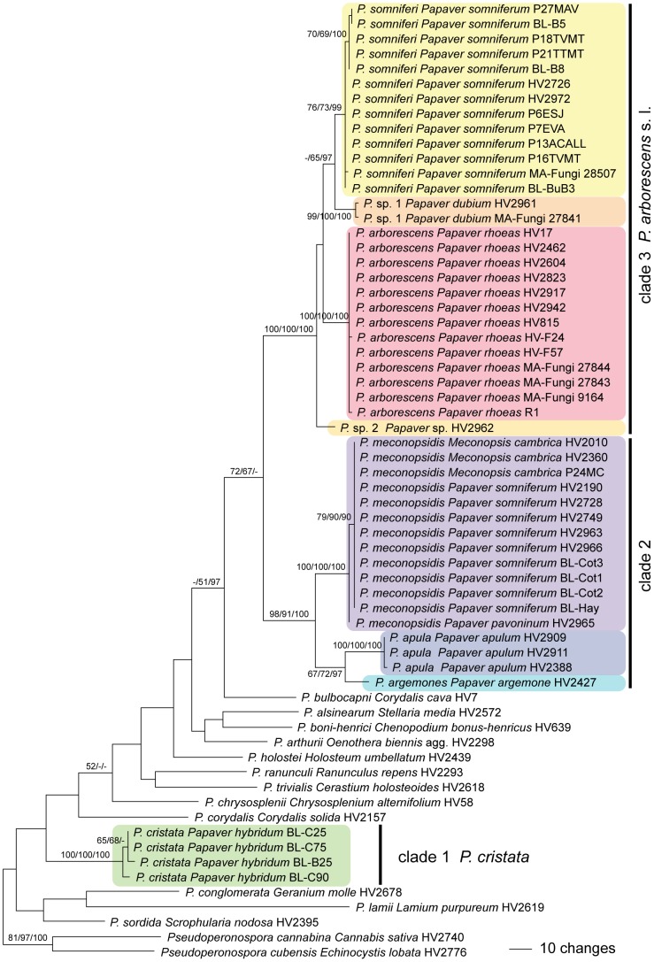
Of the 1262 characters of the combined cox1 - cox2 alignment, 234 were parsimony informative (120 in cox1, 114 in cox2). MP analyses revealed 16 MP trees 1035 steps long, one of which was selected and presented in Figure 1, with MP and ML bootstrap support above 50% and posterior probabilities above 90% given at first, second and third position, respectively, above/below the branches. Topologies of the MP trees slightly differed in the phylogenetic positions of P. sordida, P. corydalis and P. chrysosplenii. The three Bayesian runs revealed almost identical posterior probabilities (PP) and were fully compatible with the MP strict consensus tree. The Peronospora-accessions from Papaver/Meconopsis were contained within three highly supported clades, one comprising Peronospora cristata from Papaver hybridum (clade 1 in Figure 1); a second clade containing P. apula from Papaver apulum, P. argemones from Papaver argemones and P. meconopsidis from Meconopsis cambrica, Papaver pavoninum and P. somniferum (clade 2 in Figure 1); and a third clade with P. arborescens from Papaver rhoeas, P. somniferi from Papaver somniferum, P. sp. 1 from Papaver dubium and P. sp. 2 from Papaver sp. (clade 3 in Figure 1). Whereas within the Peronospora arborescens clade the ITS did not resolve accessions from the different hosts, in the cox1 - cox2 trees the accessions from Papaver dubium, P. rhoeas and P. somniferum were placed in three distinct monophyletic clades, the former two with high and the latter with medium to high support (Figure 1).
Figure 1. Phylogram showing phylogenetic relationships of Peronospora accessions from Papaver and Meconopsis.
One of 16 most parsimonious trees 1035 steps long inferred from the combined cox1-cox2 sequence data matrix; parsimony and likelihood bootstrap support above 50% and posterior probabilities above 90% are given at first, second and third position, respectively, above/below the branches. The tree was rooted with two species of Pseudoperonospora according to Göker et al. [38].
Within the Peronospora arborescens sensu lato clade (clade 3 in Figure 1), Peronospora accessions from Papaver somniferum consistently exhibited 11 and 10 common sequence substitutions in cox1 and cox2, respectively, if compared to sequences of P. arborescens from Papaver rhoeas, rendering them molecularly clearly distinct. Peronospora accessions from Papaver somniferum also differed morphologically from those from P. rhoeas in larger conidia (mean 21.1 × 17.7 µm vs. 18.3 × 16.1 µm).
Taxonomy
As a result of the morphological and molecular phylogenetic investigations, six taxa are here recognised as occurring on Papaver, two of which are described as new. In addition, detailed descriptions of the other four already described species are provided. All type specimens cited were morphologically investigated in the present study.
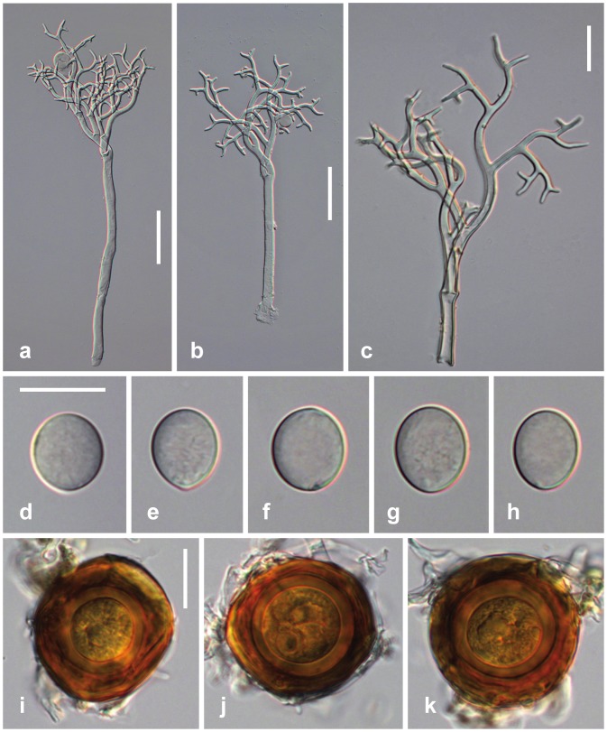
Peronospora Apula Voglmayr, sp. nov. Figure 2
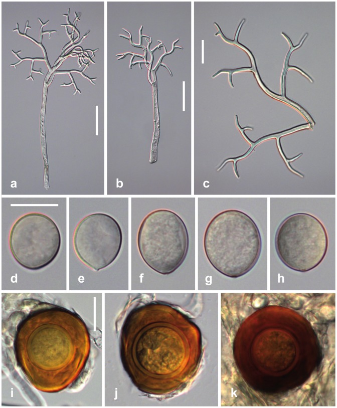
Figure 2. Morphological features of Peronospora apula.
a, b conidiophores; c ultimate branchlets; d–h conidia; i–k oogonia and oospores. Sources: a, e–k WU 32410, holotype; b WU 32408; c, d WU 32409. Scale bars a, b 50 µm, c–k 20 µm.
Mycobank: MB 808433.
Description
Infection systemic or localized, when systemic whole plants or leaves stunted, stems strongly distorted, sinuous. Down mostly hypophyllous, greyish, consisting of dense and felt-like conidiophores. Conidiophores hyaline, straight to slightly sinuous, (170–)270–430(–500) µm long; trunk straight or curved, (60–)150–280(–360) µm long (n = 25), variable in width, 4–10.5 µm wide; callose plugs absent; upper part monopodially or subdichotomously branched 5–6 times. Branches curved, sinuous. Ultimate branchlets in pairs, straight to slightly curved, (3–)5.5–11.5(–19.5) µm long, 2–2.5 µm wide at the base (n = 349), apex obtuse. Conidia subhyaline to pale brown, subglobose, ellipsoidal to obovate, (14–)16.5–20(–22) µm long, (12–)14.5–17(–19) µm wide, mean 18.3 × 15.8 µm, l/w ratio (1.01–)1.07–1.25(–1.43) (n = 214), greatest width median, base and tip round; pedicel absent in most conidia but a scar visible at the point of attachment; producing germ tubes. Oogonia globose, subglobose to irregular, yellow brown to dark reddish brown, (29–)40–48(–54) µm diam., wall smooth, 1.5–2.7 µm thick (n = 88). Oospores distinctly aplerotic, globose, (21–)25–30(–34) µm diam., wall 1.9–4.5 µm thick (n = 88), smooth.
Molecular diagnosis
Peronospora apula differs from its closest phylogenetic neighbour, P. argemones, by unique fixed alleles in two tree loci (cox1, cox2) based on alignments of the separate loci deposited in TreeBASE as study S15609: cox1 positions 343: A; 49, 205, 346, 349, 425, 640: C; 148, 610, 664: G; 193, 217, 340, 652: T; cox2 positions 154, 232, 370, 565: A; 295, 469: C; 65, 109, 187, 372, 373, 449: G; 105, 133, 262: T.
Etymology
Referring to its host, Papaver apulum Ten.
Habitat
On living leaves and stems of Papaver apulum.
Holotype
CROATIA, Istrija, SE Rovinj, ca. 800 m ESE Kamp Veštar, field, 18 May 2012, H. Voglmayr HV2911(WU 32410).
Additional specimens examined
CROATIA, Istrija, N Rovinj, E Valalta, 17 May 2012, H. Voglmayr HV2911 (WU 32409). Istrija, Mandriol ESE Bale, 14 May 2010, H. Voglmayr HV2388 (WU 32408).
Comments
Peronospora apula appears to be confined to Papaver apulum. It is closely related to P. argemones which differs by larger conidia, its host Papaver argemone and by different ITS (10 substitutions), cox1 (14 substitutions) and cox2 (15 substitutions) sequences.
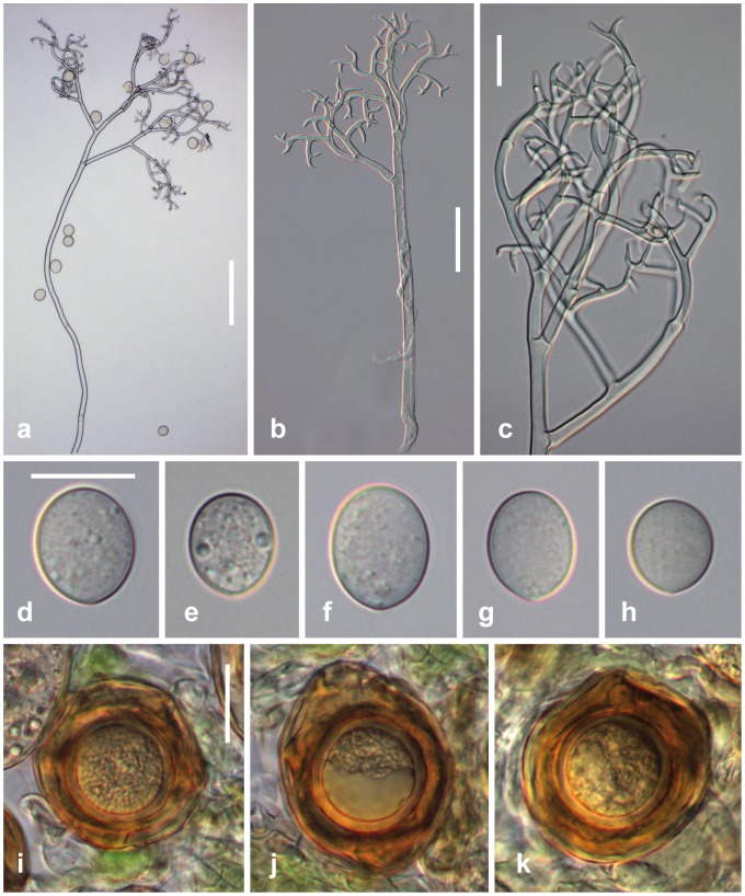
Peronospora arborescens (Berk.) de Bary, Monatsber. Königl. Preuss. Akad. Wiss. Berlin: 308-333 (1855) Figure 3 .
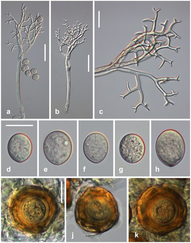
Figure 3. Morphological features of Peronospora arborescens.
a, b conidiophores; c ultimate branchlets; d–h conidia; i–k oogonia and oospores. Sources: a WU 32416; b, k WU 32411; c, f, g WU 32412; d, h WU 32418; e WU 32413. Scale bars a 100 µm, b 50 µm, c–k 20 µm.
Basionym: Botrytis arborescens Berk., J. hort. Soc., London 1∶31 (1846).
Description
Infection commonly systemic, more rarely localized, when systemic whole plants or leaves stunted, strongly distorted, chlorotic, dwarfed, when localized producing polyangular to confluent lesions with distinct margins. Down hypophyllous, greyish, consisting of dense and felt-like conidiophores. Conidiophores hyaline, straight to slightly sinuous, (290–)360–600(–720) µm long; trunk straight or curved, (110–)190–380(–460) µm long (n = 23), variable in width, 5.5–13 µm wide; callose plugs absent; upper part monopodially or subdichotomously branched 5–7 times. Branches distinctly curved, sinuous. Ultimate branchlets mostly in pairs, slightly to strongly curved, (2–)4.5–9.5(–18) µm long, 2–2.7 µm wide at the base (n = 511), apex obtuse. Conidia subhyaline to pale brown, subglobose, ellipsoidal to obovate, (14–)16.5–20(–24) µm long, (12.5–)15–17.5(–20) µm wide, mean 18.3 × 16.1 µm, l/w ratio (1.01–)1.07–1.21(–1.45) (n = 413), greatest width median, base and tip round; pedicel absent in most conidia but a scar visible at the point of attachment; producing germ tubes. Oogonia globose, subglobose to irregular, yellow brown to dark reddish brown, (37–)43–51(–59) µm diam., wall commonly wrinkled, smooth, ca. 1 µm thick (n = 193). Oospores distinctly aplerotic, globose, (21–)26–30(–33) µm diam., wall 1.5–2.7 µm thick (n = 193), smooth.
Habitat
On living leaves and stems of Papaver rhoeas L.
Typification
UK, King's Cliffe, without date and collector, ex Herb. Berkeley (K(M) 178950, holotype).
Selected additional specimens examined
AUSTRIA, Burgenland, Neusiedl/See, Kittsee, E Groβer Raubwald near Edelstal, 27 Mar 1999, H. Voglmayr HV17 (WU 22880). Niederösterreich, Baden, Bad Vöslau, 19 Apr 2011, H. Voglmayr HV2604 (WU 32411). Wien, Leopoldstadt, Praterspitz, 4 Apr 2012, H. Voglmayr HV2823 (WU 32412). CROATIA, Istrija, E Peroj, 20 May 2012, H. Voglmayr HV2917 (WU 32413). FRANCE, Herault, SW Montpellier, between Montbazin and Cournosec, 9 Apr 2001, H. Voglmayr HV-F57 (WU 32414). S Lyon, Drôme, Hameau des Balmes W Romans-s-Isère, 7 Apr 2001, H. Voglmayr HV-F24 (WU 32415). GERMANY, Baden-Württemberg, Tübingen, E Lustnau, Furtwiesen, 28 Apr 2001, H. Voglmayr HV815 (WU 32416). HUNGARY, Hajdú-Bihar, between Hajdúszoboszló and Kaba, 25 Oct 2010, H. Voglmayr HV2462 (WU 32417). ITALY, Lombardia, NW Bergamo, Barzano, near Arzenate, 28 May 2012, H. Voglmayr HV2942 (WU 32418).
Comments
Peronospora arborescens appears to be confined to Papaver rhoeas, on which it is very common. Its disease symptoms are highly distinctive, infected plants being distorted and showing light yellowish green discolouration. The various accessions included in our molecular phylogenetic analyses covered most of Europe (Austria, Croatia, France, Germany, Hungary, Italy, Romania, Spain), and were molecularly highly homogeneous. Accessions from other Papaver species were placed in distinct clades and represent genetically distinct lineages. The accessions from Spain were partly sampled within cultivated opium poppy fields.
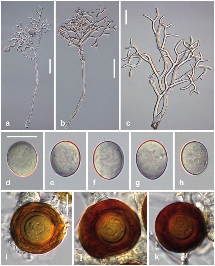
Peronospora argemones Gäum., Beitr. Kryptfl. Schweiz 5(no. 4): 72 (1923) Figure 4 .
Figure 4. Morphological features of Peronospora argemones.
a, b conidiophores; c ultimate branchlets; d–h conidia; i–k oogonia and oospores. Sources: a, c GLM 64084; b K(M) 181196, holotype. Scale bars a, b 50 µm, c-k 20 µm.
Description
Infection commonly systemic, whole plants or leaves stunted, slightly to strongly distorted. Down hypophyllous, greyish, consisting of dense and felt-like conidiophores. Conidiophores hyaline, straight to slightly sinuous, (220–)290–490(–590) µm long; trunk straight to slightly curved, (80–)150–300(–390) µm long (n = 35), variable in width, 4–13 µm wide; callose plugs absent; upper part monopodially or subdichotomously branched 5–6 times. Branches tightly intertwined, curved, sinuous. Ultimate branchlets in pairs, slightly curved, (2.5–)6–14(–23) µm long, 1.8–2.8 µm wide at the base (n = 468), apex obtuse. Conidia subhyaline to pale brown, subglobose, ellipsoidal to obovate, (16.5–)19–23.5(–26) µm long, (14.5–)16.5–20(–23) µm wide, mean 21.1 × 18.1 µm, l/w ratio (1.02–)1.1–1.23(–1.37) (n = 181), greatest width median, base and tip round; pedicel absent in most conidia but a scar visible at the point of attachment; producing germ tubes. Oogonia globose, subglobose to irregular, light to dark reddish brown, (42–)48–59(–66) µm diam., wall smooth, 1–1.7 µm thick (n = 109). Oospores distinctly aplerotic, globose, (25–)28–34(–38) µm diam., wall 1.8–4.2 µm thick (n = 109), smooth.
Habitat
On living leaves and stems of Papaver argemone L.
Typification
GERMANY, Berlin, Lichterfelde, June 1896, P. Sydow, Phycomyc. Protomyc. 5 (K(M) 181196, lectotype here designated, MBT177702; WU s.n., isotype).
Additional specimen examined
GERMANY, Sachsen-Anhalt, Bitterfeld, SW Friedersdorf, Muldeaue, 8 May 2004, H. Jage (GLM 64084).
Comments
Peronospora argemones appears to be confined to Papaver argemone. Gäumann [13] separated P. argemones from P. arborescens based on larger conidia. Our measurements fit well those reported by Gäumann [13] (21.1 × 18.1 vs. 21 × 18.6). P. argemones is not closely related to P. arborescens but to P. meconopsidis which has larger conidia. Its closest relative, however, is P. apula, which differs by slightly smaller conidia and its host Papaver apulum.
Peronospora cristata Tranzschel, Trav. Mus. Bot. Acad. Sci. St. Petersburg 1∶49 (1902) Figure 5 .
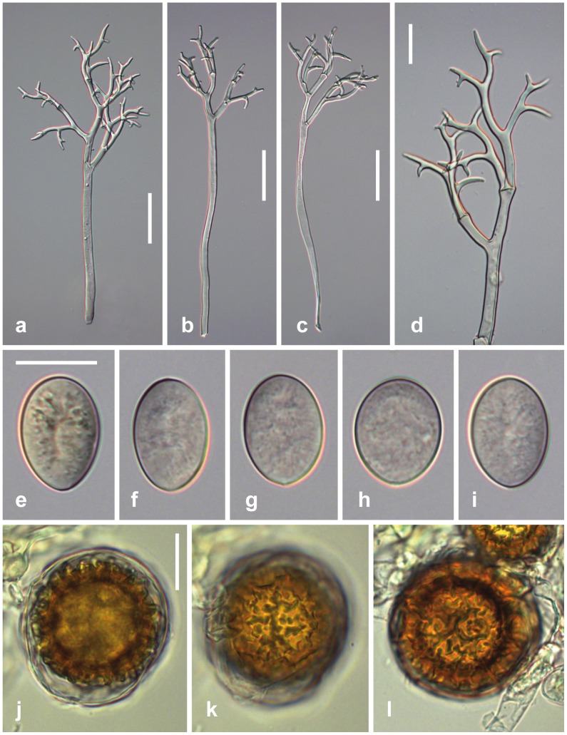
Figure 5. Morphological features of Peronospora cristata.
a-c conidiophores; d ultimate branchlets; e–i conidia; j–l oogonia and oospores. Sources: a, b, d, e, j, k LE 185561, holotype; c, h WU 32419; f, g, i, l WU 32421. Scale bars a-c 50 µm, d-l 20 µm.
Description
Infection commonly systemic, more rarely localized, when systemic whole plants or leaves stunted, slightly distorted, chlorotic. Down hypophyllous, greyish, consisting of dense and felt-like conidiophores. Conidiophores hyaline, straight or slightly sinuously curved, (200–)240–330(–380) µm long; trunk straight or curved, (95–)130–220(–280) µm long (n = 33), variable in width, 4.5–10.5 µm wide; callose plugs absent; upper part monopodially or subdichotomously branched 3–5 times. Branches straight to slightly sinuously curved. Ultimate branchlets in pairs, slightly to distinctly curved, (2.5–)5–10.5(–19) µm long, 2–3.6 µm wide at the base (n = 263), apex obtuse. Conidia pale brown to brown, broadly ellipsoidal, ellipsoidal to obovate, (21.5–)25–28(–32) µm long, (15.5–)18–21(–24) µm wide, mean 26.5 × 19.6 µm, l/w ratio (1.15–)1.27–1.43(–1.53) (n = 122), greatest width median, base and tip round; pedicel absent in most conidia but a scar visible at the point of attachment; producing germ tubes. Oogonia mostly globose, rarely subglobose to irregular, golden to reddish brown, (38–)47–57(–69) µm diam., wall smooth, ca. 1 µm thick (n = 52). Oospores almost plerotic to slightly aplerotic, globose, (34–)40–49(–54) µm diam., wall distinctly irregularly verrucose, ca. 5.5–10 µm thick including ornamentation (n = 52); verrucae 1.2–2.3 µm thick, rarely isolated, commonly confluent, forming irregular crests to an incomplete labyrinthic net.
Habitat
On living leaves and stems of Papaver hybridum L.
Typification
UKRAINE, Crimea ("Tauria"), Georgyievskij Monastery near Sevastopol', 26 Apr 1901, W. Tranzschel (LE 185561, holotype).
Additional specimens examined
SPAIN, Córdoba, at (SW) the Puente de Andalucía, IFAPA, 4 Apr 2008, B. B. Landa & M. Montes (WU 32419). Same locality, 8 Apr 2008, B. B. Landa & M. Montes (WU 32420). Same locality, 15 Apr 2008, B. B. Landa & M. Montes (WU 32421). UKRAINE, Crimea ("Tauria"), Hortus Nikitensis, 26 Apr 1902, W. Tranzschel (LE 43379). Simeiz, 30 Apr 1902, W. Tranzschel (LE 43377). Jalta, 14 May 1903, W. Tranzschel (LE 185505).
Comments
Peronospora cristata has been confirmed only from Papaver hybridum, with which it is apparently co-occurring. The type and authentic specimens of Tranzschel [23] from the Crimean Peninsula morphologically fully resemble the recent Spanish collections for which DNA data are available. The irregularly verrucose oospores are highly distinctive for the species, all other Peronospora species on Papaver having smooth oospores. In addition, it has the largest conidia of all Peronospora species on Papaver. Peronospora meconopsidis, which has conidia of similar size, differs by smooth oospores and different hosts (Papaver somniferum, Meconopsis cambrica).
Peronospora meconopsidis Mayor, Mém. Soc. neuchât. de Sc. Nat. 9(1): 34 (1958) Figure 6 .
Figure 6. Morphological features of Peronospora meconopsidis.
a, b conidiophores; c ultimate branchlets; d–h conidia; i–k oogonia and oospores. Sources: a, f, g WU 32426; b, c–e NEU, holotype; h WU 32425; i K(M) 179241; j, k K(M) 179245. Scale bars a, b 50 µm, c–k 20 µm.
Replaced synonym
Peronospora gaeumannii Mayor [as 'gaeumanni'], Ber. schweiz. bot. Ges. 59∶278 (1949), non Peronospora gaeumannii Mundk. [as 'gaeumanni'], Scientific Monogr. Coun. Agric. Res. India 12∶8 (1938).
Description
Infection localized, producing more or less distinct polyangular to confluent lesions. Down mostly hypophyllous, greyish, consisting of mostly scattered conidiophores, very rarely dense and felt-like. Conidiophores hyaline, straight, (230–)240–510(–730) µm long; trunk straight, (70–)110–330(–500) µm long (n = 39), variable in width, 5–15 µm wide; callose plugs absent; upper part monopodially or subdichotomously branched 3–5 times. Branches slightly to strongly sinuously curved. Ultimate branchlets in pairs, slightly to distinctly curved, (2.5–)7.5–17.5(–37) µm long, 1.9–3.3 µm wide at the base (n = 810), apex obtuse. Conidia pale to medium brown, subglobose, ellipsoidal to obovate, (17–)22.5–27.5(–35.5) µm long, (15–)19–22.5(–26) µm wide, mean 24.8 × 20.6 µm, l/w ratio (1.03–)1.13–1.28(–1.47) (n = 416), greatest width median, base and tip round; pedicel absent in most conidia but a scar visible at the point of attachment; producing germ tubes. Oogonia rarely globose, mostly subglobose to irregular, reddish brown, (29–)40–48(–54) µm diam., wall smooth, 1–1.5 µm thick (n = 88). Oospores distinctly aplerotic, globose, (21–)25–30(–34) µm diam., wall 1.7–3.3 µm thick (n = 88), smooth.
Habitat
On living leaves of Meconopsis cambrica Vig., Papaver pavoninum C.A. Mey. and P. somniferum L.
Typification
SWITZERLAND, Neuchâtel, Val-de-Travers, Môtiers, Gorges de la Poëta Raisse, on Meconopsis cambrica, 31 July 1945, 21 Sep 1946, 17 Sep 1947, 17 Aug 1949, 14 July 1952, E. Mayor (NEU, lectotype here designated, MBT177701). ibid., 17 Sep 1947, E. Mayor (NEU, isolectotype). Neuchâtel, Vallée des Ponts, Combe Varin, garden, 27 June 1920, E. Mayor (NEU, syntype).
Additional selected specimens examined
On Meconopsis cambrica: AUSTRIA, Styria, Graz, Botanical Garden, 14 Sep 2002, H. Voglmayr HV2010 (WU 32422). UK, London, Kew Gardens, 14 Nov 2008, H. Voglmayr HV2360 (WU 32423). On Papaver pavoninum: RUSSIA, Moskva, Principal Botanical Garden an SSSR, 1 June 1959, E. Protsenko (K(M) 179239). On Papaver somniferum: AFGHANISTAN, Jalalabad, 14 Mar 1973, M.A. Ghani 7 (K(M)179245). AUSTRIA, Niederösterreich, Gänserndorf, Weiden an der March, Dornparz ESE Zwerndorf, 6 July 2011, H. Voglmayr HV2749 (WU 32424). Austria, Oberösterreich, Schärding, St. Willibald, 31 July 2005, H. Voglmayr HV2190 (WU 32425). CZECH REPUBLIC, Morava, Hranice, between Teplice nad Bečvou and Černotin, 25 June 2011, H. Voglmayr HV2728 (WU 32426). PAKISTAN, Darra Adam Khel, 9 Mar 1975, M.A. Ghani (K(M) 179248). POLAND, Nisko, Rudnik, 2 July 1957, J. Kowalski, in J. Kochman Mycoth. Polon. 11 (K(M) 179244). ROMANIA, Transsilvania, Hunedoara, Gura Zlata, 4 Aug 1963, M. Bechet, in Borza, Gergely & Raţiu, Fl. Roman. Exs. 3006 (K(M) 179243). TURKMENISTAN, Kordon Kepelya, 18 Apr 1978, V.A. Mel'nik (K(M) 179241). THAILAND, Mae Cham, Chiang Mai ("Chiengmai"), 1 Sep 1982, P. Pitakpaivan 0472 (K(M) 179247).
Comments
Peronospora meconopsidis is commonly observed on Meconopsis cambrica and Papaver somniferum, but its disease symptoms are usually rather inconspicuous and localized compared to most other Peronospora species from Papaver. Although it has not been recorded from Papaver somniferum in Europe, it is apparently common and widespread on that host according to own observations and upon examination of herbarium specimens. It has commonly been misidentified as Peronospora arborescens, which mainly differs by conspicuous disease symptoms relating to mostly systemic infection which has never been observed for P. meconopsidis. Based on similar conidial sizes, P. meconopsidis has been referred to as Peronospora cristata in recent literature, which goes back to Reid [14] who classified accessions from Meconopsis cambrica under that species. However, P. cristata is easily distinguishable by its verrucose oospores, which are unique in Peronospora on Papaver. The Australia records of Peronospora cristata from Papaver somniferum [5] therefore actually represent Peronospora meconopsidis, which is also corroborated by sequence data (Figure 1).
Oospores of Peronospora meconopsidis are reported here for the first time, and they have only been found in young infected plants of Papaver somniferum collected in Asia. No oospores were found in specimens of Papaver somniferum from Europe or from Meconopsis cambrica despite thorough investigations.
The species was first described as Peronospora gaeumannii by Mayor [46]. However, because this is a younger homonym of P. gaeumannii Mundk., Mayor [26] proposed the new name P. meconopsidis. Three authentic specimens mentioned in the original description of P. gaeumannii Mayor are present at NEU, of which the largest, best developed and preserved is here selected as lectotype. This folder also contains the original drawings and spore statistics published in Mayor [46]. According to the herbarium label, the lectotype specimen consists of several collections from the same place collected from 1945 to 1952, which were subsequently mixed and cannot be separated any more. The mean spore sizes recorded by Mayor [46] agree well with those of the current study (23.5 × 21.15 vs. 24.8 × 20.6 µm).
Peronospora somniferi Voglmayr, sp. nov. Figure 7 .
Figure 7. Morphological features of Peronospora somniferi.
a, b conidiophores; c ultimate branchlets; d–h conidia; i–k oogonia and oospores. Sources: a, g WU 32432; b, d, i–k WU 32428, holotype; c MA 65574; e, f WU 32429; h WU 32430. Scale bars a, b 50 µm, c–k 20 µm.
Mycobank MB 808434.
Description
Infection systemic or localized, when systemic whole plants or leaves stunted, stems strongly distorted, sinuous. Down mostly hypophyllous, greyish, consisting of dense and felt-like conidiophores. Conidiophores hyaline, straight to slightly sinuous, (280–)320–510(–660) µm long; trunk straight or curved, (100–)140–330(–490) µm long (n = 33), variable in width, 5.5–17 µm wide; callose plugs absent; upper part monopodially or subdichotomously branched 4–7 times. Branches straight to sinuously curved. Ultimate branchlets in pairs, straight to slightly curved, (2–)4.5–10(–18.5) µm long, 1.9–3.2 µm wide at the base (n = 550), apex obtuse. Conidia subhyaline to pale brown, subglobose, ellipsoidal to obovate, (15.5–)19–23(–28) µm long, (14.5–)16.5–19(–22.5) µm wide, mean 21.1 × 17.7 µm, l/w ratio (1.01–)1.11–1.28(–1.48) (n = 927), greatest width median, base and tip round; pedicel absent in most conidia but a scar visible at the point of attachment; producing germ tubes. Oogonia globose, subglobose to irregular, yellow brown to dark reddish brown, (31–)40–48(–57) µm diam., wall smooth, ca. 1 µm thick (n = 157). Oospores distinctly aplerotic, globose, (19–)24–28(–34) µm diam., wall 1.6–2.8 µm thick (n = 157), smooth.
Molecular diagnosis
Peronospora somniferi differs from its closest phylogenetic neighbour, P. arborescens, by unique fixed alleles in two tree loci (cox1, cox2) based on alignments of the separate loci deposited in TreeBASE as study S15609: cox1 positions 205, 337, 418, 491, 646: A; 195: C; 229, 331, 499, 557, 589: T; cox2 positions 313, 370, 430, 523: A; 493: C; 382, 541: G; 220, 253, 427: T.
Etymology
Referring to its host, Papaver somniferum L.
Habitat
On living leaves and stems of Papaver somniferum.
Holotype
CZECH REPUBLIC, Morava, Hranice, between Teplice nad Bečvou and Černotin, 25 June 2011, H. Voglmayr HV2726 (WU 32428).
Additional specimens examined
AUSTRIA, Oberösterreich, Linz Land, Kronsdorf, Schieferegg, 25 May 2004, G. Bedlan 721 (WU 32427). IRAN, Neelabad, 10 Apr 1973, M.A. Ghani 3 (K(M) 179246). SPAIN, Albacete, Casa Arriba los Llanos, 10 June 2005, B. Landa (WU 32429). Sevilla, Écija, Casilla San José, 29 Apr 2004, B. Landa (WU 32430). Sevilla, Écija, San Rafael, 29 Apr 2004, B. Landa (WU 32431). Sevilla, Écija, Vacas, without collector and date (MA-Fungi 65574). Malaga, Antequera, Monteluna, without collector and date (MA-Fungi 65500). Marchena, Cortijo del Rio, 4 Apr 2004, B. Landa (WU 32432).
Comments
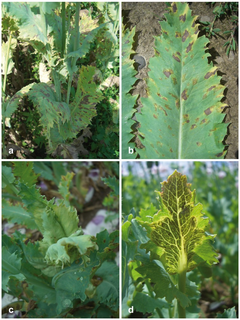
Peronospora somniferi appears to be confined to Papaver somniferum. It is closely related to P. arborescens which differs by smaller conidia and a different host, Papaver rhoeas. Peronospora somniferi cannot be reliably distinguished from P. arborescens by ITS data alone because there is only a single consistent nucleotide difference at the beginning of the ITS1, but cox1 and cox2 are distinctive and good barcode markers for the species (11 and 10 diagnostic substitutions, respectively). Peronospora somniferi has been reported as an economically important pathogen of Papaver somniferum throughout Europe [4]. Peronospora meconopsidis, which also occurs on Papaver somniferum, differs mainly by a non-systemic infection which is characterised by typical polyangular spots (see Figure 8).
Figure 8. Disease symptoms of Peronospora meconopsidis and P. somniferi on opium poppy (Papaver somniferum).
a, b polyangular leaf lesions of P. meconopsidis; c, d systemic infection of P. somniferi from above (c) and from underneath (d), showing the grey down. Sources: a WU 32424; b WU 32426; c, d WU 32428, holotype.
Key to known Species of Peronospora on Papaver and Meconopsis
Note: Morphological identification requires well developed material as well as knowledge of the host. For conidial measurements, it is crucial that a sufficient number of mature conidia are included. Because some species are highly similar, they often cannot be unequivocally identified by morphology alone, and sequence data (cox1 or cox2) are essential in case of poorly developed specimens, if the host species is unknown or represents a host species from which no sequence data are available or from which several Peronospora species are known.
1 Oospore wall irregularly verrucose, only known from Papaver hybridum............................................................................... P. cristata
Oospore wall smooth..................................................................2
2(1) Conidia in mean longer than 20 µm..................................3
Conidia in mean shorter than 20 µm........................................5
3(2) Infection local, conidia in mean longer than 22 µm, confirmed from Meconopsis cambrica, Papaver pavoninum, P. somniferum.................................................................... P. meconopsidis
Infection systemic, conidia in mean shorter than 22 µm..........4
4(1) On Papaver argemone.....................................P. argemones
On Papaver somniferum...........................................P. somniferi
5(2) On Papaver apulum, conidiophores (170–)270–430(–500) µm high...................................................................................P. apula
On Papaver rhoeas, conidiophores (290–)360–600(–720) µm high............................................................................P. arborescens
Discussion
Molecular Phylogenetic Investigations
The current investigations clearly show that the biodiversity of Peronospora on Papaver is higher than previously perceived, which is in line with other investigations on Peronosporaceae (e.g. [37], [47]–[63]), demonstrating that high biodiversity is commonly the result of high host specificity. Peronospora from Papaver are distributed amongst three clades (Figure 1), of which P. cristata from clade 1 is phylogenetically isolated from the other Peronospora species from Papaver, but clades 2 and 3 appear to be closely related (Figure 1). Of special interest is clade 3, the species of which were formerly classified under Peronospora arborescens, and which is here referred to as P. arborescens sensu lato clade. Whereas the ITS data are highly similar within this clade and therefore do not allow for unequivocal distinction (data not shown), the cox1 and cox2 data are highly distinctive for the accessions from various hosts, in which each form genetically homogeneous lineages irrespective of the geographic origins. This is evident for the Peronospora accessions from Papaver rhoeas and P. somniferum which were sampled from various regions all over Europe, and which form distinct uniform genetic lineages. In addition, also differences in conidial sizes could be documented for both lineages. Therefore, the accessions from Papaver somniferum, previously classified under P. arborescens, are here described as a distinct species, P. somniferi. Apart from these two lineages, another two genetically distinct entities were present within the P. arborescens s. l. clade (Figure 1), Peronospora sp. 1 from Papaver dubium, and Peronospora sp. 2 from an unidentified Papaver species. However, because only few accessions were available for these, we currently refrain from describing them as new taxa. Considering the numerous additional Papaver species for which Peronospora have been recorded (see [4]) but for which no accessions were available for molecular phylogenetic investigations, additional genetically distinct entities may turn up in the future.
Molecular Barcoding
cox1, chosen as barcoding locus for higher animals and considered to be the primary barcoding marker for organisms unless shown to be unsuitable (http://www.barcodeoflife.org), has been demonstrated to be an appropriate barcoding locus for oomycetes [32], which is confirmed in the current study. However, cox2 shows similarly good resolution and consequently is an equally good barcoding marker; it even has some advantages over cox1, as it usually amplifies better especially in cases of low DNA quantity or old herbarium samples (as also shown in [64]), and thus cox2 sequences are available for many more species in Peronosporales. The current study provides additional data for equally good discriminative power of cox1 and cox2; e.g. Peronospora somniferi is distinct from Peronospora arborescens by shared 11 and 10 substitutions in cox1 and cox2, respectively, clearly showing a similar significant barcode gap between both species in both markers. Therefore, both cox1 and cox2 are considered good markers for reliable identification of Peronospora species on Papaver, and it is recommended to sequence both loci in studies of Peronosporales whenever possible to obtain representative robust data for molecular barcoding. On the other hand, the ITS region does not resolve closely related species like P. arborescens and P. somniferi, which has been observed also in other groups of Peronosporaceae (e.g. [47], [54]).
Nomenclature of Peronospora on Papaver
In the recent literature, accessions infecting Papaver were mainly classified as Peronospora arborescens. However, as the molecular data clearly show, there are several species involved, six of which are recognized and treated in the current publication. However, nomenclature of these species is partly complex mainly due to misleading species concepts of the past, as shall be outlined below.
In a phytopathological perspective, an important result of recent molecular phylogenetic investigations was that downy mildew disease of the crop Papaver somniferum is caused by two distinct species, which were classified as Papaver arborescens and Papaver cristata [4], [5]. However, the current detailed investigations show that this classification cannot be retained and has to be substantially modified.
Starting from Gäumann [13], Peronospora accessions from Papaver somniferum have been classified as Peronospora arborescens, which has been consistently followed. However, the current molecular phylogenetic investigations show that these accessions are genetically distinct from accessions from the type host, Papaver rhoeas, and there are also differences in conidial sizes. Consequently, the Peronospora accessions from Papaver somniferum belonging to the Peronospora arborescens sensu lato clade (clade 3; Figure 1) are here classified as a distinct species, P. somniferi.
Recently, the name Peronospora cristata was used for a second Peronospora species on Papaver somniferum that was first recorded from that host by Scott et al. [5] from Tasmania. This name was applied because their ITS sequences matched an English accession from Meconopsis cambrica that had previously been deposited in GenBank (DQ885375) under Peronospora cristata. This naming apparently goes back to Reid [14], who compared conidial measurements of Peronospora from Meconopsis cambrica with those from Papaver argemone and considered them to be conspecific due to similar size. In addition, because conidial sizes were also similar to those reported for Peronospora cristata from Papaver hybridum, he considered accessions from these three hosts to be conspecific, and thus classified them under P. cristata due to priority. However, he ignored that the oospores of Peronospora cristata are distinctly verrucose, being significantly different from the smooth oospores of Peronospora argemones. These substantial morphological differences are also mirrored by a distant phylogenetic position of P. cristata (Figure 1), confirming that P. cristata is a clearly distinct species confined to Papaver hybridum.
In addition, also the accessions from Papaver argemone and Meconopsis cambrica are phylogenetically distinct (Figure 1) and therefore have to be classified under P. argemones and P. meconopsidis, respectively. Consequently, as the Peronospora of M. cambrica and the second Peronospora species from P. somniferum are conspecific, the correct name to be applied for this species is P. meconopsidis.
Host-parasite Relationships
The phylogenetic relationships of Peronospora on Papaver and those of their hosts are only partly congruent. For instance, within Papaver sect. Argemonidium which form a closely related group [1], the Peronospora species from Papaver argemone and P. apulum (Peronospora argemones and P. apula) are closest relatives, whereas the Peronospora from the third species of the section, Papaver pavoninum, falls within P. meconopsidis which is sister group to the former two species (Figure 1). However, Peronospora cristata, parasitising the fourth species of sect. Argemonidium, Papaver hybridum, is phylogenetically and morphologically distinct from all other Peronospora species on Papaver.
Remarkably, Peronospora meconopsidis infects hosts from two genera, Meconopsis and Papaver. However, molecular phylogenetic investigations show that Meconopsis is polyphyletic and embedded within Papaver, M. cambrica being unrelated to the other Meconopsis species [1]. Whereas the two main hosts of Peronospora meconopsidis, M. cambrica and Papaver somniferum are contained within the same clade but not closely related, the third host species, P. pavoninum, is member of the phylogenetically isolated Papaver section Argemonidium [1]. This indicates that phylogenetic radiation of Peronospora on Papaver is rather effected by host jumps.
Finally, from a pytopathological point of view the results from this study showed that wild Papaver spp. cannot play any role as primary inoculum for downy mildew epidemics in cultivated opium poppy crops since there is high host specificity. This result was corroborated when different Peronospora specimens (P. somniferi, P. cristata and P. arborescens) were sampled from the same fields of cultivated Papaver somniferum where Papaver hybridum and Papaver rhoeas were growing as weeds. Consequently, to avoid development of downy mildew epidemics in cultivated opium poppy efforts should be placed in controlling airborne P. somniferi sporangia from diseased opium poppy plants as well as avoiding the use infected seed lots [22].
Acknowledgments
We thank the herbarium curators of GLM, K, LE, MA and NEU for sending specimens for investigation and Walter Till (WU) for handling the herbarium loans. C. Cantalapiedra (IAS-CSIC) is gratefully acknowledged for technical assistance. D. E. L. Cooke (Scottish Crop Research Institute, Invergowrie, Dundee, Scotland, UK), F. S. Hay (Tasmanian Institute of Agricultural Research, University of Tasmania, Burnie, Australia) and P. J. Cotterill (GlaxoSmithKline, Latrobe, Tasmania, Australia) are gratefully acknowledged for sending material of P. meconopsidis, and G. Bedlan for contributing a specimen of P. somniferi. F.J Muñoz-Ledesma and technicians from Alcaliber, S.A., Carmona, Spain are gratefully acknowledged for providing specimens of P. meconopsidis, P. arborescens, and P. somniferi.
Funding Statement
Financial support by the Austrian Science Fund (FWF, project P22739-B20, http://www.fwf.ac.at/) to HV and by Incentivos a Proyectos de Excelencia (P10-AGR-6497), CICE-Junta de Andalucía and FEDER (EU) to MMB is gratefully acknowledged. The funders had no role in study design, data collection and analysis, decision to publish, or preparation of the manuscript.
References
- 1. Carolan JC, Hook IL, Chase MW, Kadereit JW, Hodkinson TR (2006) Phylogenetics of Papaver and related genera based on DNA sequences from ITS nuclear ribosomal DNA and plastid trnL intron and trnL–F intergenic spacers. Ann Bot 98: 141–155. [DOI] [PMC free article] [PubMed] [Google Scholar]
- 2.Kapoor LD (1995) Opium poppy: botany, chemistry and pharmacology. New York: Food Products Press.
- 3. Cotterill PJ, Pascoe IG (1998) Downy mildew of Papaver somniferum in Tasmania. Australas Plant Pathol 27: 263–264. [Google Scholar]
- 4. Landa BB, Montes-Borrego M, Muñoz-Ledesma FJ, Jiménez-Díaz RM (2007) Phylogenetic analysis of downy mildew pathogens of opium poppy and PCR-based in planta and seed detection of Peronospora arborescens . Phytopathology 97: 1380–1390. [DOI] [PubMed] [Google Scholar]
- 5. Scott JB, Hay FS, Wilson CR (2004) Phylogenetic analysis of the downy mildew pathogen of oilseed poppy in Tasmania, and its detection by PCR. Mycol Res 108: 98–295. [DOI] [PubMed] [Google Scholar]
- 6. Yossifovitch M (1929) Peronospora arborescens (Berk.) de Bary, parasite tres important de Papaver somniferum en Yougoslavie. Rev Pathol Veg Entomol Agric 16: 235–270. [Google Scholar]
- 7.Farr DF, Rossman AY (2014) Fungal Databases, Systematic Mycology and Microbiology Laboratory, ARS, USDA. Available: http://nt.ars-grin.gov/fungaldatabases/. Accessed 2014 Apr 15.
- 8. Constantinescu O (1991) An annotated list of Peronospora names. Thunbergia 15: 1–110. [Google Scholar]
- 9. Berkeley MJ (1846) Observations, botanical and physiological on the potato murrain. J Royal Hortic Soc 1: 9–34. [Google Scholar]
- 10. Maiti S, Chattopadhyay SB (1986) Studies on diseases of Indian medicinal plants. Indian J Mycol Res 24: 21–27. [Google Scholar]
- 11. Alcock NL (1933) Downy mildew of Meconopsis . New Flora Silva 5: 279–282. [Google Scholar]
- 12. Cotton AD (1929) A fungus disease of Meconopsis . Gard Chron 85: 143–144. [Google Scholar]
- 13. Gäumann E (1923) Beiträge zu einer Monographie der Gattung Peronospora Corda. Beitr Kryptogamenfl Schweiz 5(4): 1–360. [Google Scholar]
- 14. Reid DA (1969) New or interesting British plant diseases. Trans Br Mycol Soc 52: 19–38. [Google Scholar]
- 15. Behr L (1956) Der Falsche Mehltau am Mohn (Peronospora arborescens (Berk.) de Bary). Untersuchungen zur Biologie des Erregers. J Phytopathol 27: 289–334. [Google Scholar]
- 16. Francis SM (1981) Peronospora arborescens . CMI Descriptions of Pathogenic Fungi and Bacteria 686: 1–2. [Google Scholar]
- 17. Gustavsson A (1959) Studies on nordic peronosporas. I. Taxonomic revision. Opera Bot Soc Bot Lundensi 3(1): 1–271. [Google Scholar]
- 18. Gustavsson A (1991) The genus Peronospora in the Iberian peninsula, especially in northern Spain and Andorra. An Jard Bot Madrid 49: 3–38. [Google Scholar]
- 19.Kochman J, Majewski T (1970) Grzyby (Mycota). Tom IV. Glonowce (Phycomycetes), Wroslikowe (Peronosporales). Warzawa: Panstwowe Wydawnictwo Naukowe.
- 20. Landa BB, Montes-Borrego M, Muñoz-Ledesma FJ, Jiménez-Díaz RM (2005) First report of downy mildew of opium poppy caused by Peronospora arborescens in Spain. Plant Dis 89: 338. [DOI] [PubMed] [Google Scholar]
- 21. Montes-Borrego M, Muñoz-Ledesma FJ, Jiménez-Díaz RM, Landa BB (2008) Downy mildew of commercial opium poppy crops in France is caused by Peronospora arborescens . Plant Dis 92: 834. [DOI] [PubMed] [Google Scholar]
- 22. Montes-Borrego M, Landa BB, Navas-Cortés JA, Muñoz-Ledesma FJ, Jiménez-Díaz RM (2009) Role of oospores as primary inoculum for epidemics of downy mildew caused by Peronospora arborescens in opium poppy crops in Spain. Plant Pathol 58: 1092–1103. [Google Scholar]
- 23. Săvulescu T (1948) Les especes de Peronospora Corda de Roumanie. Sydowia 2: 255–307. [Google Scholar]
- 24. Muskett A, Malone J (1984) Catalogue of Irish fungi-V. Mastigomycotina and Zygomycotina . Proc R Ir Acad Sect B 84: 83–102. [Google Scholar]
- 25. Tranzschel W (1902) Contributiones ad floram mycologicam Rossiae. I. Enumeratio fungorum in Tauria a. 1901 lectorum. Trav Mus Bot Acad Imp Sci Pétersbourg 1: 47–75. [Google Scholar]
- 26. Mayor E (1958) Catalogue des Péronosporales, Taphrinales, Erysiphacées, Ustilagninales et Urédinales du canton de Neuchâtel. Mém Soc Neuchâteloise Sci Nat 9: 1–202. [Google Scholar]
- 27.Thiers B (2014) Index Herbariorum: A global directory of public herbaria and associated staff. New York Botanical Garden's Virtual Herbarium. Available: http://sweetgum.nybg.org/ih/. Accessed 2014 Apr 15.
- 28. Riethmüller A, Voglmayr H, Göker M, Weiβ M, Oberwinkler F (2002) Phylogenetic relationships of the downy mildews (Peronosporales) and related groups based on nuclear large subunit ribosomal DNA sequences. Mycologia 94: 834–849. [DOI] [PubMed] [Google Scholar]
- 29. Bonants P, Hagenaar-de Weerdt M, van Gent-Pelzer M, Lacourt I, Cooke D, et al. (1997) Detection and identification of Phytophthora fragariae Hickman by the polymerase chain reaction. Eur J Plant Pathol 103: 345–355. [Google Scholar]
- 30.White TJ, Bruns T, Lee S, Taylor JW (1990) Amplification and direct sequencing of fungal ribosomal RNA genes for phylogenetics In: Innis MA Gelfand DH, Sninsky JJ, White TJ, editors. PCR protocols, a guide to methods and application. London: Academic Press, 315–322.
- 31. Moncalvo JM, Wang HH, Hseu RS (1995) Phylogenetic relationships in Ganoderma inferred from the internal transcribed spacers and 25S ribosomal DNA sequences. Mycologia 87: 223–238. [Google Scholar]
- 32. Robideau GP, de Cock AWAM, Coffey MD, Voglmayr H, Brouwer H, et al. (2011) DNA barcoding of oomycetes with cytochrome c oxidase subunit I and internal transcribed spacer. Mol Ecol Res 11: 1002–1011. [DOI] [PMC free article] [PubMed] [Google Scholar]
- 33. Hudspeth DSS, Nadler SA, Hudspeth MES (2000) A cox2 phylogeny of the Peronosporomycetes (Oomycetes). Mycologia 92: 674–684. [Google Scholar]
- 34. Werle E, Schneider C, Renner M, Völker M, Fiehn W (1994) Convenient single-step, one tube purification of PCR products for direct sequencing. Nucleic Acids Res 22: 4354–4355. [DOI] [PMC free article] [PubMed] [Google Scholar]
- 35. Voglmayr H, Jaklitsch W (2008) Prosthecium species with Stegonsporium anamorphs on Acer. Mycol Res 112: 885–905. [DOI] [PubMed] [Google Scholar]
- 36. Edgar RC (2004) MUSCLE: multiple sequence alignment with high accuracy and high throughput. Nucleic Acids Res 32: 1792–1797. [DOI] [PMC free article] [PubMed] [Google Scholar]
- 37. Göker M, Voglmayr H, García Blázquez G, Tellería MT, Martín MP (2009) Molecular taxonomy of phytopathogenic fungi: a case study in Peronospora . PLoS ONE 4: e6319. [DOI] [PMC free article] [PubMed] [Google Scholar]
- 38. Göker M, Voglmayr H, Riethmüller A, Oberwinkler F (2007) How do obligate parasites evolve? A multi-gene phylogenetic analysis of downy mildews. Fungal Gen Biol 44: 105–122. [DOI] [PubMed] [Google Scholar]
- 39. Montes-Borrego M, Muñoz Ledesma FJ, Jiménez-Díaz RM, Landa BB (2009) A nested-polymerase chain reaction protocol for detection and population biology studies of Peronospora arborescens, the downy mildew pathogen of opium poppy, using herbarium specimens and asymptomatic, fresh plant tissues. Phytopathology 99: 73–81. [DOI] [PubMed] [Google Scholar]
- 40.Swofford DL (2002) PAUP*: phylogenetic analysis using parsimony (*and other methods) Version 4.0b10. Sinauer Associates, Sunderland, MA.
- 41. Hasegawa M, Kishino H, Yano T (1985) Dating of the human-ape splitting by a molecular clock of mitochondrial DNA. J Mol Evol 22: 160–174. [DOI] [PubMed] [Google Scholar]
- 42. Posada D, Crandall KA (1998) Modeltest: testing the model of DNA substitution. Bioinformatics 14: 817–818. [DOI] [PubMed] [Google Scholar]
- 43. Stamatakis E (2006) RAxML-VI-HPC: maximum likelihood-based phylogenetic analyses with thousands of taxa and mixed models. Bioinformatics 22: 2688–2690. [DOI] [PubMed] [Google Scholar]
- 44. Silvestro D, Michalak I (2012) raxmlGUI: a graphical front-end for RAxML. Org Div Evol 12: 335–337. [Google Scholar]
- 45. Huelsenbeck JP, Ronquist F (2001) MRBAYES: Bayesian inference of phylogenetic trees. Bioinformatics 17: 754–755. [DOI] [PubMed] [Google Scholar]
- 46. Mayor E (1949) Mélanges mycologiques III. Ber Schweiz. Bot Ges 59: 268–284. [Google Scholar]
- 47. Choi YJ, Hong SB, Shin HD (2007) Re-consideration of Peronospora farinosa infecting Spinacia oleracea as distinct species, Peronospora effusa . Mycol Res 110: 381–391. [DOI] [PubMed] [Google Scholar]
- 48. Choi YJ, Shin HD, Thines M (2009) Two novel Peronospora species are associated with recent reports of downy mildew on sages. Mycol Res 113: 1340–1350. [DOI] [PubMed] [Google Scholar]
- 49. Choi YJ, Shin HD, Voglmayr H (2011) Reclassification of two Peronospora species parasitic on Draba in Hyaloperonospora based on morphological molecular and phylogenetic data. Mycopathologia 171: 151–159. [DOI] [PubMed] [Google Scholar]
- 50. Choi YJ, Thines M, Runge F, Hong SB, Telle S, et al. (2011) Evidence for high degrees of specialisation, evolutionary diversity, and morphological distinctiveness in the genus Bremia . Fungal Biol 115: 102–111. [DOI] [PubMed] [Google Scholar]
- 51. García Blázquez G, Göker M, Voglmayr H, Martín MP, Tellería MT, et al. (2008) Phylogeny of Peronospora, parasitic on Fabaceae, based on ITS sequences. Mycol Res 112: 502–512. [DOI] [PubMed] [Google Scholar]
- 52. Göker M, Riethmüller A, Voglmayr H, Weiβ M, Oberwinkler F (2004) Phylogeny of Hyaloperonospora based on nuclear ribosomal internal transcribed spacer sequences. Mycol Prog 3: 83–94. [Google Scholar]
- 53. Rouxel M, Mestre P, Comont G, Lehman BL, Schilder A, et al. (2013) Phylogenetic and experimental evidence for host-specialized cryptic species in a biotrophic oomycete. New Phytol 197: 251–263. [DOI] [PubMed] [Google Scholar]
- 54. Runge F, Choi YJ, Thines M (2011) Phylogenetic investigations in the genus Pseudoperonospora reveal overlooked species and cryptic diversity in the P. cubensis species cluster. Eur J Plant Pathol 129: 135–146. [Google Scholar]
- 55. Schröder S, Telle S, Nick P, Thines M (2011) Cryptic diversity of Plasmopara viticola (Oomycota, Peronosporaceae) in North America. Org Div Evol 11: 3–7. [Google Scholar]
- 56. Thines M (2011) Recent outbreaks of downy mildew on grape ivy (Parthenocissus tricuspidata, Vitaceae) in Germany are caused by a new species of Plasmopara . Mycol Prog 10: 415–422. [Google Scholar]
- 57. Thines M, Kummer V (2013) Diversity and species boundaries in floricolous downy mildews. Mycol Prog 12: 321–329. [Google Scholar]
- 58. Thines M, Telle S, Ploch S, Runge F (2009) Identity of the downy mildew pathogens of basil, coleus, and sage with implications for quarantine measures. Mycol Res 113: 532–540. [DOI] [PubMed] [Google Scholar]
- 59. Thines M, Runge F, Telle S, Voglmayr H (2010) Phylogenetic investigations in the downy mildew genus Bremia reveal several distinct lineages and a species with a presumably exceptional wide host range. Eur J Plant Pathol 128: 81–89. [Google Scholar]
- 60. Voglmayr H (2003) Phylogenetic study of Peronospora and related genera based on nuclear ribosomal ITS sequences. Mycol Res 107: 1132–1142. [DOI] [PubMed] [Google Scholar]
- 61. Voglmayr H (2008) Progress and challenges in systematics of downy mildews and white blister rusts: new insights from genes and morphology. Eur J Plant Pathol 122: 3–18. [Google Scholar]
- 62. Voglmayr H, Göker M (2011) Morphology and phylogeny of Hyaloperonospora erophilae and H. praecox sp. nov., two downy mildew species co-occurring on Draba verna sensu lato. Mycol Prog 10: 283–292. [Google Scholar]
- 63. Voglmayr H, Choi YJ, Shin HD (2014) Multigene phylogeny, taxonomy and reclassification of Hyaloperonospora on Cardamine . Mycol Prog 13: 131–144. [DOI] [PMC free article] [PubMed] [Google Scholar]
- 64. Telle S, Thines M (2008) Amplification of cox2 (∼620 bp) from 2 mg of up to 129 years old herbarium specimens, comparing 19 extraction methods and 15 polymerases. PLoS ONE 3: e3584. [DOI] [PMC free article] [PubMed] [Google Scholar]


