Abstract
In human social interactions, facial emotional expressions are a crucial source of information. Repeatedly presented information typically leads to an adaptation of neural responses. However, processing seems sustained with emotional facial expressions. Therefore, we tested whether sustained processing of emotional expressions, especially threat-related expressions, would attenuate neural adaptation. Neutral and emotional expressions (happy, mixed and fearful) of same and different identity were presented at 3 Hz. We used electroencephalography to record the evoked steady-state visual potentials (ssVEP) and tested to what extent the ssVEP amplitude adapts to the same when compared with different face identities. We found adaptation to the identity of a neutral face. However, for emotional faces, adaptation was reduced, decreasing linearly with negative valence, with the least adaptation to fearful expressions. This short and straightforward method may prove to be a valuable new tool in the study of emotional processing.
Keywords: EEG, perception, emotion, adaptation, fear, face
INTRODUCTION
Facial emotional expressions are an important source of information for human social interactions. Their special role seems to be reflected in preferential processing. They are perceived even outside the center of attention (Eastwood et al., 2001), enhance spatial attention (Phelps et al., 2006), have priority over neutral faces when competing for awareness (West et al., 2009) and increase the difficulty to disengage attention (Hodsoll et al., 2011). Of all the various emotional expressions, threat-related faces seem to have an even more privileged role. It has been deemed important for survival to recognize a threat-related face (e.g. a fearful or angry face) among distracting stimuli in order to identify the threat as fast as possible and subsequently initiate an appropriate response (Mogg and Bradley, 1998). This vital role of threat-related faces is reflected in a commonly found attention bias towards threat (Öhman et al., 2001). In addition, threatening faces seem to be processed more extensively. Even 7-month-old infants exhibit prolonged dwelling times (Kotsoni et al., 2001) and delayed disengagement of attention for fearful faces compared with neutral and happy faces or control stimuli (Peltola et al., 2008, 2009). The effect that threat-related stimuli induce difficulties to disengage attention continues to exist in adults (Fox et al., 2002) and is even more enhanced in anxious individuals (Georgiou et al., 2005).
Furthermore, early visual processing seems to be modulated by the significance of threat-related facial expressions (for review, see Vuilleumier and Driver, 2007; Vuilleumier and Pourtois, 2007). A number of studies have shown that threat-related expressions lead to increased activation in the face selective fusiform cortex (Breiter et al., 1996; Morris et al., 1998; Surguladze et al., 2003) and even in early visual areas of the occipital cortex (Pessoa et al., 2002; Vuilleumier et al., 2004). Typically, threat-related faces also enhance activation of the amygdala and there is now some evidence that the observed boosting of activity in distant visual areas is the result of feedback from the amygdala to these areas (Vuilleumier et al., 2004).
One possibility to investigate the effect of emotional expressions on visual face processing is to measure neural adaptation. In adaptation paradigms, a stimulus is presented repeatedly and the neural activity evoked by this stimulus is recorded. With increasing repetitions, a reduction of neural activity can be observed. Grill-Spector and colleagues (2006) hypothesized that this reduction can be attributed to a general reduction of firing rates (neural fatigue), to an increasingly sparser representation of the stimulus in the neural population (sharpening) or to an attenuation of the duration of the firing responses over time (facilitation). The function of neural adaptation might be to reduce metabolic costs for irrelevant, already known information and allocate resources to the processing of novel or more relevant information. A simple method to study neural adaptation was recently described by Rossion and Boremanse (2011). In an electroencephalography (EEG) study, they presented the same face identity or different face identities flickering with a frequency of 3.5 Hz. The amplitude of the thereby induced steady state visual evoked potential (ssVEP) was lowered for the same face identity as compared with different identities: the areas encoding the face identity adapted to the repeated exposure of the same face and hence responded less strongly. Based on the assumption that threat-related emotional expressions enhance activity in visual cortex (Vuilleumier and Driver, 2007), we hypothesized that threat-related expressions lead to reduced neural adaptation compared with neutral expressions. For other emotional expressions, such as happy faces we expected less attenuation of adaptation.
To test this hypothesis, we adopted the method introduced by Rossion and Boremanse (2011) and induced ssVEPs by presenting same and different facial identities flickering at a frequency of 3 Hz. We investigated whether adaptation of the ssVEP amplitude to facial identity was attenuated when faces contained happy, mixed or fearful expressions. Adaptation to fearful faces was expected to be lowest.
METHODS
Participants
In this study, we tested 26 healthy participants with a mean age of 23.07 years (s.d. = 1.94). Due to gender differences in lateralization effects of emotional face processing (Rahman and Anchassi, 2011), only males were included in the study. The ethics committee of the University of Amsterdam approved the experimental procedure and written informed consent was obtained from each subject. Data from two participants were not included in the final analysis due to broken electrodes during recording, thus we analysed data from 24 subjects.
Stimuli
Eight Caucasian male faces with the expressions neutral, happy, angry, fearful, sad, disgusted, contemptuous and surprised, respectively, were chosen from the Radboud Faces Database (Langner et al., 2010). Distinct exterior features like hair, ears or skin blemishes were removed from the faces and the stimuli were presented using Presentation ® software (Version 14.9, www.neurobs.com). The processed faces were presented on a light gray background on an 18-inch computer screen with a screen resolution of 1024 × 768 pixels and a refresh rate of 60 Hz. To avoid low-level adaptation, the size of the face stimuli (visual angle: 5.7° × 8°) varied randomly with each presentation between 75% and 100% of the initial size. A fixation bar was superimposed between the eyes of the presented faces at the point where face identification is optimal (Hsiao and Cottrell, 2008).
Design
The amount of adaptation served as the dependent variable and was measured as the difference between the amplitude of the ssVEP evoked by one face identity subtracted from the amplitude evoked by different face identities. This difference was calculated for neutral, happy, fearful and mixed (i.e. all) expressions each, so that in total eight blocks of visual stimulation were presented. In four blocks the same face identity was presented with a neutral, happy, fearful or mixed expression, respectively. In the remaining four blocks, different face identities were presented with neutral, happy, fearful or mixed expressions, respectively.
Procedure
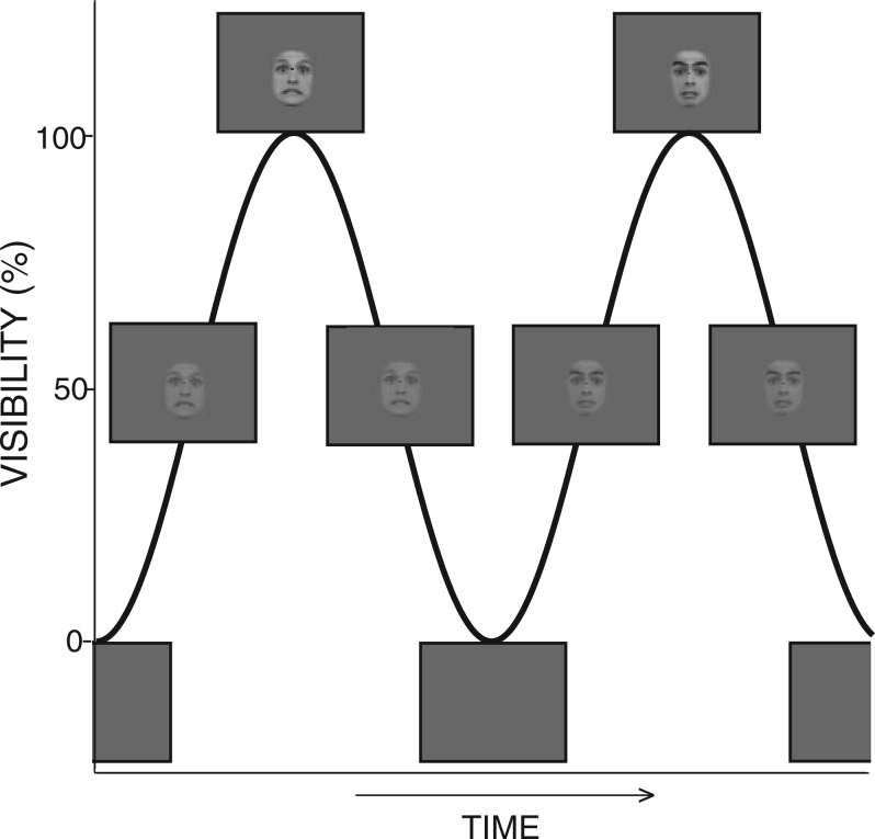
After electrode cap placement, participants were seated with a viewing distance of 100 cm in front of the computer screen. Every block started with the presentation of a black fixation bar for 2.5 s followed by visual stimulation of the face stimuli at 3 Hz for 90 s. Picture presentation was sinusoidal, i.e. starting from a grey background the pixels reached full visibility after 166.5 ms and lost visibility for the following 166.5 ms until they vanished completely into a grey background. Then the cycle started anew (Figure 1). Between conditions, there was a break of 90 s. The order of the conditions was randomized for each participant. Face identity and emotional expression were also pseudo-randomized between conditions and within conditions of different identities or mixed emotional expressions, such that a face with same identity and expression was not presented twice in a row. To ensure fixation and attention throughout the presentation period, participants were asked to indicate a brief colour change of the fixation bar by pressing a button with the right index finger. Colour changes took place on average seven times per condition.
Fig. 1.
Two cycles of the visual stimulation. The face stimuli were presented in a sinusoidal manner with 3 Hz, i.e. one face every 333 ms. Stimulation started with a grey background and the face stimulus was fully visible after 166.5 ms.
EEG recording and analysis
EEG was recorded and sampled at 1024 Hz using the BioSemi Active Two 64-channel active EEG system (BioSemi, Amsterdam, The Netherlands). Vertical and horizontal eye movements were recorded from electrooculography (EOG) electrodes below and next to the right eye. All analyses were performed in Matlab. Data was re-referenced to both earlobes. EEG data was high-pass filtered offline at 0.5 Hz with a two-way least squares linear finite impulse response (FIR) filter. A Fast Fourier Transformation by means of the fft(Matlab) command (Cooley–Tukey algorithm) was applied to an 85 s epoch starting 5 s after the onset of visual stimulation. We did not apply any windowing before and did not use a normalization of the frequency spectrum. The first 5 s after stimulation onset were not included to assure the ssVEP effect to establish itself. Thus, we have a high frequency resolution with a frequency bin width of 0.012 Hz (1024/85 000 = 0.012 Hz). The statistical analysis was conducted over log-transformed data (base 10) as the power amplitude varied strongly between individuals. Statistical comparisons between different neutral identities and same neutral identity at each individual electrode were performed to detect adaptation to face identity. To correct for the Type-I error induced by multiple comparisons, we only considered differences relevant when they concerned at least three neighbouring electrodes with P-values<0.05. Electrodes showing an adaptation effect to face identity in the neutral condition were then pooled for all conditions and the adaptation effects (different–same identity) on these electrodes for the emotional conditions were calculated.
RESULTS
Behavioural results
The mean detection rate of the red bar was 96% (s.d. = 0.03; range: 87.5–100%) indicating that subjects attended the fixation bar throughout stimulation time.
EEG results
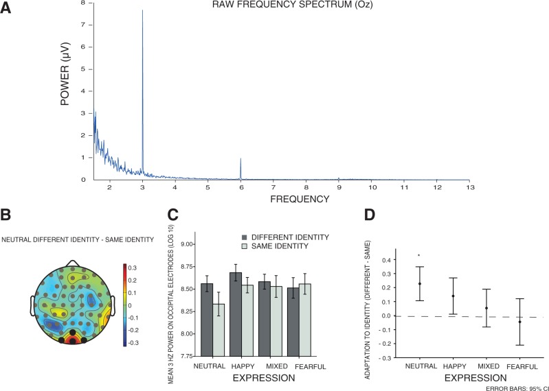
Visual stimulation resulted in a clear peak on 3 Hz as well as on the first (6 Hz) and second (9 Hz) harmonic (Figure 2A). Statistical comparisons (one-sided paired t-tests) revealed a significant adaptation effect of the 3 Hz amplitude in the neutral condition on a cluster of occipital electrodes, namely Oz, Iz, I1 and I2 (Figure 2B), that is, 3 Hz amplitude on these electrodes was lower when the same neutral face was presented compared with when different neutral faces were presented [all t(23) ≥ 1.83 and P-values < 0.05]. On the second harmonic (i.e. 6 Hz) we did not find a significant difference between amplitude evoked by different and same neutral faces on any electrode position [all t(23) ≤ 1.103, P-values > 0.05]. Next, we were interested in the influence of emotional expressions on the adaptation of the 3 Hz ssVEP amplitude. Therefore, we subtracted the mean 3 Hz amplitude for this cluster of occipital electrodes (i.e. Oz, Iz, I1 and I2) for the same face identity from the mean 3 Hz amplitude for different face identities for all four expression conditions, respectively. Figure 2C illustrates the means of all conditions. Based on our hypothesis, we performed a repeated measures ANOVA and tested for a linear effect of emotion on adaptation to face identity. We expected to find the largest adaptation effect for neutral faces and reduced adaptation for emotional expressions, especially the threat-related emotion, fear. We indeed found that adaptation was largest for neutral faces and decreased significantly and linearly for happy, mixed expressions and fearful faces [F(1) = 7.963, P < 0.05; Figure 2D]. Subsequent t-tests showed that the adaptation effect was significantly different from zero for the neutral condition [t(23) = 2.527, P < 0.05], but not for the happy [t(23) = 1.925, P > 0.05], mixed [t(23) = 0.620, P > 0.05] or fearful condition [t(23) = −0.499, P > 0.05]. Comparing the conditions with each other revealed that only the adaptation to neutral faces was significantly different from the adaptation to fearful faces [t(23) = 2.36, P < 0.05].
Fig. 2.
(A) Raw frequency spectrum from 1.5 to 13 Hz recorded on electrode Oz. (B) Topographical map of EEG power at 3 Hz for the same identity condition subtracted from the EEG power at 3 Hz for the different face identities condition with neutral expression. (C) 3 Hz power for different and same face identity with neutral, happy, mixed and fearful expression averaged over all participants. Error bars represent 95% confidence interval. (D) Adaptation effect (different–same identity) for neutral, happy, mixed and fearful facial expressions. Only adaptation to the neutral face identity is significantly different from zero. Error bars represent 95% confidence interval.
DISCUSSION
The present results demonstrate that emotional facial expressions lead to sustained activity in the visual cortex and reduce neural adaptation to face identity. When presenting neutral faces, we observed adaptation to face identity as shown before (Rossion and Boremanse, 2011; Rossion et al., 2012). This adaptation to neutral faces cannot be explained by low-level adaptation because the low-level features of the stimuli changes constantly. Interestingly, we found a reduction of adaptation to face identity for emotional expressions. Adaptation was modulated by the valence of the facial expression and decreased linearly from neutral to happy, mixed and fearful expressions. Positive emotional expressions were less strong in reducing adaptation to face identity than mixed (positive and negative) and fearful expressions. As hypothesized, the ssVEP amplitude evoked by fearful faces demonstrated the least adaptation to face identity. The threat associated with fearful stimuli seems to prevent a reduction of neural activity over time, even if the face identity remains identical.
Our findings are in line with EEG studies showing that the amplitude over occipital electrodes is sensitive to emotional content. An enhanced ssVEP amplitude was found for emotionally arousing compared to neutral content (Keil et al., 2003) and adaptation of occipital event-related potentials (ERPs) was attenuated in response to emotionally loaded stimuli (Schupp et al., 2006). Furthermore, similar results were obtained in functional magnetic resonance imaging (fMRI) studies, namely attenuated adaptation to emotional expressions in visual cortex (Rotshtein et al., 2001; Suzuki et al., 2011; note that the latter study finds greater attenuation of adaptation for happy compared with angry faces). However, some fMRI studies do not find reduced or attenuated adaptation to emotional expressions. Winston et al. (2004) showed that adaptation to same face identity across different emotional expressions does occur. There are several important differences between both studies, such as number of stimulus repetitions (2 vs 270) and employed measurement (BOLD response vs ssVEP amplitude). Most importantly, Winston et al. (2004) pooled responses over all expressions (i.e. angry, disgusted, fearful, sad and happy) to investigate which brain areas show adaptation to face identity in particular. By pooling over different emotional expressions, possible reductions of adaptation to face identity for fearful expressions might simply be attenuated by the presence of happy, sad and disgusted faces. Another fMRI study (Ishai et al., 2004) also suggests that adaptation to fearful facial expressions occurs. In their study, a target face had to be remembered and detected within a stream of distractor faces. For fearful target faces, BOLD response indeed adapted with repetition. However, when a fearful face was repeated as a distractor face—more similar to our task that did not involve any active identification of face identity—adaptation was attenuated.
The finding that the negative valence of emotional expressions prevents adaptation in visual cortex is hard to explain by bottom–up explanations of adaptation that assume neural fatigue or sharpening of the representation of the repeated feature (Grill-Spector et al., 2006). However, it can be understood in the context of recent studies on adaptation in fMRI paradigms (Ewbank et al., 2013, 2012) suggesting that adaptation can also occur as a consequence of ‘top–down modulation’. These studies were based on predictive coding models hypothesizing that repetition leads to a reduction in prediction error between bottom–up (stimulus driven) and top–down (prediction driven) inputs. When incoming information and the predicted cause do not match, the lower level areas produce a prediction error, which is then re-used by higher level areas to update the perceptual cause. However, when input and prediction do line up, which is the case with repetition, the prediction error diminishes and neural activity adapts. Therefore, less adaptation to face identity for emotional and particularly threat-related expressions could indicate a sustained prediction error in the visual areas. One possible higher level area causing this sustained prediction error signalling by the visual areas might be the amygdala, whose top–down projections were shown to be involved in modulating responses to emotional stimuli in cortical visual areas (Vuilleumier et al., 2004). Alternatively, the amygdala response to emotional stimuli could lead to attentional modulation of the activation in visual areas. However, the current task required the participants to attend and respond to the colour of a bar superimposed on the faces and thus, effectively ignore the faces. Moreover, the modulatory effect of threat-related emotional expressions on activation in the face processing visual areas has shown to be additive to the effects of attention on activity in these areas (Vuilleumier et al., 2001). Still, the exact influence of amygdala activation on adaptation in the visual cortex, the role of multiple levels of visual attention and their contribution to a possibly sustained prediction error in threat-related face processing remain to be clarified in more detail.
In general, it should be noted that the attenuating effect of emotional expressions on adaptation might be limited to adaptation to face identity and not to adaptation to emotional stimuli per se. Furthermore, it is possible that the attenuating effect of emotional expressions on neural adaptation is limited to a short time frame (e.g. 90 s) and that with longer visual stimulation adaptation to threat-related expressions would equalize adaptation to neutral facial expressions. Additionally, a recent study showed that adaptation to neutral face identity does also occur at higher frequency rates (i.e. 4 and 8 Hz; Rossion et al., 2012). In our study, we presented face stimuli study with one frequency rate only (i.e. 3 Hz). Thus, future studies are needed to determine whether the influence of emotional expressions on ssVEP adaptation also holds at lower and higher frequency rates, whether it still holds with longer stimulation duration and whether this finding can be extended to emotional stimuli in general.
Other lines of research suggest that processing of emotional facial expressions is dependent on individual differences in anxiety levels. A recent study by Wieser et al., (2011) has shown that the ssVEP response evoked by emotional expressions is related to social anxiety. They presented their subjects with neutral, happy and angry faces and measured the evoked ssVEPs amplitude. Individuals with high social anxiety showed enhanced ssVEP amplitude on occipital electrodes elicited by threat-related compared with happy or neutral facial expressions. This sensitivity of the ssVEP amplitude to emotional valence of faces could, however, not be found in a control group with low social anxiety. In the present study, we showed that the adaptation of ssVEP amplitude is a measurement showing sensitivity to emotional valence even in a healthy group of participants. It is likely that the amount of adaptation to different emotional expressions is indeed also modulated by individual differences and might serve as an index for anxiety sensitivity. As the above described method has short recording times (90 s per block) and is independent of task-relevant behavioural measures, it might be an objective and reliable diagnostic tool for anxiety vulnerability in experimental and even clinical settings that could overcome the shortcomings of self-report questionnaires. Further research is needed to examine to what extent the amount of ssVEP adaptation to threat-related expressions is based on individual anxiety levels.
Here, we could demonstrate that emotions reduce neural adaptation. Additionally, we could show that neural adaptation is lowest for threat-related stimuli. This short and straightforward task may prove to be a useful new tool in studying visual processing of emotional stimuli.
Conflict of Interest
None declared.
Acknowledgments
The authors would like to thank Elexa St. John-Saaltink for her assistance in collecting the data and Mike X Cohen for helpful advice.
This work was supported by an advanced investigator grant from The European Research Council (ERC) to V.A.F.L. [230355].
REFERENCES
- Breiter HC, Etcoff NL, Whalen PJ, et al. Response and habituation of the human amygdala during visual processing of facial expressions. Neuron. 1996;17:875–87. doi: 10.1016/s0896-6273(00)80219-6. [DOI] [PubMed] [Google Scholar]
- Ewbank MP, Henson RN, Rowe JB, Stoyanova RS, Calder AJ. Different neural mechanisms within occipitotemporal cortex underlie repetition suppression across same and different-size faces. Cerebral Cortex. 2013;23(5):1073–84. doi: 10.1093/cercor/bhs070. [DOI] [PMC free article] [PubMed] [Google Scholar]
- Ewbank MP, Lawson RP, Henson RN, Rowe JB, Passamonti L, Calder AJ. Changes in “top down” connectivity underlie repetition suppression in the ventral visual pathway. Journal of Neuroscience. 2011;31:5635–42. doi: 10.1523/JNEUROSCI.5013-10.2011. [DOI] [PMC free article] [PubMed] [Google Scholar]
- Eastwood JD, Smilek D, Merikle PM. Differential attentional guidance by unattended faces expressing positive and negative emotion. Perception and Psychophysics. 2001;63:1004–13. doi: 10.3758/bf03194519. [DOI] [PubMed] [Google Scholar]
- Fox E, Russo R, Dutton K. Attentional bias for threat: Evidence for delayed disengagement from emotional faces. Cognition and Emotion. 2002;16:355–79. doi: 10.1080/02699930143000527. [DOI] [PMC free article] [PubMed] [Google Scholar]
- Georgiou GA, Bleakley C, Hayward J, et al. Focusing on fear: Attentional disengagement from emotional faces. Visual Cognition. 2005;12:145–58. doi: 10.1080/13506280444000076. [DOI] [PMC free article] [PubMed] [Google Scholar]
- Grill-Spector K, Henson R, Martin A. Repetition and the brain: neural models of stimuls-specific effects. Trends in Cognitive Science. 2006;10:14–23. doi: 10.1016/j.tics.2005.11.006. [DOI] [PubMed] [Google Scholar]
- Hodsoll S, Viding E, Lavie N. Attentional capture by irrelevant emotional distractor faces. Emotion. 2011;2:346–53. doi: 10.1037/a0022771. [DOI] [PubMed] [Google Scholar]
- Hsiao J-H, Cottrell G. Two fixations suffice in face recognition. Psychological Science. 2008;19:998–1006. doi: 10.1111/j.1467-9280.2008.02191.x. [DOI] [PMC free article] [PubMed] [Google Scholar]
- Ishai A, Pessoa L, Bikle PC, Ungerleider LG. Repetition suppression of faces is modulated by emotion. Proceedings of the National Academy of Science USA. 2004;101:9827–32. doi: 10.1073/pnas.0403559101. [DOI] [PMC free article] [PubMed] [Google Scholar]
- Keil A, Gruber T, Müller MM, et al. Early modulation of visual perception by emotional arousal: Evidence from steady-state visual evoked brain potentials. Cognitive, Affective and Behavioral Neuroscience. 2003;3:195–206. doi: 10.3758/cabn.3.3.195. [DOI] [PubMed] [Google Scholar]
- Kotsoni E, de Haan M, Johnson MH. Categorical perception of facial expressions by 7-month old infants. Perception. 2001;30:1115–25. doi: 10.1068/p3155. [DOI] [PubMed] [Google Scholar]
- Langner O, Dotsch R, Bijlstra G, Wigbouldus DHJ, Hawk ST, van Knippenberg A. Presentation and validation of the Radboud Face Database. Cognition and Emotion. 2010;24:1377–88. [Google Scholar]
- Mogg K, Bradley BP. A cognitive-motivational analysis of anxiety. Behavior Research and Therapy. 1998;36:809–48. doi: 10.1016/s0005-7967(98)00063-1. [DOI] [PubMed] [Google Scholar]
- Morris JS, Friston KJ, Bücherl C, et al. A neuromodulatory role for the human amygdala in processing emotional facial expressions. Brain. 1998;121:47–57. doi: 10.1093/brain/121.1.47. [DOI] [PubMed] [Google Scholar]
- Öhman A, Lundqvist D, Esteves F. The face in the crowd revisited: A threat advantage with schematic stimuli. Journal of Personality and Social Psychology. 2001;80:381–96. doi: 10.1037/0022-3514.80.3.381. [DOI] [PubMed] [Google Scholar]
- Peltola MJ, Lepäänen JM, Palokangas T, Hietanen JK. Fearful faces modulate looking duration and attention disengagement in 7-month-old infants. Developmental Science. 2008;11:60–8. doi: 10.1111/j.1467-7687.2007.00659.x. [DOI] [PubMed] [Google Scholar]
- Peltola MJ, Leppänen JM, Vogel-Farley VK, Hietanen JK, Nelson CA. Fearful faces but not fearful eyes alone delay attention disengagement in 7-month-old infants. Emotion. 2009;9:560–5. doi: 10.1037/a0015806. [DOI] [PMC free article] [PubMed] [Google Scholar]
- Phelps E, Ling S, Carrasco M. Emotion facilitates perception and potentiates the perceptual benefits of attention. Psychological Science. 2006;17:292–9. doi: 10.1111/j.1467-9280.2006.01701.x. [DOI] [PMC free article] [PubMed] [Google Scholar]
- Pessoa L, McKenna M, Gutierrez E, Ungerleider LG. Neural processing of emotional faces requires attention. Proceedings of the National Academy of Science USA. 2002;99:11458–63. doi: 10.1073/pnas.172403899. [DOI] [PMC free article] [PubMed] [Google Scholar]
- Rahman Q, Anchassi T. Men appear more lateralized when noticing emotion in male faces. Emotion. 2011;12:174–9. doi: 10.1037/a0024416. [DOI] [PubMed] [Google Scholar]
- Rossion B, Alonso Prieto E, Boremanse A, Kuefner D, Van Belle G. A steady-state visual evoked potential approach to individual face perception: Effect of inversion, contrast-reversal and temporal dynamics. NeuroImage. 2012;63:1585–600. doi: 10.1016/j.neuroimage.2012.08.033. [DOI] [PubMed] [Google Scholar]
- Rossion B, Boremanse A. Robust sensitivity to facial identity in the right human occipito-temporal cortex as revealed by steady-state visual evoked potentials. Journal of Vision. 2011;11:1–21. doi: 10.1167/11.2.16. [DOI] [PubMed] [Google Scholar]
- Rotshtein P, Malach R, Hadar U, Graif M, Hendler T. Feeling or features: Different sensitivity to emotion in high-order visual cortex and amygdala. Neuron. 2001;32:747–57. doi: 10.1016/s0896-6273(01)00513-x. [DOI] [PubMed] [Google Scholar]
- Schupp HT, Stockburger J, Codispoti M, Junghöfer M, Weike AI, Hamm AO. Stimulus novelty and emotion perception: the near absence of habituation in the visual cortex. Neuroreport. 2006;17:365–9. doi: 10.1097/01.wnr.0000203355.88061.c6. [DOI] [PubMed] [Google Scholar]
- Surguladze SA, Brammer MJ, Young AW, et al. A preferential increase in the extrastriate response to signals of danger. NeuroImage. 2003;19:1317–28. doi: 10.1016/s1053-8119(03)00085-5. [DOI] [PubMed] [Google Scholar]
- Suzuki A, Goh JOS, Hebrank A, Sutton BP, Jenkins L, Flicker BA, Park DC. Sustained happiness? Lack of repetition suppression in right-ventral visual cortex for happy faces. Social, Cognitive and Affective Neuroscience. 2011;6:434–41. doi: 10.1093/scan/nsq058. [DOI] [PMC free article] [PubMed] [Google Scholar]
- Vuilleumier P, Armony JL, Driver J, Dolan RJ. Effects of attention and emotion on face processing in the human brain: An event-related fMRI study. Neuron. 2001;30:829–41. doi: 10.1016/s0896-6273(01)00328-2. [DOI] [PubMed] [Google Scholar]
- Vuilleumier P, Driver JL. Modulation of visual processing by attention and emotion: windows on causal interactions between human brain regions. Philosophical Transactions of the Royal Society London, B: Biological Science. 2007;362:837–55. doi: 10.1098/rstb.2007.2092. [DOI] [PMC free article] [PubMed] [Google Scholar]
- Vuilleumier P, Pourtois G. Distributed and interactive brain mechanisms during emotion face perception: Evidence from functional neuroimaging. Neuropsychologia. 2007;45:174–94. doi: 10.1016/j.neuropsychologia.2006.06.003. [DOI] [PubMed] [Google Scholar]
- Vuilleumier P, Richardson MP, Armony JL, Driver J, Dolan RJ. Distant influences of amygdala lesion on visual cortical activation during emotional face processing. Nature Neuroscience. 2004;7:1271–8. doi: 10.1038/nn1341. [DOI] [PubMed] [Google Scholar]
- West GL, Anderson AAK, Pratt J. Motivationally significant stimuli show visual prior entry: Evidence for attentional capture. Journal of Experimental Psychology: Human Perception and Performance. 2009;35:1032–42. doi: 10.1037/a0014493. [DOI] [PubMed] [Google Scholar]
- Wieser MJ, McTeague LM, Keil A. Sustained preferential processing of social threat cues: Bias without competition? Journal of Cognitive Neuroscience. 2011;23:1973–86. doi: 10.1162/jocn.2010.21566. [DOI] [PMC free article] [PubMed] [Google Scholar]
- Winston JS, Henson RNA, Fine-Goulden MR, Dolan RJ. fMRI adaptation reveals dissociable neural representations of identity and expression in face perception. Journal of Neurophysiology. 2004;92: 1830–9. doi: 10.1152/jn.00155.2004. [DOI] [PubMed] [Google Scholar]


