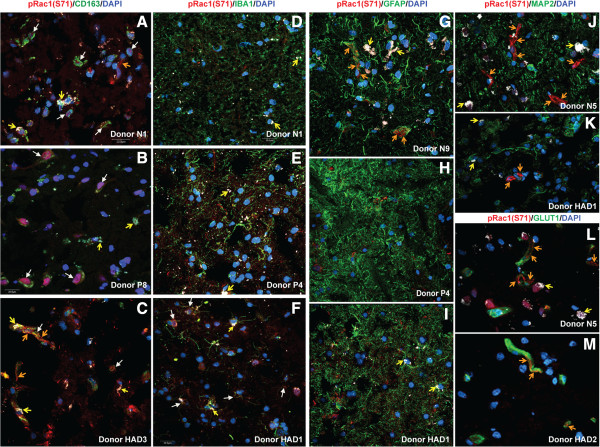
Figure 5.
Expression of pRac1 (S71) in human brain immune cells. Confocal microscopy shows expression of pRac1(S71) in human brain macrophages (A-C, white arrows) and blood vessels tight junction strands (A, C, G, J-M orange arrows). Most brain tissues samples did not show significant expression of pRac1 (S71) in microglia (D, E), but brain tissues of some HIVE patients showed pRac1(S71) in microglia (F, white arrows). Data showed no expression of pRac1 (S71) in astrocytes (G-I) or neurons (J, K). For all experiments, a 4th laser line was used to differentiate lipofuscin-like autofluorescent pigments from antibody staining (yellow arrows). Scale bar for all panels: 20 μM.

