Abstract
Nanotechnology is the engineering of functional systems at the molecular scale which may exert a revolutionary impact on cancer diagnosis and therapy. Nanotechnology is being applied to cancer in two broad areas: i) the development of nanovectors such as nanoparticles which can be loaded with drugs or imaging agents and then targeted to tumours, and ii) high-throughput nanosensor devices for detecting the biological signatures of cancer. Combined, such technologies could lead to earlier diagnosis and better treatment for cancer patients.
Keywords: Nanotechnology, Cancer, Nanovectors, Cantilevers
Introduction
Nanotechnology is a multidisciplinary field. It covers a vast array of devices that are derived from the areas of engineering, biology, physics and chemistry. These devices include vectors for the targeted delivery of anticancer drugs, imaging contrast agents and arrays for the early detection of tumors (1). These, and other nano-devices, may support the future diagnosis and treatment of common conditions such as cancer (2–4).
Nanotechnology was first proposed in 1867 by James Clarke Maxwell, a Scottish physicist who developed electromagnetic theory and statistical physics. He suggested the concept of “Maxwell’s Demon”, an entity able to manipulate individual molecules. Nanotechnology was predicted by the Nobel Prize winner physicist Richard Feynman. In his talk “There is plenty of room at the bottom”, given at Caltech in 1959, he spoke of building factories that maneuvered things atom by atom. Later in 1974, this concept was defined by Professor Norio Taniguchi of Tokyo University of Science as “the separation, consolidation, and deformation of materials by one atom or one molecule”. The idea was then popularized in the 1980’s by Dr. K. Eric Drexler, who suggested that it would be possible to build computers and robots far smaller than a single cell (5, 6).
Nanotechnology refers to structures that have been engineered at an atomic scale. Nanotechnology typically refers to structures up to 200 nanometers in size. As a system’s size diminishes to nano-scale it leaves a Newtonian world and enters a ‘quantum realm’. In this realm, particle behavior and processes are governed by quantum and statistical mechanics and atoms are manipulated by covalent, electromagnetic and Van der Walls forces. Structures made up of a monolayer (a sheet that is one atom thick), have properties that differ markedly from conventional experience. Opaque and stable metals, such as gold, are transformed into monolayers that are transparent, inflammable catalysts. Nanotechnology is being used not only in the development of such catalysts, but additionally in semi-conductor technologies, such as memory storage, construction and household products such as sunglasses (7).
Relating to cancer, nanotechnology is being applied in the following two broad areas:
Nanovectors
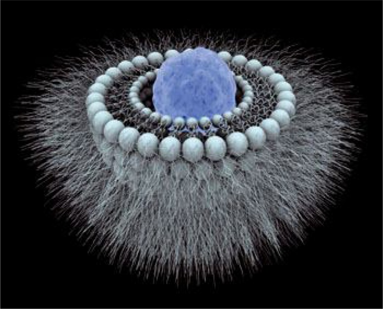
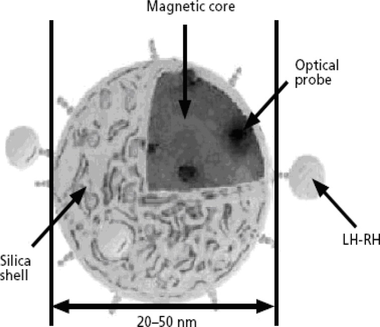
Nanovectors may eventually become a powerful tool for the delivery of anticancer drugs. Usually have a tripartite constitution; a core constituent material, a therapeutic and / or imaging payload, and biological surface modifiers which enhance the bio-distribution and tumor targeting of the nanoparticle (10).
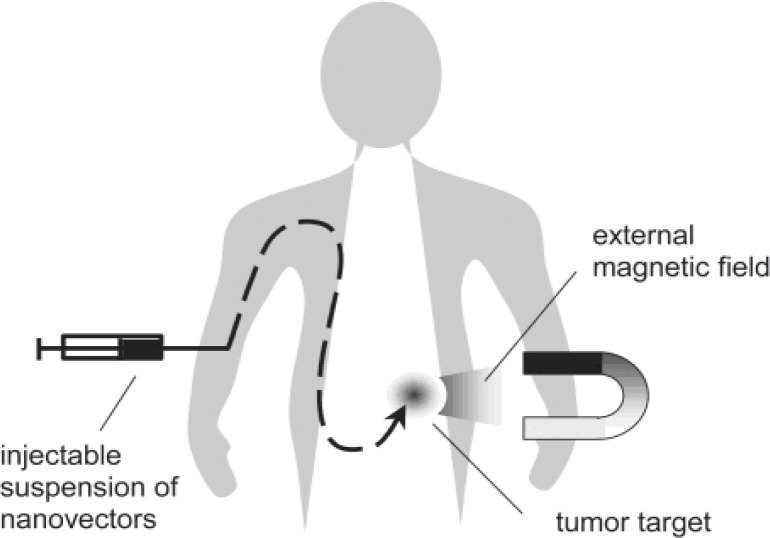
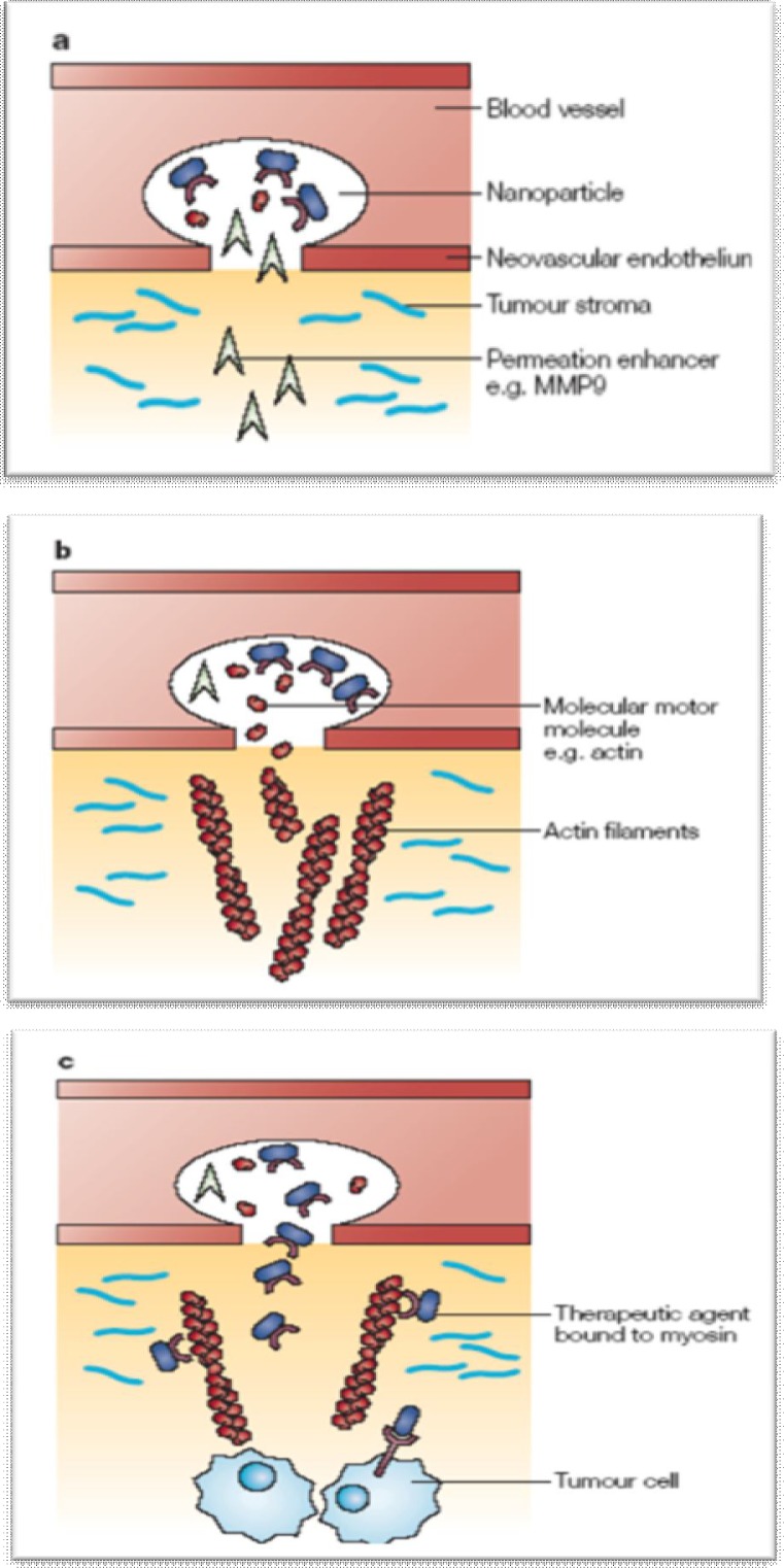
Through the process of targeted biorecognition, nanovectors have the potential to deliver large amounts of a therapeutic or imaging agent to a tumor. For example, a nanovector may selectively bind to the neovascular endothelium of a tumor and release a penetration enhancer. This may then deploy a track of molecular motor molecules such as actin (11). The nanovector, or another, can then release a therapeutic agent, bound to a conjugate molecule, such as myosin. This agent could then travel along the molecular track, reaching deep into the cancer lesion and overcoming opposing oncotic and osmotic pressures. In addition to the delivery of therapeutic agents, nanovector’s formulations can be designed to reduce the clearance time of small peptide drugs by providing protection of active agents from enzymatic or environmental degradation and avoiding obstacles to the targeting of the active moiety (12).
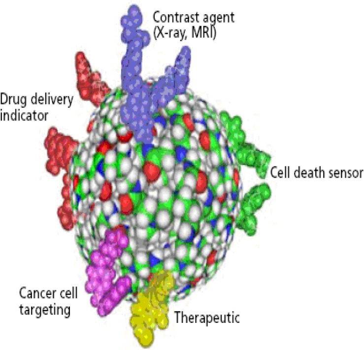
Nanovectors can also act as carriers for the delivery of targeted imaging payloads. Their constituent materials might possess image-enhancement properties. This includes gadolinium or iron oxide-based nanoparticles and multiple-mode imaging contrast nano-agents, which combine magnetic resonance with biological targeting and optical detection. Low-density lipid nanoparticles also have been used to enhance ultrasound imaging (13).
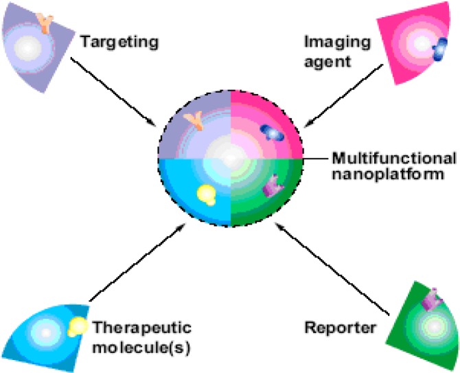
Many polymer-based nanovectors have been investigated and some appear more promising for clinical translation. For different imaging modalities, it is possible to develop nanoparticles, which can provide signal enhancement combined with biomolecular targeting capabilities (14). These nanovectors have typically been developed as components of a multi-functional unit with generic core components, a nanoplatform. A nanoplatform consists of a targeting device that provides specificity of binding. This is coupled to a generic image enhancing agent, a generic therapeutic agent (such as a cell toxin) and a generic reporter agent which enables sensors to detect the successful delivery of the therapeutic agent to its target (15–17).
Multifunctional nanoparticles can be targeted to cancer cells using receptor ligands. In this diagram a vector has been engineered at the atomic level to deliver a magnetic core and an optical probe to cells with receptors for the luteinizing hormone (LH-RH) (18).
Dendrimers
Dendrimers are larger and more complex molecules with well-defined chemical structures. They consist of three major architectural components: core, branches, and end groups.
One of the most appealing aspects of technologies based on dendrimers is that it is relatively easy to precisely control their size, composition, and chemical reactivity. Dendrimers can serve as versatile nanoscale platforms for creating multifunctional devices capable of detecting cancer and drug delivery (19, 20).
Multicomponent targeting strategies
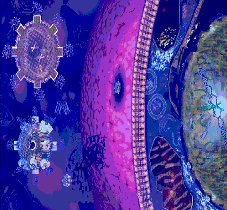
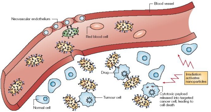
Nanoparticles can be designed to extravasate into the tumour stroma through the fenestrations of the angiogenic vasculature and demonstrate targeting by enhanced permeation and retention. The particles carry multiple antibodies, which direct antitumor action by targeting epitopes on cancer cells. When irradiated by external energy, nanoparticles become activated and release their cytotoxic action. Through this, they could provide anti-angiogenic therapy by preferentially adhering to cancer neovasculature and causing it to collapse (21, 22).
A nanodevice with multiple-barrier-avoidance capability:
Nanoscale cantilevers
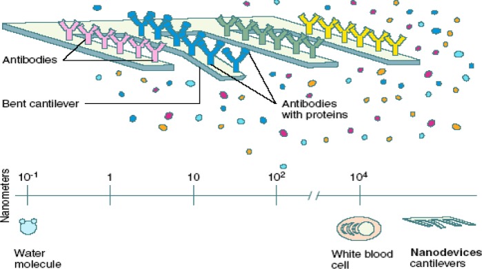
Cantilevers are constructed as parts of a larger diagnostic device and can provide rapid and selective detection of cancer-related molecules. Cancer specific molecules bind to molecules that act as sensors. The binding of a cancer molecule causes the sensor to change conformational shape. This movement is exaggerated as the sensor molecule acts like a single lever cantilever bridge (23–25).
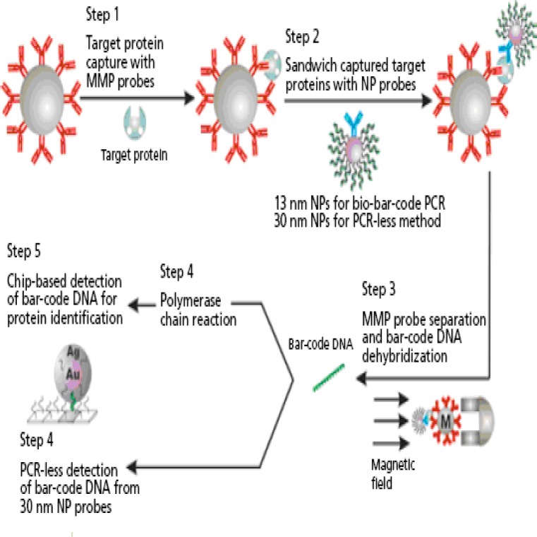
DNA-coated gold nanoparticles (NPs) form the basis of a system that also uses larger magnetic microparticles (MMPs) to detect femtomolar (10 -15 Molar) concentrations of serum proteins. In this case, a monoclonal antibody to prostate specific antigen (PSA) is attached to the MMP, creating a reagent to capture free PSA. A second antibody to PSA is attached to the NPs and two particles create a sandwich of the captured protein which can be easily separated using a magnetic field. The bar code can then be amplified using PCR. This technique has the potential to quantify an antigen at concentrations that would not be possible using a conventional enzyme linked imunosorbant assay (ELISA) (26–28).
Challenges in cancer nanotechnology
In an ideal scenario, the onset of the transformational processes leading towards malignancy would be detected early. This could exist as routine screening by non-invasive means such as proteomic pattern analysis from blood samples, or the in vivo imaging of molecular profiles and evolving lesion contours. The biology of the host and the disease would be accurately determined, and dictate choices for targeting and barrier-avoiding strategies for an intervention plan. Transforming cellular populations would be eradicated or at least contained, without collateral effects on healthy tissues, in a routine that could be repeated many times. Treatment efficacy would be monitored in real time. Therapeutics would be supplanted by personalized prevention. If fully integrated with the established cancer research enterprise, nanotechnology might help this vision become reality. Some of the principal challenges along this path are as follows:
Developing approaches for the in vivo detection and monitoring of cancer markers
Improvement of the targeting efficacy of therapeutic or imaging agents to cancer lesions and their microenvironment
Refining technology platforms for early detection of cancer biomarkers ex vivo
Engineering nanoparticles to avoid biological and biophysical barriers (29–31).
Concluding remarks
Nanotechnology is expected to play an important role in the early detection of transforming cell populations by in vivo imaging or ex vivo analysis. This new and highly sophisticated technology allows the appropriate combination of agents, based on accurate tumour specific biological information, targeting them to the cancer cells, whilst avoiding biological barriers. In addition, the technology permits monitoring of the treatment effect. Nanovector delivery systems are also showing promise. However, their approval perspectives, biodistribution, reliability of production protocols, as well as safety for patients and the health-care workers should not be ignored.
(Please cite as: Hassanzadeh P, Fullwood I, Sothi S, Aldulaimi D. Cancer nanotechnology. Gastroenterology and Hepatology From Bed to Bench 2011;4(2):63-69).
References
- 1.McNeil SE. Unique benefits of nanotechnology to drug delivery and diagnostics. Methods Mol Biol. 2011;697:3–8. doi: 10.1007/978-1-60327-198-1_1. [DOI] [PubMed] [Google Scholar]
- 2.Langer R. Drug delivery and targeting. Nature. 1998;392:5–10. [PubMed] [Google Scholar]
- 3.Menciassi A, Sinibaldi E, Pensabene V, Dario P. From miniature to nano robots for diagnostic and therapeutic applications. Conf Proc IEEE Eng Med Biol Soc. 2010;2010:1954–57. doi: 10.1109/IEMBS.2010.5627629. [DOI] [PubMed] [Google Scholar]
- 4.Leucuta SE. Nanotechnology for delivery of drugs and biomedical applications. Curr Clin Pharmacol. 2010;5:257–80. doi: 10.2174/157488410793352003. [DOI] [PubMed] [Google Scholar]
- 5.Jain RK. The next frontier of molecular medicine: delivery of therapeutics. Nature Med. 1998;4:655–57. doi: 10.1038/nm0698-655. [DOI] [PubMed] [Google Scholar]
- 6.Srinivas PR, Barker P, Srivastava S. Nanotechnology in early detection of cancer. Lab Invest. 2002;82:657–62. doi: 10.1038/labinvest.3780460. [DOI] [PubMed] [Google Scholar]
- 7.Safron NS, Brewer AS, Arnold MS. Semiconducting two-dimensional graphene nanoconstriction arrays. Small. 2011;7:492–98. doi: 10.1002/smll.201001193. [DOI] [PubMed] [Google Scholar]
- 8.Veiseh O, Kievit FM, Fang C, Mu N, Jana S, Leung MC, et al. Chlorotoxin bound magnetic nanovector tailored for cancer cell targeting, imaging, and siRNA delivery. Biomaterials. 2010;31:8032–42. doi: 10.1016/j.biomaterials.2010.07.016. [DOI] [PMC free article] [PubMed] [Google Scholar]
- 9.Chaudhuri P, Soni S, Sengupta S. Single-walled carbon nanotube-conjugated chemotherapy exhibits increased therapeutic index in melanoma. Nanotechnology. 2010;21:025102. doi: 10.1088/0957-4484/21/2/025102. [DOI] [PubMed] [Google Scholar]
- 10.Benyettou F, Lalatonne Y, Sainte-Catherine O, Monteil M, Motte L. Superparamagnetic nanovector with anti-cancer properties: gamma Fe2O3@Zoledronate. Int J Pharm. 2009;379:324–27. doi: 10.1016/j.ijpharm.2009.04.010. [DOI] [PubMed] [Google Scholar]
- 11.Ciofani G, Riggio C, Raffa V, Menciassi A, Cuschieri A. A bi-modal approach against cancer: magnetic alginate nanoparticles for combined chemotherapy and hyperthermia. Med Hypotheses. 2009;73:80–82. doi: 10.1016/j.mehy.2009.01.031. [DOI] [PubMed] [Google Scholar]
- 12.Pugno NM. A new concept for smart drug delivery: adhesion induced nanovector implosion. Open Med Chem J. 2008;2:62–65. doi: 10.2174/1874104500802010062. [DOI] [PMC free article] [PubMed] [Google Scholar]
- 13.Schlupp P, Blaschke T, Kramer KD, Höltje HD, Mehnert W, Schäfer-Korting M. Drug Release and Skin Penetration from Solid Lipid Nanoparticles and a Base Cream: A Systematic Approach from a Comparison of Three Glucocorticoids. Skin Pharmacol Physiol. 2011;24:199–209. doi: 10.1159/000324053. [DOI] [PubMed] [Google Scholar]
- 14.Leonarduzzi G, Testa G, Sottero B, Gamba P, Poli G. Design and development of nanovehicle-based delivery systems for preventive or therapeutic supplementation with flavonoids. Curr Med Chem. 2010;17:74–95. doi: 10.2174/092986710789957760. [DOI] [PubMed] [Google Scholar]
- 15.Kerr D. Sensor-augmented insulin-pump therapy in type 1 diabetes. N Engl J Med. 2010;363:2070–71. doi: 10.1056/NEJMc1009685. [DOI] [PubMed] [Google Scholar]
- 16.Frasconi M, Tortolini C, Botrè F, Mazzei F. Multifunctional au nanoparticle dendrimer-based surface plasmon resonance biosensor and its application for improved insulin detection. Anal Chem. 2010;82:7335–42. doi: 10.1021/ac101319k. [DOI] [PubMed] [Google Scholar]
- 17.Banerjee SS, Chen DH. Multifunctional pH-sensitive magnetic nanoparticles for simultaneous imaging, sensing and targeted intracellular anticancer drug delivery. Nanotechnology. 2008;19:505104. doi: 10.1088/0957-4484/19/50/505104. [DOI] [PubMed] [Google Scholar]
- 18.Minko T, Patil ML, Zhang M, Khandare JJ, Saad M, Chandna P, et al. LHRH-targeted nanoparticles for cancer therapeutics. Methods Mol Biol. 2010;624:281–94. doi: 10.1007/978-1-60761-609-2_19. [DOI] [PubMed] [Google Scholar]
- 19.Huißmann S, Wynveen A, Likos CN, Blaak R. The effects of pH, salt and bond stiffness on charged dendrimers. J Phys Condens Matter. 2010;22:232101. doi: 10.1088/0953-8984/22/23/232101. [DOI] [PubMed] [Google Scholar]
- 20.Suehiro T, Tada T, Waku T, Tanaka N, Hongo C, Yamamoto S, et al. Temperature-dependent higher order structures of the (Pro-Pro-Gly)(10) -modified dendrimer. Biopolymers. 2011;95:270–77. doi: 10.1002/bip.21576. [DOI] [PubMed] [Google Scholar]
- 21.Liu XQ, Song WJ, Sun TM, Zhang PZ, Wang J. Targeted delivery of antisense inhibitor of miRNA for antiangiogenesis therapy using cRGD-functionalized nanoparticles. Mol Pharm. 2011;8:250–59. doi: 10.1021/mp100315q. [DOI] [PubMed] [Google Scholar]
- 22.Danhier F, Feron O, Préat V. To exploit the tumor microenvironment: Passive and active tumor targeting of nanocarriers for anti-cancer drug delivery. J Control Release. 2010;148:135–46. doi: 10.1016/j.jconrel.2010.08.027. [DOI] [PubMed] [Google Scholar]
- 23.Labuda A, Grütter PH. Exploiting cantilever curvature for noise reduction in atomic force microscopy. Rev Sci Instrum. 2011;82:013704. doi: 10.1063/1.3503220. [DOI] [PubMed] [Google Scholar]
- 24.Sansoz F, Gang T. A force-matching method for quantitative hardness measurements by atomic force microscopy with diamond-tipped sapphire cantilevers. Ultramicroscopy. 2010;111:11–19. doi: 10.1016/j.ultramic.2010.09.012. [DOI] [PubMed] [Google Scholar]
- 25.Lee H, Yu A, Flores-Mir C. Limited evidence that cantilevers are associated with slightly lower survival rates of implant-supported fixed partial dentures. J Am Dent Assoc. 2010;141:1371–72. doi: 10.14219/jada.archive.2010.0083. [DOI] [PubMed] [Google Scholar]
- 26.Privorotskaya N, Liu YS, Lee J, Zeng H, Carlisle JA, Radadia A, et al. Rapid thermal lysis of cells using silicon-diamond microcantilever heaters. Lab Chip. 2010;10:1135–41. doi: 10.1039/b923791g. [DOI] [PubMed] [Google Scholar]
- 27.Chen X, Lee DW. Design and optimization of inplane actuator driven cantilever with high sensitivity sensors. J Nanosci Nanotechnol. 2010;10:3236–40. doi: 10.1166/jnn.2010.2257. [DOI] [PubMed] [Google Scholar]
- 28.Koeser J, Bammerlin M, Battiston FM, Hubler U. Nanomechanical cantilever sensors as a novel tool for real-time monitoring and characterization of surface layer formation. J Nanosci Nanotechnol. 2010;10:2578–82. doi: 10.1166/jnn.2010.1412. [DOI] [PubMed] [Google Scholar]
- 29.Cassol L, Silveira Graudenz M, Zelmanowicz A, Cancela A, Werutsky G, Rovere RK, et al. Basal-like immunophenotype markers and prognosis in early breast cancer. Tumori. 2010;96:966–70. [PubMed] [Google Scholar]
- 30.Govindarajan A, Paty PB. Predictive markers of colorectal cancer liver metastases. Future Oncol. 2011;7:299–307. doi: 10.2217/fon.10.184. [DOI] [PubMed] [Google Scholar]
- 31.Shah S, Liu Y, Hu W, Gao J. Modeling Particle Shape-Dependent Dynamics in Nanomedicine. J Nanosci Nanotechnol. 2011;11:919–928. doi: 10.1166/jnn.2011.3536. [DOI] [PMC free article] [PubMed] [Google Scholar]


