Abstract
Background:
We determined the blood lipid-lowering effects of eicosapentaenoic acid (EPA) on hypertriglyceridemic subjects with Leu162/Val in exon 5 and G/C in intron7 polymorphism of peroxisome proliferator-activated receptor alpha (PPARα)genotypes that, to our knowledge, have not been previously studied.
Methods:
A total of 170 hypertriglyceridemic subjects were enrolled and genotyped for Ala54Thr, Leu162Val, and intron7 polymorphism by the use of a polymerase chain reaction–restriction fragment length polymorphism method. After determination of their genotypes, the first 23 eligible subjects who were found as Ala54 carriers and the first 23 eligible Thr54 carriers were enrolled in the study and stratified for PPARα genotypes. Participants took 2 g of pure EPA daily for 8 weeks. Fasting blood lipid and lipoprotein profiles were determined and changes from baseline were measured.
Results:
We observed significant difference between EPA supplementation and Leu162 and Val162, Interon 7 (GG and GC) carriers (P < 0.001). We did not observe significant associations between the PPARα L162V single nucleotide polymorphism and multiple lipid and lipoprotein measures. Although EPA consumption lowered lipid and lipoprotein concentrations in Leu162 and Val162 carriers and Interon 7 CC and GC carriers, these differences between the studied groups were not statistically significant.
Conclusions:
EPA consumption has a lipid-lowering effect in hypertriglyceridemic subjects in both Leu162 and Val162 carriers. But there was no significant interaction between EPA supplementation and PPARα genotypes. Thus, genetic variation within the PPARα Leu162/Val cannot modulate the association of EPA intakes with lipid and lipoprotein profile. However, we must note that the sample size in this study was small.
Keywords: Eicosapentaenoic acid, hypertriglyceridemic, lipid profile, peroxisome proliferator-activated receptor alpha, polymorphism
INTRODUCTION
Cardiovascular diseases (CVD) are the major cause of death in adults in several countries.[1] The development of CVD is multifactorial, with risk factors that are modifiable, such as diet, exercise, and smoking, and others that are not, such as gender, age, and genetics. The behavioral factors might modify a number of biochemical markers involved in dyslipidemia, hypertension, inflammation, and insulin resistance.[2,3]
Alterations in lipid and lipoprotein metabolisms have been investigated as important CVD risk factors. High triglycerides (TG) and low high-density lipoprotein (HDL) concentration are strong predictable factors of cardiac events.[1,4] Either environmental or genetic factors can affect the lipoproteins concentration, which may affect the risk of developing CVD. Thus, key metabolism regulators such as the peroxisome proliferator-activated receptor (PPAR) family are the candidate genes to investigate genetic predisposition of this complex disease.[1]
PPAR family members are nuclear receptors involved in lipid and carbohydrate metabolisms, adipogenesis, and insulin sensitivity.[5] PPARα is highly expressed in tissues with high fatty acid catabolism and integrates several metabolic pathways, including fatty acid β-oxidation, lipoprotein synthesis, and amino acid catabolism.[5] PPARα is located on 22q13.3, and it consists of eight exons. Single nucleotide polymorphisms (SNPs) were described in this gene and were found to be associated with dyslipidemia, insulin resistance, diabetes, and CVD.[1,5,6] Hypertriglyceridemia (HTG) is a common and heterogeneous metabolic disorder that represents a risk factor for premature coronary heart disease. HTG can be caused by various interactions between environmental and genetic factors.[6]
In humans, several variants in PPARα have been identified. One variant in exon 5, Leu162Val, has been studied extensively and has been associated with body mass index (BMI), fasting concentration of total cholesterol (TC), HDL-cholesterol (HDL-C), low density lipoprotein-cholesterol (LDL-C), apolipoprotein B (ApoB), apolipoprotein A-I (ApoA-I), and the progression of atherosclerosis.[7,8,9,10,11,12] Furthermore, this variant has been shown to have an effect on transactivation efficiency in vitro.[8,9,10,11,12,13] In addition to its effect on fasting lipid parameters, PPARα variants may exert their effect on atherosclerosis progression via the modulation of postprandial lipid metabolism.[12]
L162V polymorphism represents a C to G substitution leading to a leucine to valine amino acid exchange in codon 162.[8] Studies evaluating the association between this polymorphism and lipid profile reported controversial results.[5] The intron7 G/C polymorphism leads to a G to C substitution on intron7. An association between this SNP and lower TG levels was observed in diabetic patients. Conversely, the C allele of intron7 G/C was associated with atherosclerosis progression[14] and left ventricular growth induced by exercise.[15] The V162 allele was also associated with higher levels of TC,[8,11] LDL-C,[11] and ApoB levels[11,16] in other studies. Previous studies demonstrated that although the V162 allele was associated with lipid abnormalities, carriers of the less common allele were not at increased risk of developing cardiovascular disease or diabetes.[8,11,12,17]
Activation of PPARα by agonists leads to reduced adiposity and lowered triglyceride levels by reduced food intake.[18]
Prospective cohort studies and secondary prevention trials indicate that n-3 polyunsaturated fatty acids (PUFAs) from fish or plant sources lower the risk of CVD.[19,20,21,22,23,24] Furthermore, since fatty acids are ligands for PPARα, we conducted this study to determine the lipid lowering effects of eicosapentaenoic acid (EPA) supplementation in PPARα polymorphism.
METHODS
Subjects
The participants were selected from the hypertriglyceridemic subjects referred from the Tehran Central Laboratories to the Endocrinology and Metabolism Research Center (EMRC). The inclusion criteria were serum TG level higher than 200 mg/dL (>2.3 mmol/L), and fasting blood glucose of less than 110 mg/dL (<6.2 mmol/L). Those who received lipid lowering agents, oral contraceptive pills, diuretics, sex hormones, thyroid medications, omega-3 supplement, patients with a history of gastrointestinal diseases, and smokers were excluded from the study.
In total, 170 hypertriglyceridemic subjects were selected and genotyped for Ala54Thr and PPARα genotypes, using a polymerase chain reaction–restriction fragment length polymorphism (PCR–RFLP) method.[25] After determination of their genotypes, the first 23 eligible subjects who were found to be Ala54 carriers and the first 23 eligible Thr54 carriers were enrolled in the study, and stratified for PPARα genotypes.
Participants took two grams per day of pure EPA for 8 weeks (four gel caps, each containing 500 mg ethyl ester EPA 90%, a gift from Minami Nutrition, Edegem, Belgium). Two capsules were taken in the morning and two in the evening. The participants were followed-up weekly at the EMRC; a checklist for weekly consumption of capsules was filled and capsules for the next week were given to them.
A blood sample was drown from each participant following a 14 h overnight fasting at the baseline and after 8 weeks of EPA supplementation.
To determine the plasma fatty acid composition, fatty acid was extracted and methylated by Folsh's method,[26] then the fatty acids methyl esters were measured by gas chromatography.
The study was approved by the Ethics Committee of EMRC, Tehran University of Medical Sciences (TUMS). All the participants were informed of the nature of the study and gave a written consent. The biochemical analyses were carried out at the EMRC Laboratory, TUMS, Tehran, Iran. Genetic studies were conducted at the Department of Medical Genetics, TUMS, Tehran, Iran. The determination of composition of plasma fatty acids was carried out at the Department of Medicinal Chemistry and Pharmaceutical Sciences Laboratory, TUMS, Tehran, Iran.
Laboratory analyses
Plasma and sera were separated from the blood samples by centrifuging at 4°C and 1800g for 15 min and were stored in 1-ml aliquots in sterile tubes at −80°C until used. Serum and plasma lipid and lipoprotein levels were measured as described previously.[25]
Genotyping
Leu162Val (gene ID55465)
The Leu162Val mutation of the PPARα gene is caused by a C to G transversion at nucleotide 484 in exon 5. The PCR–RFLP method was performed as described previously.[27]
Intron7
The PCR–RFLP method was performed to determine intron7 polymorphism (mutation) as described previously.[27]
Statistical analyses
The normality of distribution of continuous variables was tested by one-sample Kolmogorov–Smirnov test. To normalize the continuous variables not normally distributed, a log transformation was applied. Differences between serum lipid levels and fatty acids concentration between the two study groups with different PPARα genotypes were tested separately by analysis of covariance, and baseline levels of lipids, gender, BMI, and age were considered covariates. Because only a few subjects with Val/Val were found among the participants, they were pooled with Leu162/Val subjects and analyses were carried out on the pooled data. Results are presented as mean ± standard deviation unless otherwise noted. Analyses were performed by SPSS for Windows (SPSS Inc., Chicago, IL, USA, Version 11.5). P < 0.05 was considered statistically significant.
RESULTS
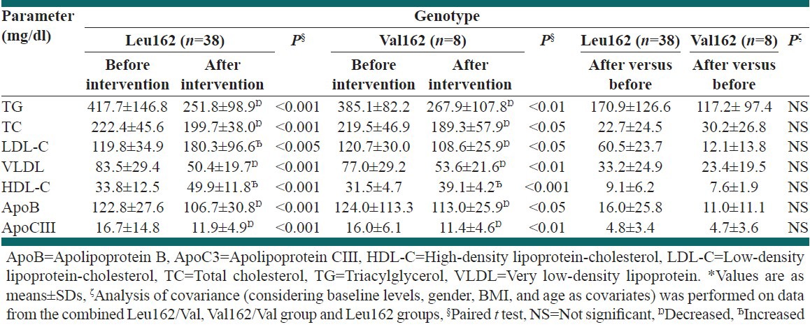
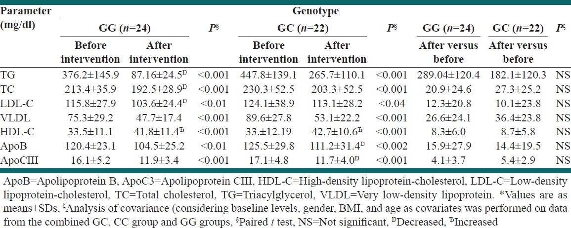
The baseline characteristics of subjects were described previously.[25,27] For comparison of groups with different PPARα genotypes, the data obtained from Val162/Val and Leu162/Val subjects were combined. EPA supplementation decreased the levels of serum lipid and lipoprotein in the two study groups. No interaction was observed between PPARα genotypes and the degree of changes in plasma lipids and lipoproteins after EPA consumption [Table 1]. EPA supplementation decreased levels of serum lipid and lipoprotein in the two study groups. No interaction was observed between CC and GC carriers and the degree of changes in plasma lipids and lipoproteins after EPA consumption [Table 2]. Although EPA consumption lowered the lipid and lipoprotein concentrations in Leu162 and Val162 carriers and CC and GC carriers, the observed difference between the studied groups was not statistically significant [Tables 1 and 2]. EPA supplementation increased the plasma EPA in both Leu162 and Val162 carriers [Table 3] (P = 0.01 and P = 0.05, respectively), but it was observed that it has been significantly increased more in Val162 carriers than Leu162 (P = 0.001) [Table 3].
Table 1.
Changes in serum lipids and lipoproteins of hypertriglyceridemic subjects at baseline and after 8-weeks eicosapentaenoic acid supplementation in the studied groups

Table 2.
Changes in serum lipids and lipoproteins of hypertriglyceridemic subjects at baseline and after 8-weeks eicosapentaenoic acid supplementation in the studied groups

Table 3.
Changes in plasma eicosapentaenoic acid (EPA) levels in hypertriglyceridemic subjects after 8-week EPA supplementation stratified by Leu162 and Val162 peroxisome proliferator-activated receptor alpha genotypes*

Supplementation increased plasma EPA in GG and GC carriers (P = 0.001 and P =0.001) [Table 4], but it was observed that it has been significantly increased more in GC carriers than in GG carriers (P = 0.001) [Table 4].
Table 4.
Changes in plasma eicosapentaenoic acid levels in hypertriglyceridemic subjects after 8-weeks EPA supplementation stratified by GG and GC polymorphism of peroxisome proliferator-activated receptor alpha genotypes*

DISCUSSION
As PPARα has a role in lipid homeostasis, we were therefore interested in their variable plasma lipid levels that often account for the independent increased risk of atherosclerosis[28,16] and cardiovascular risk factors.[29]
The major aim of this study was to identify the role that PPARα Leu162Val polymorphism may play in the variability of serum lipid levels, and changes in lipids and lipoproteins level in response to EPA supplementation.
Among the reported polymorphisms for the PPARα gene, Leu162Val polymorphism is the most prevalent with a frequency of 10.45% in diabetic and non-diabetic patients.[13] It is also associated with alterations in lipoprotein concentrations in both diabetic and non-diabetic subjects.[16] We found a prevalence of 21.8% for the Leu162/Val polymorphism in these Iranian hypertriglyceridemic subjects.[27] There was no difference in the frequency of Leu162/Val between men and women, and there were no significant differences between lipid and lipoprotein in Leu162 and Val162 carriers except for TC. In our study, a comparison between Val162 carries and Leu carriers has indicated a significant association between PPARα Leu162Val polymorphism and blood lipids, where the Val162 carriers showed higher concentrations of TC.[27]
This polymorphism has been previously found to be associated with greater blood TC,[16] LDL-C,[8,11,12] TG levels,[9,30] and lower HDL-C concentration[8] depending upon the population studied. Most studies that have found an association between the Leu162Val polymorphism and greater blood lipid concentrations were conducted in diabetic subjects; few studies have found a positive association in healthy subjects.[11,31]
The intron7 allele has been shown to be associated with increased progression of atherosclerosis. The Val162 allele and intron7 allele are in strong allelic association, such that 78% of Val162 carriers are found in combination with the intron7.[14] In our study the frequency of the intron7 polymorphism was 55.3%, which is much greater than the frequency of Leu162Val polymorphism in the studied subjects, and all of the subjects who were Leu162/Val carriers also had an intron7 allele. Except for blood TC level, there were no significant associations between intron7 allele and blood lipid, ApoB, or apolipoprotein CIII (ApoCIII) concentrations. Similar results have been observed in Foucher's study in which intron7 polymorphism was only associated with blood TC levels.[32]
Frequency of intron7 polymorphism is greater than Leu162Val and all hypertriglyceridemic subjects with Leu162 polymorphism might be carriers of the intron7 polymorphism.[31]
The expected changes in post-supplementation plasma lipids and lipoproteins were observed with EPA intervention. Results of the present study clearly show that two carriers of PPARα genotypes can influence the lipid-lowering effects of EPA supplementation in hypertriglyceridemic subjects.
Although EPA supplementation decreased serum lipids and lipoproteins and increased serum HDL-C in Leu162, Val162 carriers and GG, GC carriers, there was no significant interaction between the two carriers after EPA consumption.
We believe that these data provide some insight into the pathways that could be involved in generating the association between LDL cholesterol concentration and the Leu162Val polymorphism at the PPARα locus.[11] There was no association between the Leu162Val polymorphism at the PPARα locus and TG, HDL-C, very low-density lipoprotein, ApoB, and ApoCIII concentrations. Several possible reasons exist for these results. Intrer-individual variation in the plasma lipid and lipoprotein. This phenomenon will reduce the chance of finding an association between a genetic variant and plasma lipid and lipoprotein concentrations, especially when the changes are small, as in this case. Alternatively, additional gene–gene or gene–diet interactions may exist that modulates the effect of the L162V polymorphism, and this will need to be addressed in future studies. Potential gene–environment interactions may be particularly relevant to this polymorphism. The level of exposure to known endogenous (such as non-esterified fatty acids) or exogenous ligands (such as dietary PUFAs) may therefore be important in determining the biochemical phenotype observed with this polymorphism.[11]
Previous studies have shown the PPARα L162V SNP to be associated with multiple lipid and lipoprotein measures, with one study finding the effect of the L162V SNP on triacylglycerol and ApoCIII concentrations to be dependent on PUFA intake.[9,10,11,12,33]
In the current study, we did not observe significant associations between the PPARα L162V SNP and multiple lipid and lipoprotein measures. However, we did not observe a significant interaction between the Leu162, Val162 and EPA consumption with regards to lipid and lipoprotein concentrations in hypertriglyceridemic subjects. But we could observe a significant difference between EPA supplementation and Leu162 and Val162, GG and GC polymorphism. This finding suggests that genetic variation within the PPARα Leu162/Val cannot modulate the association of EPA intakes with lipid and lipoprotein profiles. It is possible that the sample of study is not high enough to affect the association between PPARα genotype and lipid after EPA consumption However, previous studies of genetic variation in the PPARα gene have found gene-by-diet interactions specific to different polymorphisms in different racial-ethnic groups (i.e., L162V in whites and V227A in Asians).[33]
The role of dietary fatty acids in regulating plasma lipoprotein and lipid concentrations is well documented.[34] Compared with SFA intake, PUFA intake has been shown to lower LDL-C and HDL-C concentrations. However, the great inter-individual lipoprotein and lipid response to such dietary changes is not well understood. Genetic factors are likely to play a significant role, because polymorphisms in several genes seem to modulate lipoprotein and lipid responses to dietary modifications.[35] This study is the first in hypertriglyceridemic subjects to examine whether plasma lipid and lipoprotein responsiveness to EPA supplementation are influenced by the PPARα Leu162Val gene polymorphism or not.
In vitro studies have shown that the Val162 allele has a greater transactivation activity when treated with a PPARα agonist.[8] Previous studies showed the increasing effect of the Leu162Val polymorphism on TC, LDL-C, HDL-C,[8] ApoB,[11,12,16] apo A-1,[16] and ApoCIII.[11] This polymorphism was also found to be associated with decreased concentrations of fasting serum triacylglycerol among white subjects with normal glucose tolerance.[36,37] Their investigation of the gene-by-EPA consumption interaction effects on the metabolic profile led us to several interesting observations. They found that EPA consumption interacts with the Leu162/Leu and Leu162Val polymorphism to modulate the TC concentrations These gene-by-EPA supplementation interaction effects may then modulate plasma lipid concentrations and need further replication in different intervention studies and later in a meta- analysis.[37] Although we cannot interaction between EPA consumption and PPARα genotypes. One study of the gene-by-diet interaction effects on the metabolic profile led to several interesting observations. They found that diet interacts with the Leu162Val polymorphism to modulate TC concentrations, plasma ApoA-1 concentrations, and cholesterol in small LDL particles. This gene-by diet interaction effects may then modulate plasma lipid concentrations and need further replication in different intervention studies and later in a meta-analysis. In addition, independent of the genotype, diet had an effect on HDL.[36,38]
CONCLUSIONS
EPA consumption has lipid-lowering effect in hypertriglyceridemic subjects in both Leu162 and Val162, GG, and GC carriers. But there was no significant interaction between EPA supplementation and PPARα genotypes. Thus, genetic variation within the PPARα Leu162/Val cannot modulate the association of EPA intakes with lipid and lipoprotein profiles. However, we must note the sample size in this study.
ACKNOWLEDGMENTS
The authors thank all the subjects who participated in this study, the staff of the Danesh, Kach, Masood, and EMRC laboratories, and Shariaty Hospital Heart Diseases Center, Tehran. The EPA caps were a kind gift from Minami Nutrition, Belgium.
Footnotes
Source of Support: Nil
Conflict of Interest: None declared
REFERENCES
- 1.Chen ES, Mazzotti DR, Furuya TK, Cendoroglo MS, Ramos LR, Araujo LQ, et al. Association of PPARα gene polymorphisms and lipid serum levels in a Brazilian elderly population. Exp Mol Pathol. 2010;88:197–201. doi: 10.1016/j.yexmp.2009.10.001. [DOI] [PubMed] [Google Scholar]
- 2.Tanaka T, Ordovas JM, Delgado-Lista J, Perez-Jimenez F, Marin C, Perez- Martinez P, et al. Peroxisome proliferator-activated receptor alpha polymorphisms and postprandial lipemia in healthy men. J Lipid Res. 2007;48:1402–8. doi: 10.1194/jlr.M700066-JLR200. [DOI] [PubMed] [Google Scholar]
- 3.Karpe F. Postprandial lipoprotein metabolism and atherosclerosis. J Intern Med. 1999;246:341–55. doi: 10.1046/j.1365-2796.1999.00548.x. [DOI] [PubMed] [Google Scholar]
- 4.Cullen P. Evidence that triglycerides are an independent coronary heart disease risk factor. Am J Cardiol. 2000;86:943–9. doi: 10.1016/s0002-9149(00)01127-9. [DOI] [PubMed] [Google Scholar]
- 5.Yong EL, Li J, Liu MH. Single gene contributions: Genetic variants of peroxisome proliferator-activated receptor (isoforms alpha, beta/delta and gamma) and mechanisms of dyslipidemias. Curr Opin Lipidol. 2008;19:106–12. doi: 10.1097/MOL.0b013e3282f64542. [DOI] [PubMed] [Google Scholar]
- 6.Burch LR, Donnelly LA, Doney AS, Brady J, Tommasi AM, Whitley AL, et al. Peroxisome proliferator-activated receptor-delta genotype influences metabolic phenotype and may influence lipid response to statin therapy in humans: A genetics of diabetes audit and research Tayside study. J Clin Endocrinol Metab. 2010;95:1830–7. doi: 10.1210/jc.2009-1201. [DOI] [PubMed] [Google Scholar]
- 7.Evans D, Aberle J, Wendt D, Wolf A, Beisiegel U, Mann WA. A polymorphism, L162V, in the peroxisome proliferator-activated receptor alpha (PPARα) gene is associated with lower body mass index in patients with non-insulin-dependent diabetes mellitus. J Mol Med (Berl) 2001;79:198–204. doi: 10.1007/s001090100189. [DOI] [PubMed] [Google Scholar]
- 8.Flavell DM, Pineda Torra I, Jamshidi Y, Evans D, Diamond JR, Elkeles RS, et al. Variation in the PPARα gene is associated with altered function in vitro and plasma lipid concentrations in Type II diabetic subjects. Diabetologia. 2000;43:673–80. doi: 10.1007/s001250051357. [DOI] [PubMed] [Google Scholar]
- 9.Robitaille J, Brouillette C, Houde A, Lemieux S, Pérusse L, Tchernof A, et al. Association between the PPARα-L162V polymorphism and components of the metabolic syndrome. J Hum Genet. 2004;49:482–9. doi: 10.1007/s10038-004-0177-9. [DOI] [PubMed] [Google Scholar]
- 10.Tai ES, Corella D, Demissie S, Cupples LA, Coltell O, Schaefer EJ, et al. Polyunsaturated fatty acids interact with the PPARA-L162V polymorphism to affect plasma triglyceride and apolipoprotein C-III concentrations in the Framingham Heart Study. J Nutr. 2005;135:397–403. doi: 10.1093/jn/135.3.397. [DOI] [PubMed] [Google Scholar]
- 11.Tai ES, Demissie S, Cupples LA, Corella D, Wilson PW, Schaefer EJ, et al. Association between the PPARA L162V polymorphism and plasma lipid levels: The Framingham Offspring Study. Arterioscler Thromb Vasc Biol. 2002;22:805–10. doi: 10.1161/01.atv.0000012302.11991.42. [DOI] [PubMed] [Google Scholar]
- 12.Vohl MC, Lepage P, Gaudet D, Brewer CG, Bétard C, Perron P, et al. Molecular scanning of the human PPARα gene: Association of the L162v mutation with hyperapobetalipoproteinemia. J Lipid Res. 2000;41:945–52. [PubMed] [Google Scholar]
- 13.Sapone A, Peters JM, Sakai S, Tomita S, Papiha SS, Dai R, et al. The human peroxisome proliferator-activated receptor alpha gene: Identification and functional characterization of two natural allelic variants. Pharmacogenetics. 2000;10:321–33. doi: 10.1097/00008571-200006000-00006. [DOI] [PubMed] [Google Scholar]
- 14.Flavell DM, Jamshidi Y, Hawe E, Pineda Torra I, Taskinen MR, Frick MH, et al. Peroxisome proliferator-activated receptor alpha gene variants influence progression of coronary atherosclerosis and risk of coronary artery disease. Circulation. 2002;105:1440–5. doi: 10.1161/01.cir.0000012145.80593.25. [DOI] [PubMed] [Google Scholar]
- 15.Jamshidi Y, Montgomery HE, Hense HW, Myerson SG, Torra IP, Staels B, et al. Peroxisome proliferator-activated receptor alpha gene regulates left ventricular growth in response to exercise and hypertension. Circulation. 2002;105:950–5. doi: 10.1161/hc0802.104535. [DOI] [PubMed] [Google Scholar]
- 16.Lacquemant C, Lepretre F, Pineda Torra I, Manraj M, Charpentier G, Ruiz J, et al. Mutation screening of the PPARα gene in type 2 diabetes associated with coronary heart disease. Diabetes Metab. 2000;26:393–401. [PubMed] [Google Scholar]
- 17.Garenc C, Aubert S, Laroche J, Girouard J, Vohl MC, Bergeron J, et al. Population prevalence of APOE, APOC3 and PPAR-alpha mutations associated to hypertriglyceridemia in French Canadians. J Hum Genet. 2004;49:691–700. doi: 10.1007/s10038-004-0208-6. [DOI] [PubMed] [Google Scholar]
- 18.Wagener A, Goessling HF, Schmitt AO, Mauel S, Gruber AD, Reinhardt R, et al. Genetic and diet effects on PPAR-α and PPAR-γ signaling pathways in the Berlin Fat Mouse Inbred line with genetic predisposition for obesity. Lipids Health Dis. 2010;9:99. doi: 10.1186/1476-511X-9-99. [DOI] [PMC free article] [PubMed] [Google Scholar]
- 19.Erkkilä A, de Mello VD, Risérus U, Laaksonen DE. Dietary fatty acids and cardiovascular disease: An epidemiological approach. Prog Lipid Res. 2008;47:172–87. doi: 10.1016/j.plipres.2008.01.004. [DOI] [PubMed] [Google Scholar]
- 20.Jacobson TA. Role of n-3 fatty acids in the treatment of hypertriglyceridemia and cardiovascular disease. Am J Clin Nutr. 2008;87:1981S–90. doi: 10.1093/ajcn/87.6.1981S. [DOI] [PubMed] [Google Scholar]
- 21.Wang C, Harris WS, Chung M, Lichtenstein AH, Balk EM, Kupelnick B, et al. n-3 Fatty acids from fish or fish-oil supplements, but not alpha-linolenic acid, benefit cardiovascular disease outcomes in primary- and secondary-prevention studies: A systematic review. Am J Clin Nutr. 2006;84:5–17. doi: 10.1093/ajcn/84.1.5. [DOI] [PubMed] [Google Scholar]
- 22.Mozaffarian D. Does alpha-linolenic acid intake reduce the risk of coronary heart disease? A review of the evidence. Altern Ther Health Med. 2005;11:24–30. [PubMed] [Google Scholar]
- 23.Brouwer IA, Katan MB, Zock PL. Dietary alpha-linolenic acid is associated with reduced risk of fatal coronary heart disease, but increased prostate cancer risk: A meta-analysis. J Nutr. 2004;134:919–22. doi: 10.1093/jn/134.4.919. [DOI] [PubMed] [Google Scholar]
- 24.Egert S, Stehle P. Impact of n-3 fatty acids on endothelial function: Results from human interventions studies. Curr Opin Clin Nutr Metab Care. 2011;14:121–31. doi: 10.1097/MCO.0b013e3283439622. [DOI] [PubMed] [Google Scholar]
- 25.Pishva H, Mahboob SA, Mehdipour P, Eshraghian MR, Mohammadi-Asl J, Hosseini S, et al. Fatty acid-binding protein-2 genotype influences lipid and lipoprotein response to eicosapentaenoic acid supplementation in hypertriglyceridemic subjects. Nutrition. 2010;26:1117–21. doi: 10.1016/j.nut.2009.09.028. [DOI] [PubMed] [Google Scholar]
- 26.Folch J, Lees M, Sloane Stanley GH. A simple method for the isolation and purification of total lipides from animal tissues. J Biol Chem. 1957;226:497–509. [PubMed] [Google Scholar]
- 27.Pishva H, Mahboob SA, Mehdipour P, Eshraghian MR, Mohammadi-Asl J, Hosseini S, et al. Association between the FABP2 Ala54Thr, PPARα Leu162/Val, and PPARα intron7 polymorphisms and blood lipids ApoB and ApoCIII in hypertriglyceridemic subjects in Tehran. J Clin Lipidol. 2009;3:187–94. doi: 10.1016/j.jacl.2009.04.001. [DOI] [PubMed] [Google Scholar]
- 28.Genest JJ, Jr, Martin-Munley SS, McNamara JR, Ordovas JM, Jenner J, Myers RH, et al. Familial lipoprotein disorders in patients with premature coronary artery disease. Circulation. 1992;85:2025–33. doi: 10.1161/01.cir.85.6.2025. [DOI] [PubMed] [Google Scholar]
- 29.DeFronzo RA, Bonadonna RC, Ferrannini E. Pathogenesis of NIDDM. A balanced overview. Diabetes Care. 1992;15:318–68. doi: 10.2337/diacare.15.3.318. [DOI] [PubMed] [Google Scholar]
- 30.Sparsø T, Hussain MS, Andersen G, Hainerova I, Borch-Johnsen K, Jørgensen T, et al. Relationships between the functional PPARα Leu162Val polymorphism and obesity, type 2 diabetes, dyslipidaemia, and related quantitative traits in studies of 5799 middle-aged white people. Mol Genet Metab. 2007;90:205–9. doi: 10.1016/j.ymgme.2006.10.007. [DOI] [PubMed] [Google Scholar]
- 31.Pishva H, Mahboob SA, Mehdipour P, Eshraghian MR, Mohammadi-Asl J, Hosseini S, et al. Association between the FABP2 Ala54Thr, PPARα Leu162/Val, and PPARα intron7 polymorphisms and blood lipids ApoB and ApoCIII in hypertriglyceridemic subjects in Tehran. J Clin Lipidol. 2009;3:187–94. doi: 10.1016/j.jacl.2009.04.001. [DOI] [PubMed] [Google Scholar]
- 32.Foucher C, Rattier S, Flavell DM, Talmud PJ, Humphries SE, Kastelein JJ, et al. Response to micronized fenofibrate treatment is associated with the peroxisome- proliferator-activated receptors alpha G/C intron7 polymorphism in subjects with type 2 diabetes. Pharmacogenetics. 2004;14:823–9. doi: 10.1097/00008571-200412000-00005. [DOI] [PubMed] [Google Scholar]
- 33.Volcik KA, Nettleton JA, Ballantyne CM, Boerwinkle E. Peroxisome proliferator- activated receptor alpha genetic variation interacts with n-6 and long-chain n-3 fatty acid intake to affect total cholesterol and LDL-cholesterol concentrations in the Atherosclerosis Risk in Communities Study. Am J Clin Nutr. 2008;87:1926–31. doi: 10.1093/ajcn/87.6.1926. [DOI] [PMC free article] [PubMed] [Google Scholar]
- 34.Mensink RP, Katan MB. Effect of dietary fatty acids on serum lipids and lipoproteins. A meta-analysis of 27 trials. Arterioscler Thromb. 1992;12:911–9. doi: 10.1161/01.atv.12.8.911. [DOI] [PubMed] [Google Scholar]
- 35.Ordovas JM. The genetics of serum lipid responsiveness to dietary interventions. Proc Nutr Soc. 1999;58:171–87. doi: 10.1079/pns19990023. [DOI] [PubMed] [Google Scholar]
- 36.Paradis AM, Fontaine-Bisson B, Bossé Y, Robitaille J, Lemieux S, Jacques H, et al. The peroxisome proliferator-activated receptor alpha Leu162Val polymorphism influences the metabolic response to a dietary intervention altering fatty acid proportions in healthy men. Am J Clin Nutr. 2005;81:523–30. doi: 10.1093/ajcn.81.2.523. [DOI] [PubMed] [Google Scholar]
- 37.Nielsen EM, Hansen L, Echwald SM, Drivsholm T, Borch-Johnsen K, Ekstrøm CT, et al. Evidence for an association between the Leu162Val polymorphism of the PPARα gene and decreased fasting serum triglyceride levels in glucose tolerant subjects. Pharmacogenetics. 2003;13:417–23. doi: 10.1097/01.fpc.0000054105.48725.5c. [DOI] [PubMed] [Google Scholar]
- 38.Halliwell B, Chirico S. Lipid peroxidation: Its mechanism, measurement, and significance. Am J Clin Nutr. 1993;57:715S–24. doi: 10.1093/ajcn/57.5.715S. [DOI] [PubMed] [Google Scholar]


