Abstract
Botulinum neurotoxin type F (BoNT/F) may be produced by Clostridium botulinum alone or in combination with another toxin type such as BoNT/A or BoNT/B. Type F neurotoxin gene sequences have been further classified into seven toxin subtypes. Recently, the genome sequence of one strain of C. botulinum (Af84) was shown to contain three neurotoxin genes (bont/F4, bont/F5, and bont/A2). In this study, eight strains containing bont/F4 and seven strains containing bont/F5 were examined. Culture supernatants produced by these strains were incubated with BoNT/F-specific peptide substrates. Cleavage products of these peptides were subjected to mass spectral analysis, allowing detection of the BoNT/F subtypes present in the culture supernatants. PCR analysis demonstrated that a plasmid-specific marker (PL-6) was observed only among strains containing bont/F5. Among these strains, Southern hybridization revealed the presence of an approximately 242-kb plasmid harboring bont/F5. Genome sequencing of four of these strains revealed that the genomic backgrounds of strains harboring either bont/F4 or bont/F5 are diverse. None of the strains analyzed in this study were shown to produce BoNT/F4 and BoNT/F5 simultaneously, suggesting that strain Af84 is unusual. Finally, these data support a role for the mobility of a bont/F5-carrying plasmid among strains of diverse genomic backgrounds.
INTRODUCTION
Botulism caused by type F botulinum neurotoxin (BoNT/F) is relatively rare and accounts for ∼1% of the botulism cases reported to the Centers for Disease Control and Prevention (CDC) annually (1). Nonetheless, genes encoding BoNT/F diverge by up to 25% and are the most diverse of the BoNT serotypes (BoNT/A to -G) (2). Phylogenetic analysis of these genes identified a total of seven toxin subtypes (F1 to F7) (2). Moreover, the organisms that produce BoNT/F are also diverse. Subtypes F1 to F5 are produced by Clostridium botulinum group I strains, which digest a variety of proteins (i.e., they are proteolytic). Subtype F6 is produced by C. botulinum group II strains, which are nonproteolytic. Finally, subtype F7 is produced by rare strains of C. baratii.
The predicted amino acid sequence of BoNT/F5 is highly divergent from other BoNT/F subtypes (2). Compared to the other subtypes, BoNT/F5 contains a unique light chain region (i.e., the enzymatic portion of the neurotoxin, which among BoNT/F serotypes, functions to cleave the synaptic vesicle protein synaptobrevin 2 [also termed VAMP-2]). Kalb et al. (3) demonstrated that BoNT/F5 cleaves synaptobrevin 2 at a location different from that of the other BoNT/F subtypes. BoNT/F is the only serotype where different toxin subtypes have been shown to display differential enzymatic cleavage.
In previous work, we examined the neurotoxin gene sequences among a panel of 15 strains of C. botulinum harboring either bont/F4 or bont/F5, and a subset of these strains also contained bont/A2 (2). Nearly all of these strains were isolated from various sources (soil, stool, etc.) in Argentina. Recent work by Dover et al. (4) showed that C. botulinum strain Af84, also isolated in Argentina, contains three botulinum neurotoxin genes. Both bont/A2 and bont/F4 are present on the chromosome, and bont/F5 is present on a large plasmid (pCLQ, ∼246 kb). None of the strains that we previously examined was shown to contain all three bont genes, In this study, we used mass spectral analysis to confirm that these 15 strains produce the expected type F toxin subtypes, assessed their genetic diversity, and determined the genomic location of either bont/F4 or bont/F5 in order to better understand the genetic relationships among these strains.
MATERIALS AND METHODS
Strains used in this study.
The C. botulinum strains shown in Table 1 were recovered from bovine brain medium (5) and grown in cooked meat glucose starch (CMGS) medium (Remel, Lenexa, KS) at 35°C under anaerobic conditions for 24 to 48 h. CMGS cultures were streaked on egg yolk agar (5), and single colonies were selected and grown in Trypticase-peptone-glucose-yeast extract (TPGY) medium (Remel). Culture supernatants for mass spectral analysis were prepared from TPGY medium cultures incubated for 5 days. Cultures were centrifuged at 20,000 × g and filtered through 0.45-μm syringe filters.
TABLE 1.
Strains examined in this study
| Strain | Toxin serotype | Toxin subtype(s) | Reference |
|---|---|---|---|
| CDC 49930 | F | F4 | 2 |
| CDC 54074 | F | F5 | 2 |
| CDC 54075 | F | F5 | 2 |
| CDC 54076 | Af | A2, F4 | 2 |
| CDC 54078 | F | F4 | 2 |
| CDC 54079 | Af | A2, F5 | 2 |
| CDC 54084 | Af | A2, F5 | 2 |
| CDC 54085 | F | F5 | 2 |
| CDC 54087 | F | F4 | 2 |
| CDC 54088 | F | F4 | 2 |
| CDC 54089 | F | F4 | 2 |
| CDC 54090 | F | F5 | 2 |
| CDC 54091 | Af | A2, F4 | 2 |
| CDC 54093 | Af | A2, F4 | 2 |
| CDC 54096 | Af | A2, F5 | 2 |
| CDC 40234 (CDC A3) | A | A3 | 7 |
| CDC 40234a | Nontoxic | NA | 13 |
| ATCC 3502 | A | A1 | 16 |
| Loch Maree | A | A3 | 12 |
| 657 | Ba | B5, A4 | 12 |
| Af84 | Af | A2, F4, F5 | 4 |
| Okra | B | B1 | 16 |
| Langeland | F | F1 | 14 |
| 12F | F | F1 | 2 |
| 4VI | F | F1 | 2 |
| 4VII | F | F1 | 2 |
| Pasteurized crab | F | F1 | 2 |
| 8g | F | F1 | 2 |
Plasmid cured.
Mass spectral analysis of culture supernatants.
Monoclonal antibody 6F5 was used for BoNT/F extraction (3). Dynabeads (M-280 streptavidin purchased from Invitrogen, Carlsbad, CA) were used at 1.3 g/cm3 in phosphate-buffered saline (PBS), pH 7.4, containing 0.1% Tween 20 and 0.02% sodium azide. The beads were rinsed three times with HBS-EP buffer (GE Healthcare, Piscataway, NJ). Monoclonal antibody 6F5 (2 μg) was immobilized on streptavidin Dynabeads (100 μl), and a standard orbital shaker was used to bind the antibody to the beads for 1 h.
An aliquot of 20 μl of antibody-coated beads was added to 200 μl of HBS-EP buffer, 100 μl of 10× PBS–0.1% Tween 20, and 500 μl of culture supernatant. Negative controls consisted of TPGY medium with no toxin. After mixing for 1 h with constant agitation at room temperature, the beads were removed from the mixture and washed once in 1 ml of HBS-EP buffer. Positive controls consisted of TPGY medium spiked with 1 50% lethal dose of BoNT/F.
The beads were reconstituted in 18 μl of reaction buffer consisting of 0.05 M HEPES (pH 7.3), 25 mM dithiothreitol, 20 μM ZnCl2, and 2 μl of the BoNT/F-specific peptide substrate TSNRRLQQTQAQVDEVVDIMRVNVDKVLERDQKLSELDDRADAL (Midwest Bio-tech Inc., Fishers, IN). The final concentration of substrate was 50 pmol/μl. All samples then were incubated at 37°C for 4 h with no agitation. A 2-μl aliquot of each reaction supernatant was mixed with 18 μl of a matrix solution consisting of α-cyano-4-hydroxycinnamic acid at 5 mg/ml in 50% acetonitrile, 0.1% trifluoroacetic acid, and 1 mM ammonium citrate. A 0.5-μl aliquot of this mixture was pipetted onto one spot of a 384-spot matrix-assisted laser desorption/ionization plate (Applied Biosystems, Framingham, MA). Mass spectra of each spot were obtained by scanning from 1,100 to 4,800 m/z in MS-positive ion reflector mode on an Applied Biosystems 5800 Proteomics Analyzer (Applied Biosystems). The instrument uses an Nd-YAG laser at 355 nm, and each spectrum is an average of 2,400 laser shots.
Genomic DNA extraction.
Genomic DNA was extracted from cultures grown in TPGY medium with the MasterPure kit (Epicenter, Madison, WI) with modifications previously described (2). For genome sequencing experiments, DNA was further purified and concentrated with the DNA Clean & Concentrator-5 kit (Zymo Research, Irvine, CA). DNA was eluted in 10 mM Tris (pH 8.0).
DNA microarrays and analysis.
The C. botulinum group I subtyping microarray was designed as described elsewhere (6). Briefly, the microarray featured 225 probes representing selected regions from various complete C. botulinum group I genome sequences, the bont/A to -G genes, and plasmid-specific regions. Microarray spotting was performed by ArrayIt (Sunnyvale, CA), and hybridizations were carried out as previously described (7).
The log of the ratio of the mean fluorescence signal at 635 nm for triplicate probes compared to background fluorescence (locations spotted with buffer alone) was calculated. Log ratios were converted to binary data as follows. Log ratios of ≥1.0 were assigned a value of 1, and log ratios of <1.0 were assigned a value of 0. The binary hybridization profiles were compared by using DendroUPGMA (http://genomes.urv.cat/UPGMA/) and selecting the Jaccard coefficient. The resulting distance matrix was used to construct a dendrogram by the unweighted-pair group method using average linkages (UPGMA), which was rendered in FigTree v1.4.0 (http://tree.bio.ed.ac.uk/software/figtree/).
Genome sequencing and analysis.
Genomic DNA isolated from two representative strains producing BoNT/F4 (CDC 54088, CDC 54091) and two representative strains producing BoNT/F5 (CDC 54075, CDC 54085) were sequenced with the Ion Torrent Personal Genome Machine by using the manufacturer's 200-bp template (version 2) and sequencing kits. Sequence reads were de novo assembled with the Assembler plug-in (which runs the MIRA assembly program) in the Torrent Suite software package. Draft genome statistics are shown in Table 2.
TABLE 2.
Draft genome characteristics
| Strain | No. of reads assembled | Genome size (Mb) | No. of contigs | N50 (kb) | Fold coverage |
|---|---|---|---|---|---|
| CDC 54075 | 628,393 | 4.26 | 542 | 13.3 | 28 |
| CDC 54085 | 481,081 | 4.09 | 345 | 19.8 | 25 |
| CDC 54088 | 351,287 | 3.87 | 469 | 12.9 | 16 |
| CDC 54091 | 300,302 | 4.08 | 721 | 8.5 | 14 |
Draft and complete genomes sequences from this study and those selected from the NCBI genome database where aligned by using Gegenees (8). This software aligns fragments of each genome sequence by using BLAST. The resulting similarity matrix (based on average similarity scores of pairwise alignments) was used to generate a neighbor-joining tree in SplitsTree 4 (9), and the final tree was rendered in FigTree v1.4.0. Additional comparisons of draft genome sequences generated in this study with the pCLQ plasmid from strain Af84 were performed with BRIG version 0.95 (10).
Toxin gene localization by PCR analysis.
Genomic DNA was used to perform PCR for either pulE (with primers PUL-F [5′-ATTCTTTGGATTTCACTAAAGGATG-3′] and PUL-R [5′-AGATTTAAAGGAAACTGTACAAAAC-3′]) or PL-6 (with primers PL-6F [5′-CTAATTGCTATTACTCCTCTTTC-3′] and PL-6R [5′-CAGGAGCAGGAAGTCC-3′]).
PCR was performed with iQ Supermix (Bio-Rad, Hercules, CA) under the following conditions: 1 cycle of 95°C for 3 min; 35 cycles of 95°C for 30 s, 50°C for 30 s, and 72°C for 60 s; and 1 cycle of 72°C for 5 min.
PFGE and Southern hybridization.
C. botulinum strains were inoculated into 10 ml of TPGY medium and incubated anaerobically at 37°C to an optical density at 600 nm of 0.6. One milliliter of formaldehyde (Fisher Scientific, Hampton, NH) was added, and the cultures were placed on ice for 30 min to inhibit nuclease activity. Pulsed-field gel electrophoresis (PFGE) plugs were prepared as described by Johnson et al. (11).
PFGE was carried out with a clamped homogeneous electric field system (CHEF-DRII; Bio-Rad, Hercules CA) at 14°C in 0.5× TBE or in 16 mM HEPES–16 mM sodium acetate–0.8 mM EDTA (pH 7.5) at 6 V/cm for 24 h with 1- to 20-s pulse switching. The gels were stained with ethidium bromide (5 mg/ml), destained in water, and photographed with a Gel Imaging System (Fotodyne, Inc.). The DNA samples separated by PFGE were transferred to a positively charged nylon membrane (Immobilon-NY+; Millipore, Bedford, MA) overnight by downward capillary transfer in 0.4 M NaOH–1.5 M NaCl. The membranes were neutralized in 2 M Tris-HCl (pH 7.0) for 15 min and rinsed with 2× SSC (3 M NaCl, 0.3 M sodium citrate), and the DNA was fixed to the membrane by heating at 80°C for 30 min under vacuum on a gel dryer.
Hybridization probes were generated by PCR amplification with AmpliTaq High Fidelity DNA polymerase, buffer, and deoxynucleoside triphosphates (Applied Biosystems, Foster City, CA) with an Applied Biosystems GeneAmp PCR System 9700 according to the manufacturer's instructions. The bont/F5 probe (820 bp) was generated with primers 1000F (5′-GTAGACTTAGATAAATTTAATAAATTATATG-3′) and 1820R (5′-CTTTTTTGTGTAGCTTCAGTGGTAAAATC-3′). The bont/A probe (1.3 kb) was generated with primers LC/A1F (5′-ATGCCATTTGTTAATAAACAATTTAATTATAAAGATCC-3′) and LC/A1R (5′-GAAGTTATTATCCCTCTTACACATAGCAAC-3′).
The PCR products were purified from agarose gels with the Qiagen gel extraction kit (Qiagen, Valencia, CA) and radioactively labeled with [α-32P]ATP with the Megaprime DNA labeling system (GE Healthcare Bio-Sciences, Piscataway, NJ).
Hybridizations were performed at 42°C for 16 h in a solution containing 5× Denhardt's solution, 6× SSPE (1× SSPE is 0.18 M NaCl, 10 mM NaH2PO4, and 1 mM EDTA [pH 7.7]), 50% formamide, 0.1% SDS, 100 mg/ml herring sperm DNA (Promega, Madison, WI), and 32P-labeled probes at ∼2 × 10⁶ cpm/ml. After hybridizations, the membranes were washed twice with 2× SSPE–0.1% SDS for 5 min each time at room temperature and twice with 0.1× SSPE–0.1% SDS for 30 min each time at 42°C. Autoradiography of the membranes was performed for 6 to 24 h at −70°C with Kodak BioMax MS film with a BioMax intensifying screen (Eastman Kodak, Rochester, NY).
Microarray data and nucleotide sequence accession numbers.
Microarray data were deposited in the Gene Expression Omnibus with series accession number GSE53456. Assembled genome sequences were deposited at DDBJ/EMBL/GenBank under accession numbers AZQW01000000 (CDC 54075), AZRQ01000000 (CDC 54085), AZRR01000000 (CDC 54091), and AZRS01000000 (CDC 54088).
RESULTS
Characterization of toxins produced by C. botulinum strains containing bont/F4 or bont/F5.
BoNT/F5 cleaves synaptobrevin 2 at an amino acid position distinct from that of other BoNT/F subtypes (3). Therefore, BoNT/F5-producing strains can be distinguished from BoNT/F4-producing strains by examination of the mass spectra of their substrate cleavage products. As shown in Fig. 1A, a culture supernatant from a representative strain (CDC 54078) containing bont/F4 and incubated with the type F peptide substrate yielded a peak at m/z 1,345.5. The N-terminal product located at m/z 3,782.6 ionizes poorly because of optimization of the instrument for smaller peptide detection and is not visible in the spectrum shown in Fig. 1A. The peaks at m/z 1,873.7 and 3,254.4 were observed when culture supernatant from a representative strain (CDC 54075) containing bont/F5 was used (Fig. 1B). As expected, strains previously shown to contain bont/F4 (CDC 49930, CDC 54076, CDC 54087, CDC 54088, CDC 54089, CDC 54091, and CDC 54093) produced culture supernatants that cleaved the type F substrate, yielding a peak at m/z 1,345.5 (data not shown). Strains previously shown to contain bont/F5 (CDC 54074, CDC 54079, CDC 54084, CDC 54085, CDC 54090, and CDC 54096) produced culture supernatants that cleaved the type F substrate, yielding peaks at m/z 1,873.7 and 3,254.4 (data not shown). None of the culture supernatants examined in this study yielded substrate cleavage products associated with both BoNT/F4 and BoNT/F5.
FIG 1.

Representative mass spectra of substrate cleavage products produced by incubation with culture supernatants. Culture supernatants were incubated with a type F-specific peptide substrate. (A) Culture supernatant generated by BoNT/F4-producing strain CDC 54078. (B) Culture supernatant generated by BoNT/F5-producing strain CDC 54075.
C. botulinum strains containing the bont/F4 or bont/F5 gene are diverse.
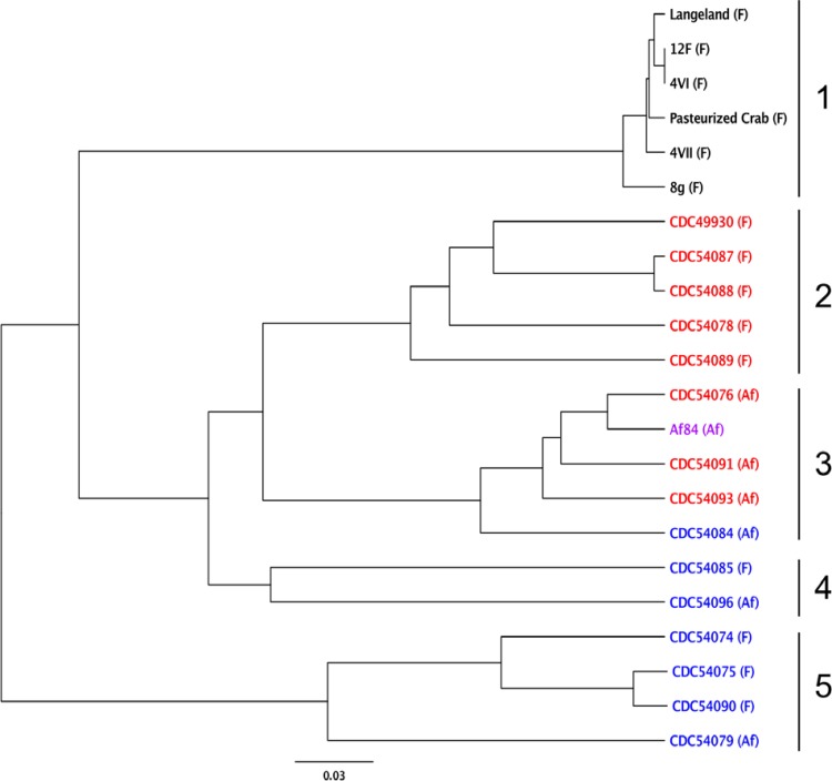
A 225-probe DNA microarray was used to determine the relative similarity of the strains examined in this study to each other on the basis of their hybridization profiles (Fig. 2). Cluster 1 contained highly similar strains and consisted entirely of strains harboring the bont/F1 gene. Cluster 2 consisted of strains harboring the bont/F4 gene, while cluster 3 consisted of bivalent (Af) strains. Interestingly, cluster 3 contained three strains harboring the bont/F4 gene (CDC 54076, CDC 54091, and CDC 54093), one strain harboring the bont/F5 gene (CDC 54084), and strain Af84, which has been shown to contain both the bont/F4 and bont/F5 genes. Clusters 4 and 5 both contained strains harboring bont/F5. Both clusters also contained monovalent (type F only) and bivalent (Af) strains. These results indicate that, as a group, the strains containing the subtype F4 and F5 neurotoxin genes are diverse in gene content.
FIG 2.
DNA microarray analysis. A dendrogram based on a distance matrix of the group I C. botulinum DNA microarray hybridization profiles of the strains indicated was generated by UPGMA. The bont/F subtype harbored by each strain is indicated by color (black, strain containing bont/F1; red, strain containing bont/F4; blue, strain containing bont/F5; purple, strain containing both bont/F4 and bont/F5). The toxin serotype(s) produced by each strain is in parentheses. Clusters of strains with a node in common are numbered 1 to 5.
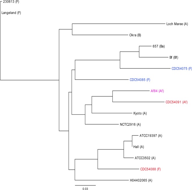
In order to examine the diversity among these strains in greater detail, four strains were selected for genome sequencing, two strains harboring bont/F4 (CDC 54088 and CDC 54091) and two strains harboring bont/F5 (CDC 54075 and CDC 54085). The entire genome sequences of these strains and additional publically available reference sequences were compared by fragmented alignment (Fig. 3). Consistent with the microarray data, the genome sequences of these strains are divergent. Strain CDC 54075 (which contains bont/F5) forms a clade with bivalent strains 657 (Ba) and Bf. Strain CDC 54085 also contains bont/F5 and is more distantly related to CDC 54075. Two strains that contain the bont/F4 gene (CDC 54088 and CDC 54091) were also distantly related to each other; however, bivalent (Af) strain CDC 54091 formed a clade with the previously sequenced genome of strain Af84.
FIG 3.
Phylogenomic analysis of C. botulinum strains. A neighbor-joining tree based on a distance matrix resulting from a fragmented alignment of various C. botulinum genome sequences is shown. Strain names are color coded as follows. Red indicates strains containing bont/F4, blue indicates strains containing bont/F5, and purple indicates a strain that carries both bont/F4 and bont/F5. The toxin serotype(s) produced by each strain is shown in parentheses. The accession numbers of sequences (separate accession numbers representing complete chromosomal and plasmid sequences are indicated when data were concatenated for analysis) selected from the NCBI genome database are CP002011 and CP002012 (230613F); CP000728 and CP000729 (Langeland); NC_010516 and NC_010379 (Okra); NC_012658, NC_012654, and NC_012657 (657); NZ_ABDP00000000 (Bf); NZ_AOSX00000000 (Af84); NC_012563 (Kyoto); NZ_ABDO00000000 (NCTC2916); NC_009697 (ATCC 19397); NC_009698 (Hall); NC_009495 and NC_009496 (ATCC 3502); NC_017299 (H04402065); and NC_010520 and NC_010418 (Loch Maree).
Genomic localization of bont/F4 or bont/F5.
In strain Af84, bont/F4 is chromosomally located and inserted into the pulE gene, which encodes a protein that is likely to be involved in pilus formation (4). Consistent with this finding, all eight strains containing bont/F4 (as well as strain Af84) failed to produce a PCR product with primers targeting the pulE gene (Fig. 4A), suggesting that the gene is not intact. All seven of the bont/F5-containing strains examined produced a PCR product (∼1.5 kb) with these primers, indicating that pulE remains intact. Alignment of the pulE gene in C. botulinum type A2 strain Kyoto (GenBank accession number: CP001581.1) with draft sequences from this study revealed that pulE remained intact in bont/F5-containing strains CDC 54075 and CDC 54085, while only the 5′ end (∼850 bp) of pulE aligned with a corresponding sequence in bont/F4-containing strains CDC 54088 and CDC 54091 (data not shown).
FIG 4.
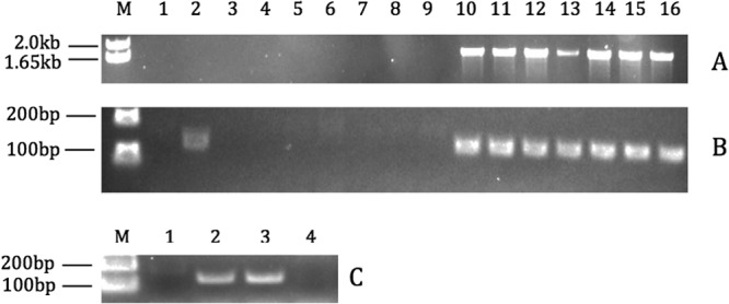
Toxin gene localization. PCR analysis was performed to determine the genomic location of the bont/F gene among C. botulinum strains. In panel A, an ∼1.5-kb PCR product from strains containing an intact pulE gene was amplified. In panel B, a 126-bp PCR product targeting a marker (PL-6) for plasmids carrying bont genes was amplified. In panel C, PCR products targeting the PL-6 plasmid marker from control strains were amplified. In Panels A and B, samples are identified as follows: 1-kb plus marker, lane M; CDC 49930, lane 1; Af84, lane 2; CDC 54076, lane 3; CDC 54078, lane 4; CDC 54087, lane 5; CDC 54088, lane 6; CDC 54089, lane 7; CDC 54091, lane 8; CDC 54093, lane 9; CDC 54074, lane 10; CDC 54075, lane 11; CDC 54079, lane 12; CDC 54084, lane 13; CDC 54085, lane 14; CDC 54090, lane 15; CDC 54096, lane 16. In panel C, samples are identified as follows: 1-kb plus marker, lane M; ATCC 3502, lane 1; Okra, lane 2; CDC 40234, lane 3; plasmid-cured CDC 40234, lane 4.
A PCR with primers targeting a DNA microarray probe (PL-6) was designed. This region targets a putative DNA primase gene (locus CLD_A0039; GenBank accession number CP000940.1) found in several sequenced C. botulinum plasmids that carry bont genes (including strains Okra, Loch Maree, and 657). As expected, a PL-6 PCR product (126 bp) was observed with genomic DNA from strain Af84, which contains bont/F5 on an approximately 246-kb plasmid (pCLQ) (Fig. 4B). PCR products were also observed among all seven of the BoNT/F5-encoding strains examined in this study. As a control, the PCR assay targeting PL-6 was performed with genomic DNA isolated from known reference strains ATCC 3502 (which carries bont/A1 on its chromosome) and Okra (with plasmid-borne bont/B1) (12). As expected, PL-6 was present in Okra but not ATCC 3502 (Fig. 4C). In addition, PL-6 was present in strain CDC 40324, which carries bont/A3 on a plasmid, but not in the identical strain cured of this plasmid (13).
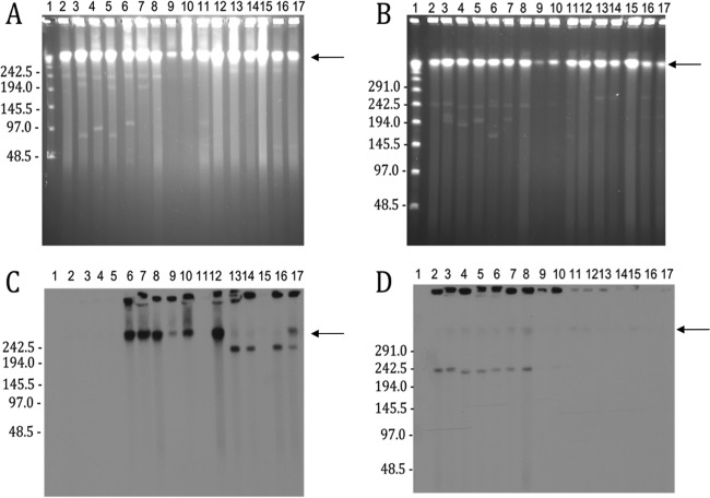
Southern hybridization with a bont/F5-specific probe demonstrated that the bont/F5 gene was indeed carried by large plasmids (∼242 kb) among the strains examined in this study (Fig. 5). The bont/A2 gene present in three of these strains (CDC 54079, CDC 54084, and CDC 54096) was localized to the chromosome. As controls, strains Langeland and ATCC 3502 were used because their toxin genes are located on the chromosome (14). Since the bont/F probe was designed to a highly divergent region of bont/F5, little hybridization with bont/F1-containing strain Langeland was observed. Strains Loch Maree and 657 contain the bont/A gene on a plasmid (12, 15). C. botulinum strain CDC 40234 has been shown to be genetically identical to strain Loch Maree by PFGE, toxin gene cluster sequencing, and multilocus sequence typing according to studies performed previously in E. A. Johnson's laboratory at the University of Wisconsin—Madison (unpublished results). Notably, strain CDC 40234 (A3) cured of its plasmid failed to hybridize with the bont/A probe (13). As expected, strain Af84 demonstrated hybridization of bont/A to the chromosome and bont/F5 hybridization to a large plasmid. Similar to the results obtained with strain Langeland, the chromosomally located bont/F4 gene in strain Af84 did not hybridize with the bont/F5-specific probe.
FIG 5.
PFGE and Southern blot assay of C. botulinum strains. PFGE was performed with either HEPES buffer (panel A) or 0.5× TBE (panel B) and undigested agarose plugs containing C. botulinum genomic DNA. Panel C is a Southern blot assay of the gel shown in panel A and probed with 32P-labeled bont/A. Panel D is a Southern blot assay of the gel shown in panel B and probed with 32P-labeled bont/F5. Samples are identified as follows: New England BioLabs λ marker, lanes 1; CDC 54074, lanes 2; CDC 54075, lanes 3; CDC 54085, lanes 4; CDC 54090, lanes 5; CDC 54079, lanes 6; CDC 54084, lanes 7; CDC 54096, lanes 8; Af84-1, lanes 9; Af84-2, lanes 10; Langeland, lanes 11; ATCC 3502, lanes 12; CDC 40234, lanes 13; Loch Maree, lanes 14; plasmid-cured CDC 40234, lanes 15; 657Ba-1, lanes 16; 657Ba-2, lanes 17. Molecular mass marker sizes are shown in kilobases to the left of each panel. The location of the chromosomal band is indicated by an arrow to the right of each panel.
Finally, draft genome sequences of CDC 54075 and CDC 54085 (both of which contain bont/F5) were compared to the complete sequence of pCLQ (which carries bont/F5 in strain Af84) by fragmented alignment. This analysis demonstrated that strains CDC 54075 and CDC 54085 share a high degree of gene content with pCLQ (Fig. 6). Two regions in pCLQ (located at kb ∼36 to 40 and ∼144 to 148) appear to be specific to strain Af84.
FIG 6.
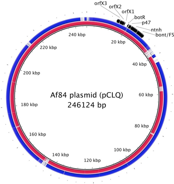
Comparison of strain and pCLQ gene contents. Draft genome sequences of strains CDC 54075 and CDC 54085 were compared to the reference plasmid pCLQ sequence. Reference sequence locations are shown on the innermost ring. The outer rings show BLAST comparisons of the pCLQ sequence and the two draft sequences. Regions with ≥70% nucleotide identity between pCLQ and strain CDC 54075 or CDC 54085 are in red or blue, respectively. The positions of the genes (bont/F5, ntnh, p47, botR, and orxX1 to -3) that make up the toxin gene cluster are also indicated.
DISCUSSION
Compared to the genes encoding other botulinum toxin serotypes, genes encoding BoNT/F have been shown to be extraordinarily diverse and can be distinguished into seven toxin subtypes (F1 to F7) (2). One of the most divergent of these toxin subtype genes (bont/F5) was shown to encode a light-chain region with little similarity to the genes encoding other subtypes. The BoNT/F light chain contains endopeptidase activity required for cleavage of synaptobrevin 2 between 58Q and 59K. Kalb et al. (3) identified a novel cleavage location for the BoNT/F5 subtype (between 54L and 55E). More recently, Dover et al. (4) demonstrated that strain Af84 carries both bont/F4 and bont/F5 in addition to bont/A2. In this study, we confirmed that the 15 previously analyzed strains harboring either bont/F4 or bont/F5 produced only one BoNT/F subtype. More importantly, this study provided an opportunity to examine the genetic relationships and possible evolutionary dynamics among this population of strains.
The ability of mass spectral analysis to distinguish unique substrate cleavage products of BoNT/F5 compared to other BoNT/F subtypes was used to examine culture supernatants generated by the panel of strains containing either bont/F4 or bont/F5. Only strains containing bont/F4 produced neurotoxins that cleaved synaptobrevin 2 at the “traditional” location, whereas only strains containing bont/F5 produced neurotoxins that cleaved synaptobrevin 2 at the novel BoNT/F5 cleavage site. None of the 15 strains examined in this study produced both toxin subtypes BoNT/F4 and BoNT/F5, suggesting that strain Af84 was indeed unique in its ability to harbor more than a single toxin subtype of the same serotype.
We also examined the strains containing bont/F4 or bont/F5 by using DNA microarrays and sequenced the genomes of two bont/F4-harboring and two bont/F5-harboring strains. While the low-density DNA microarray used in this study generally discriminated the strains containing bont/F4 and bont/F5 into separate clusters, it is also apparent that strains sharing the same type F toxin subtype gene are also diverse. Bivalent strain CDC 54091 (which contains bont/A2 and bont/F4) clustered with strain Af84 in the DNA microarray analysis, and these strains formed a clade in a phylogenetic analysis of their genome sequences. Another strain (CDC 54088) containing bont/F4 was found in a separate cluster in the DNA microarray analysis, and genome sequencing revealed that it is distantly related to strain CDC 54091. Similarly, bont/F5-containing strains CDC 54075 and CDC 54085 were found in separate clusters by DNA microarray analysis and their genome sequences are also more distantly related to each other than to several other reference sequences. Notably, the DNA microarray contains probes for only 225 genomic regions while alignment of the draft genome sequences (∼4 Mb) represented a comparison including far more genomic content. Nonetheless, both the microarray analysis and phylogenetic analysis of genome sequences support a high degree of genetic diversity among the strains of this set.
The recent publication of a complete plasmid sequence (pCLQ) present in strain Af84 (4) allowed us to compare a bont/F5-carrying plasmid with draft genome sequences generated in this study. The gene content of bont/F5-harboring strains (CDC 54075 and CDC 54085) demonstrated significant similarity to that of pCLQ. However, two regions of pCLQ appear to be strain specific, one of which (located at kb ∼36 to 40) contains several transposon-associated genes. Strain CDC 54085 displays some additional differences in gene content from pCLQ, suggesting that plasmids carrying bont/F5 may not be identical. Consistent with this possibility is the slight difference in size observed between the plasmids found in CDC 54075 and those found in CDC 54085 (Fig. 5D). It should also be noted that the Southern hybridization used in this study confirmed the presence of bont/F5 on an extrachromosomal element in all of the BoNT/F5-encoding strains examined (including Af84). Interestingly, the analysis of undigested genomic DNA samples by PFGE revealed the presence of additional smaller extrachromosomal elements that do not hybridize with bont/F5. The gene content of these putative plasmids remains unknown.
We previously showed that the bont/F5 nucleotide sequences of a group of seven C. botulinum strains were identical; however, this study demonstrates that such strains are actually genetically diverse. The presence of highly similar plasmids carrying bont/F5 among strains with genomic background differences suggests that such plasmids may be mobile. One way that strain Af84 could have arisen is the acquisition of a plasmid such as pCLQ (which harbors bont/F5) by a bivalent Af strain with chromosomally located bont/F4 and bont/A2 genes similar to strain CDC 54091. Alternatively, loss of a bont/F5-carrying plasmid by a strain similar to Af84 could have resulted in a type Af strain similar to CDC 54091.
This study underscores the importance of genomic sequence analysis of unusual strains of C. botulinum. Caution should be used in selecting appropriate reference sequences for analysis of this polyphyletic species, as strains harboring identical bont subtype genes may contain significant genomic background differences. Additional genomic studies aimed at populations of C. botulinum strains may help answer important questions relating to the acquisition and movement of various bont genes among clostridial strains.
ACKNOWLEDGMENTS
This publication was supported by funds made available from the Centers for Disease Control and Prevention Office of Public Health Preparedness and Response. The work in the laboratory of E.A.J. was sponsored by the NIH/NIAID Regional Center for Excellence for Biodefense and Emerging Infectious Diseases Research (RCE) Program. E.A.J. and M.B. acknowledge membership in and support from the Pacific Southwest RCE (NIH award U54-AI-065359). The findings and conclusions in this report are ours and do not necessarily represent the official position of the Centers for Disease Control and Prevention.
We thank James Marks at the University of California at San Francisco for monoclonal antibodies used in this study.
Footnotes
Published ahead of print 14 March 2014
REFERENCES
- 1.Gupta A, Sumner CJ, Castor M, Maslanka S, Sobel J. 2005. Adult botulism type F in the United States, 1981-2002. Neurology 65:1694–1700. 10.1212/01.wnl.0000187127.92446.4c [DOI] [PubMed] [Google Scholar]
- 2.Raphael BH, Choudoir MJ, Lúquez C, Fernández R, Maslanka SE. 2010. Sequence diversity of genes encoding botulinum neurotoxin type F. Appl. Environ. Microbiol. 76:4805-4812. 10.1128/AEM.03109-09 [DOI] [PMC free article] [PubMed] [Google Scholar]
- 3.Kalb SR, Baudys J, Webb RP, Wright P, Smith TJ, Smith LA, Fernández R, Raphael BH, Maslanka SE, Pirkle JL, Barr JR. 2012. Discovery of a novel enzymatic cleavage site for botulinum neurotoxin F5. FEBS Lett. 586:109–115. 10.1016/j.febslet.2011.11.033 [DOI] [PMC free article] [PubMed] [Google Scholar]
- 4.Dover N, Barash JR, Hill KK, Davenport KW, Teshima H, Xie G, Arnon SS. 2013. Clostridium botulinum strain Af84 contains three neurotoxin gene clusters: bont/A2, bont/F4 and bont/F5. PLoS One 8:e61205. 10.1371/journal.pone.0061205 [DOI] [PMC free article] [PubMed] [Google Scholar]
- 5.Dowell VR, Hawkins TM. 1990. Media for isolation and characterization of anaerobic bacteria, p 53–59 In Dowell VR, Hawkins TM. (ed), Laboratory methods in anaerobic bacteriology: CDC laboratory manual. U.S. Department of Health and Human Services, CDC, Atlanta, GA [Google Scholar]
- 6.Raphael BH. 2012. Exploring genomic diversity in Clostridium botulinum using DNA microarrays. Botulinum J. 2:99–108. 10.1504/TBJ.2012.050195 [DOI] [PMC free article] [PubMed] [Google Scholar]
- 7.Raphael BH, Joseph LA, McCroskey LM, Lúquez C, Maslanka SE. 2010. Detection and differentiation of Clostridium botulinum type A strains using a focused DNA microarray. Mol. Cell. Probes 24:146–153. 10.1016/j.mcp.2009.12.003 [DOI] [PubMed] [Google Scholar]
- 8.Agren J, Sundström A, Håfström T, Segerman B. 2012. Gegenees: fragmented alignment of multiple genomes for determining phylogenomic distances and genetic signatures unique for specified target groups. PLoS One 7:e39107. 10.1371/journal.pone.0039107 [DOI] [PMC free article] [PubMed] [Google Scholar]
- 9.Huson DH, Bryant D. 2006. Application of phylogenetic networks in evolutionary studies. Mol. Biol. Evol. 23:254–267. 10.1093/molbev/msj030 [DOI] [PubMed] [Google Scholar]
- 10.Alikhan NF, Petty NK, Ben Zakour NL, Beatson SA. 2011. BLAST Ring Image Generator (BRIG): simple prokaryote genome comparisons. BMC Genomics 12:402. 10.1186/1471-2164-12-402 [DOI] [PMC free article] [PubMed] [Google Scholar]
- 11.Johnson EA, Tepp WH, Bradshaw M, Gilbert RJ, Cook PE, McIntosh ED. 2005. Characterization of Clostridium botulinum strains associated with an infant botulism case in the United Kingdom. J. Clin. Microbiol. 43:2602–2607. 10.1128/JCM.43.6.2602-2607.2005 [DOI] [PMC free article] [PubMed] [Google Scholar]
- 12.Smith TJ, Hill KK, Foley BT, Detter JC, Munk AC, Bruce DC, Doggett NA, Smith LA, Marks JD, Xie G, Brettin TS. 2007. Analysis of the neurotoxin complex genes in Clostridium botulinum A1-A4 and B1 strains: BoNT/A3, /Ba4 and /B1 clusters are located within plasmids. PLoS One 2:e1271. 10.1371/journal.pone.0001271 [DOI] [PMC free article] [PubMed] [Google Scholar]
- 13.Marshall KM, Bradshaw M, Johnson EA. 2010. Conjugative botulinum neurotoxin-encoding plasmids in Clostridium botulinum. PLoS One 5:e11087. 10.1371/journal.pone.0011087 [DOI] [PMC free article] [PubMed] [Google Scholar]
- 14.Hill KK, Xie G, Foley BT, Smith TJ, Munk AC, Bruce D, Smith LA, Brettin TS, Detter JC. 2009. Recombination and insertion events involving the botulinum neurotoxin complex genes in Clostridium botulinum types A, B, E and F and Clostridium butyricum type E strains. BMC Biol. 7:66. 10.1186/1741-7007-7-66 [DOI] [PMC free article] [PubMed] [Google Scholar]
- 15.Marshall KM, Bradshaw M, Pellett S, Johnson EA. 2007. Plasmid encoded neurotoxin genes in Clostridium botulinum serotype A subtypes. Biochem. Biophys. Res. Commun. 361:49–54. 10.1016/j.bbrc.2007.06.166 [DOI] [PMC free article] [PubMed] [Google Scholar]
- 16.Hill KK, Smith TJ, Helma CH, Ticknor LO, Foley BT, Svensson RT, Brown JL, Johnson EA, Smith LA, Okinaka RT, Jackson PJ, Marks JD. 2007. Genetic diversity among botulinum neurotoxin-producing clostridial strains. J. Bacteriol. 89:818–832. 10.1128/JB.01180-06 [DOI] [PMC free article] [PubMed] [Google Scholar]


