Abstract
We report a case of a 50-year old homeless male who presented with pellagra and pellagrous encephalopathy. The characteristic - if not pathognomonic - skin lesions of pellagra support its diagnosis on solely clinical grounds. Clinicians should keep a high index of suspicion, in certain patient groups, in order to early diagnose and cure this potentially fatal but treatable disease.
Key words: pellagra, niacin, vitamin B3 deficiency, pellagrous encephalopathy
Introduction
Pellagra is a nutritional disorder caused by niacin (vitamin B3) deficiency, which leads to systemic disease with clinical manifestations from the skin, gastrointestinal tract and the nervous system.1-3 Niacin forms into nicotinic acid and nicotinamide, which serve as precursors of two important coenzymes of cellular metabolism, namely nicotinamide adenine dinucleotide (NAD) and NAD-phosphate.1
The term pellagra - from pelle agra, Italian for rough skin - was first used by Frappoli in 1771 due to its dermatologic manifestations.3 However, since Goldberger implicated vitamin B deficiency as the cause of pellagra in 1926,3 the pellagra syndrome has historically been characterized by the 4 D’s: Dermatitis, Diarrhea, and Dementia leading to Death.1,3
Pellagra was a major widespread cause of death until the early 20th century, but fortification of flour with niacin has led practically to its eradication in developed countries. However, several recent reports suggest that pellagra may even be underdiagnosed.3-6
Case Report
A 50-year old man was brought to the emergency department due to disturbed mental status. The patient was reported to be homeless, but no other information about his medical status or living conditions was available. The patient was unreliable in providing history of alcohol abuse or describing symptoms like fever in his recent past.
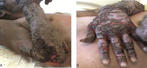
Clinical examination revealed normal vital signs, cachexia and dehydration, accompanied by extensive pigmentation and scaling eruptions on a red basis along the dorsa of his hands and the sun-exposed surface of his arms (Figure 1). The patient was confused and disorientated but responded sufficiently to simple commands. Neurological examination revealed no abnormalities concerning his cranial nerves and tendon reflexes. No flapping tremor was observed. Laboratory investigation showed mild elevation of total white blood cell count, slightly elevated creatinine and low serum albumin. The remaining of the routine laboratory tests, including glucose, electrolytes and aminotransferases, were found within normal range.
Figure 1.

Skin lesions of pellagra on the extensor surface of the patient’s forearm (A) and dorsa of his hand (B).
The patient was admitted for further investigation. Clinical follow-up did not reveal fever or other symptoms or signs indicative of an underlying infection. Intravenous fluids and high-caloric nutrition was administered. During hospitalization the patient was subjected to computed tomography imaging which excluded any brain or other pathology. Following neurological assessment and the negative imaging studies, the disturbed mental status was attributed to metabolic factors. Furthermore, as the characteristic skin lesions in an obviously malnourished homeless man were compatible with pellagra, pellagrous encephalopathy was the preferable diagnosis. Although serologic and urinary assays confirming niacin deficiency were unavailable in our hospital, treatment with nicotinamide 300 mg orally in a daily basis was started. The skin lesions improved significantly but did not resolve completely. Eighteen days later the patient died due to hospital-acquired pneumonia, without showing any remarkable improvement in his mental status.
Discussion
Patients with pellagra have a brown discoloration of the skin, especially in sun-exposed areas. In advanced stages, increased pigmentation usually leads to thin varnish-like eruptive scales. The characteristic skin rash has a typical photosensitive distribution with welldefined borders, and is mostly observed on the face, the neck - forming the so-called Casal’s necklace -, the dorsa of the hands and the extensor surface of the forearms.1-3 In addition to skin changes, gastrointestinal involvement may lead to intractable diarrhea, stomatitis and glossitis, while the neurocognitive impairment - pellagrous encephalopathy - may present as apathy, memory loss, disorientation, depression or delirium. Stupor and death result if pellagra is left untreated.3,4
Niacin deficiency should especially be suspected in the following conditions:1,3,4 i) malnutrition (homelessness, anorexia nervosa or severe comorbid conditions like end-stage malignancy or HIV); ii) malabsorption (e.g. Crohn’s or Hartnup disease); iii) chronic alcoholism; iv) hemodialysis or peritoneal dialysis; v) drugs like isoniazid, ethionamide, 6-mercaptopurine and estrogens; vi) carcinoid syndrome (due to excess turnover of tryptophan, precursor of niacin, to serotonin).
Conclusions
In conclusion, pellagra is a historically old but certainly not completely eradicated disease.5 The characteristic - if not pathognomonic - skin lesions support its diagnosis on solely clinical grounds.1,3 Clinicians should keep a high index of suspicion, in certain patient groups, in order to early diagnose and cure this potentially fatal but treatable disease.
Acknowledgements
The author would like to thank Dr. Sotiriadis D, Professor of Dermatology and head of 2nd Dermatology Department, Papageorgiou University Hospital of Thessaloniki, for his significant contribution.
References
- 1.Russel RM. Vitamin and trace mineral deficiency and excess. : Kasper DL, Braunwald E, Fauci AS, et al., Harrison’s principles of internal medicine. 16th ed New York: McGraw-Hill; 2005. pp 404-405 [Google Scholar]
- 2.MacDonald A, Forsyth A. Nutritional deficiencies and the skin. Clin Exp Dermatol 2005;30:388-90 [DOI] [PubMed] [Google Scholar]
- 3.Oldham MA, Ivkovic A. Pellagrous encephalopathy presenting as alcohol withdrawal delirium: a case series and literature review. Addict Sci Clin Pract 2012;7: 12 [DOI] [PMC free article] [PubMed] [Google Scholar]
- 4.Pipili C, Cholongitas E, Ioannidou D. The diagnostic importance of photosensitivity dermatoses in chronic alcoholism: report of two cases. Dermatol Online J 2008; 14 :15. [PubMed] [Google Scholar]
- 5.Brown TM. Pellagra: an old enemy of timeless importance. Psychosomatics 2010;51: 93-7 [DOI] [PubMed] [Google Scholar]
- 6.Okan G, Yaylaci S, Alzafer S. Pellagra: will we see it more frequently? J Eur Acad Dermatol Venereol 2009;23:365-6 [DOI] [PubMed] [Google Scholar]


