Abstract
The epithelial ovarian carcinoma is one of the most fatal gynecological cancers. Etoposide is used in treating platinum-resistant ovarian cancer. Sodium hyaluronate is a substance that binds to the CD44 receptors overexpressed in SK-OV-3 cells of epithelial ovarian carcinoma. The aim of the present work was to study the cytotoxicity effect of hyaluronate targeted solid lipid nanoparticles (SLNs) of etoposide on SK-OV-3 cells. The cytotoxicity of the targeted and nontargeted SLNs of etoposide was compared to free drug on the SK-OV-3 cells by MTT assay method. The cellular uptake of the targeted and nontargeted nanoparticles containing sodium fluorescein was also studied. The difference of cell vitality between nontargeted nanoparticles and also targeted nanoparticles with free drug was significant. Targeted nanoparticles also caused more toxicity than nontargeted nanoparticles (P < 0.05). After 4 hours of incubating, the fluorescence was remarkably higher in the cells treated by targeted SLNs rather than nontargeted ones, and there was no observable fluorescence in cells incubated with pure sodium fluorescein. Hyaluronate targeted SLNs containing etoposide increased the cytotoxicity of etoposide on SK-OV-3 cells which may be a worthwhile potential method for reducing the prescribed dose and systemic side effects of this drug in epithelial ovarian carcinoma.
1. Introduction
The epithelial ovarian carcinoma is one of the most fatal gynecological cancers across the globe. In spite of early recovery by surgical and chemotherapy treatments, the 5-year survival rate for the patients is only 13 percent. The database GLOBCAN related to the World Health Organization (WHO) has reported incidence of about 192000 cases in the world, in the year of 2000. 6000 cases of the mentioned cases have occurred in the UK, and 21000 cases in the U.S. For treating the disease, the tumor will be removed by surgical procedures and then chemotherapy would be started with platinum-based chemotherapy (cisplatin and carboplatin), which treating regime includes cisplatin and carboplatin with the drugs such as paclitaxel, docetaxel, cyclophosphamide, and doxorubicin. In some of the patients, the disease relapses after 6 months of chemotherapy; this condition is defined as platinum resistant, in which treatment would be continued with drugs such as topotecan and etoposide [1].
Etoposide, as other chemotherapy agents, has many side effects such as bone marrow suppression, granulocytopenia, thrombocytopenia [2], mucositis, moderate to severe esophagitis, hepatotoxicity, metabolic acidosis, and anemia [3].
The complications of anticancer drugs have caused scientists to try two approaches to solve the problem: developing new drugs with fewer side effects and application of new drug delivery systems with high specificity to cancerous tissues; the second approach has lower costs and more attention nowadays. Solid lipid nanoparticles (SLNs) are one of the most important nanosized drug delivery systems that were introduced about two decades ago [4].
SLNs that are often considered for intravenous application are colloidal submicron carriers sized 50 to 1000 nm and composed of solid lipids dispersed in water or surfactant aqueous solution. These nanoparticles have particular features like small size, high surface area, and high loading of drug that makes them potent and beneficial carriers for improving drug efficacy [5, 6]. SLNs are similar to o/w emulsions used for total parenteral nutrition; the difference is that emulsion liquid lipid has been replaced with a solid lipid. SLNs have advantages such as controlled drug release in considered site, excellent biocompatibility, increase in drug stability, high drug content, easy industrialization and sterilization, better control of drug release kinetics, high bioavailability for bioactive drugs, chemical protection of sensitive drugs, easier producing rather than biopolymeric nanoparticles, producible by common emulsification methods, long-time stability, and various applications [4, 7, 8].
For parenteral administration, SLN dispersions must be sterile. SLNs with appropriately small particle size less than 200 nm can be sterilized using filtration. Autoclaving the finished dispersion is not practical as the lipids melt at sterilizing temperatures and the molten lipid droplets coalesce. Therefore just aseptic manufacturing processes following sterilization of the starting materials by gamma irradiation of the final dispersion or exposure to ethylene oxide (EO) gas are applicable for their sterilization. Bacterial endotoxins in raw materials need to be monitored, especially when raw materials are of natural origin. It may be possible to lyophilize the SLN dispersions, and this lyophile can be irradiated or exposed to EO.
SLNs are used in transdermal applications, as gene vector carriers, for topical uses, as cosmeceuticals, as targeted carriers of anticancer drugs to solid tumors, in breast cancer and lymph node metastases and in antitubercular chemotherapy.
So far successful studies have been performed upon nanoparticles containing etoposide. For example, the study of Yadav et al. [9] was performed in the survey of poly(lactic-coglycolic acid)-monomethoxy-poly(polyethylene glycol) and poly(lactic-coglycolic acid)-Pluronic block copolymers and the study of Reddy et al. [10] on nanoparticles produced by tripalmitin could be mentioned.
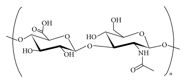
Hyaluronan (Figure 1), available in the market as sodium hyaluronate (HA), is a high molecular weight glycosaminoglycan present in extracellular matrix and is necessary for cellular growth and structural stability of organs and tissue structure.
Figure 1.

Chemical structure of hyaluronan: polymeric repeat of D-glucuronic acid and N-acetylglucosamine.
HA regulates cell proliferation and movements by interacting with CD44 receptors and receptor for HA mediated motility (RHAMM). Because of overexpression of CD44 receptors by cancer cells, interfering in CD44-HA interaction by targeting drugs at CD44 is an effective strategy to treat cancers. HA bound to nanoparticles, in addition to its targeting role, may act as a protecting agent of nanoparticles against body phagocytosis system [11–13]. The mentioned method has been used to deliver agents such as doxorubicin [14], epirubicin [15], paclitaxel [16], mitomycin C [17], SiRNA [18], and DNA [19].
To our knowledge there is not any report on the application of the hyaluronate targeted SLNs in drug delivery of etoposide in SK-OV-3 cells although there are some studies on the hyaluronate targeted SLNs. This study alongside with thousands of similar ones could help to introduce new clinically applicable drug delivery systems with appropriate physicochemical properties, successful targeting, and enhanced cytotoxicity in the future. This study was performed in order to evaluate cytotoxicity of HA targeted SLNs containing etoposide, prepared and optimized in our previous study [20] in SK-OV-3 cells.
2. Materials and Methods
2.1. Materials
Stearylamine (SA), dodecylamine (DDA), cetyl alcohol, dialysis bags with molecular weight cut-off of 12400 Da, and thiazolyl blue tetrazolium bromide (MTT) were from Sigma-Aldrich Company (US). Acetone, dichloromethane, and Tween 80 were from Merck Chemical Company (Germany). RPMI 1640 culture medium, penicillin-streptomycin, and fetal bovine serum were from PAA Company, Austria. Etoposide was a gift from Nippon Kayaku Co, Ltd. (Tokyo, Japan). Sodium hyaluronate (Mw = 6,400 Da) was from Lifecore Biomedical (US) and SK-OV-3 cells were from Pasteur Institute (Iran).
2.2. Preparing Nanoparticles
SLNs were produced by emulsification-solvent evaporation method. According to the results of our previous study [20], the lipid phase including 30 mg etoposide, 30 mg cetyl alcohol, and 30 mg SA was dissolved in 1.8 mL of 1 : 1 mixture of acetone-dichloromethane. Then the mentioned solution was added during 3 minutes to the 18 mL of Tween 80 solution (1% w/v) in deionized water, while stirring in 1200 rpm. Ultimately, produced nanoemulsion was stirred in 600 rpm in room temperature for 75 minutes to evaporate the solution [21]. The blank nanoparticles were produced by the same method but without etoposide.
2.3. Physical Binding of HA to the SLNs Surface
After 15 minutes of adding organic phase to aqueous phase, HA dissolved in deionized water containing Tween 80 (1% w/v) was added to nanoparticles mixture during 5 minutes, while stirring at 600 rpm, in order to produce targeted nanoparticles [22].
Nonbound HA was separated from nanoparticles mixture by dialyzing versus 100 mL deionized water containing Tween 80 (1% w/v) using dialysis bag with molecular weight cut-off of 12,400 Da for 40 minutes so that the deionized water containing Tween 80 (1% w/v) was replaced every 10 minutes. To determine the amount of HA bounded to SLNs after separation of unbound HA, some part of the targeted nanoparticles mixture was dried under vacuum and subjected to elemental analysis (CHN) (CHNS-932, Leco, USA) and, by subtracting the total amount of HA from gaining value, the amount of HA bound on the SLNs surface was calculated.
2.4. Measuring Particle Size, Polydispersity Index, and Zeta Potential
The particle size, polydispersity index, and zeta potential of nanoparticles were measured by a Zetasizer (Zetasizer 3000; Malvern Instruments, Malvern, UK), after 1 : 10 diluting the samples with deionized water.
2.5. Determining Drug Loading and Release
The loading efficiency percent was determined by centrifugation (Eppendorf 5430 centrifuge, Germany). The dispersion of nanoparticles was poured in centrifugal filter tubes (Amicon Ultra, Ireland) with a 10 kDa molecular weight cutoff to separate the aqueous medium [23]. The concentration of free etoposide in the filtrate was determined by measuring its absorption in 276.4 nm (UV-VIS spectrophotometer, Shimadzu Scientific Instruments, Japan) and converting the absorbance to concentration using the calibration equation of etoposide in aqueous phase containing 1% w/v of Tween 80. The amount of encapsulated drug was computed indirectly by calculating the difference between the total amounts of drug used in preparation of nanoparticles and the free drug. Ultimately, loading efficiency percent was computed by the following equation:
| (1) |
Drug release profiles from the NPLs were determined in phosphate buffer saline (PBS, 0.01 M, pH 7.4 containing 1% w/v Tween 80) at 37°C. A total of 2 mL of NPLs suspension was placed in dialysis bag with molecular weight cut-off of 12,400 Da and suspended in a beaker containing 50 mL of PBS on a magnetic stirrer with a speed of 200 rpm. Samples were withdrawn periodically and replaced with the same volume of PBS at the same temperature. The content of etoposide in the samples was determined spectrophotometrically at 268.7 nm.
2.6. MTT Colorimetric Cytotoxicity Assay
To determine cell proliferation, an MTT assay was carried out. A total of 180 μL of the cell suspension (5 × 10⁴ cells/mL) were placed in each well of a 96-well plate except for one row for blank that was filled by an equal amount of medium. After a 24 h period of incubation at 37°C in a CO2 incubator with 5% CO2 and 95% humidity, all 4 wells of cells were treated with 20 μL of one of the concentrations of etoposide as much as 0.475, 0.95, 1.9, and 3.8 μM of etoposide. The IC50 of etoposide for SK-OV-3 cells was determined to be 1.9 μM [24]. In order to assure that microorganisms would not be able to contaminate the SLNs and interfere with cytotoxicity results, preparation of solution of free drug and also preparation and dilution of SLNs suspensions were carried out in aseptic conditions under a laminar flow hood. It should be pointed out that solutions of organic and aqueous phases were presterilized by ultraviolet germicidal irradiation method.
Treated groups included either a solution of free drug in 1 w/v% aqueous solution of Tween 80 or encapsulated drug in nontargeted and targeted nanoparticles, with blanks of nontargeted and targeted nanoparticles, while culture medium and Tween 80 1 w/v% (each one in 8 wells) serve as control groups. The cells were incubated for further 48 h. After the treatment, 20 μL/well of the MTT solution (5 mg/mL of PBS) was added to the cells and incubated for 3 h; then the supernatant was removed carefully and the formazan crystals were dissolved by adding 150 μL of DMSO. Finally, the absorbance of each well was measured at 570 nm by an ELIZA plate reader (STAT FAX 2100 Microplate Reader, Awareness Technology, Inc., US). The effect of each treatment on cell viability was calculated by comparing the relative absorbance of treated cells against the respective controls, using the following equation [25]:
| (2) |
2.7. Qualitative Comparison of Drug Uptake from Nanoparticles by Fluorescence Imaging
First, 2700 μL of the cellular suspension with the concentration of 105 cells/mL was poured into 10 wells of a 12-well plate containing lamels at the bottom and then incubated for 48 h in CO2 incubator. Then the nontargeted and targeted nanoparticles were loaded with sodium fluorescein instead of etoposide by the same method as mentioned above for drug-loaded SLNs. The final concentration of loaded sodium fluorescein in nanoparticles was 1 mg/mL. Blank nanoparticles were also prepared but without sodium fluorescein. To prepare free sodium fluorescein solution, 10 μL of stock solution (100 mg/mL) was diluted to 1 mL to provide the final concentration of 1 mg/mL.
Finally, 300 μL of each sample was added to 2 wells (one for imaging in the 1st hour and the other for imaging in 4th hour) and was incubated. Lamels were withdrawn and imaging was performed by visible fluorescence microscope (Olympus, IX71, Japan) [11].
2.8. Statistical Analysis
All data are the results of three separate experiments, and the results are expressed as the mean ± standard deviation (n = 3). Statistical analysis was performed using one-way analysis of variance (ANOVA) and an independent Student's t-test with the SPSS software (version 18, US). A P value of less than 0.05 was considered significant.
3. Results and Discussion
3.1. Physicochemical Properties of Nanoparticles
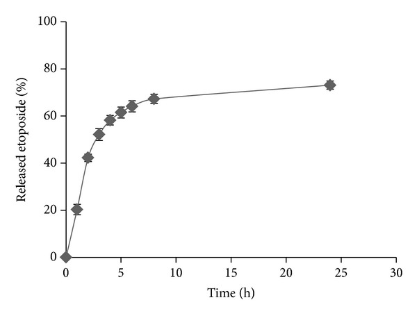
Table 1 represents properties of nanoparticles. The particle size of nontargeted and targeted SLNs was 179.6 ± 16.31 and 416.42 ± 31.85, respectively. Zeta potential of nontargeted SLNs was 11.82 ± 0.52 that changed to −12.65 ± 0.49 after coating with HA. Drug loading efficiency was about 64.92 ± 3.76% and release efficiency percent in 24 h was 65.47 ± 4.68% which is an acceptable value. HA was coated as much as 55.89 percent on the SLNs. Figure 2 represents drug release profile from HA targeted nanoparticles.
Table 1.
Properties of solid lipid nanoparticles of etoposide.
| SLNs type | Particle size (nm) | pdI | zeta potential (mV) | Drug loading efficiency (%) | RE in 24 h (%) |
|---|---|---|---|---|---|
| Non-targeted SLNs | 179.6 ± 16.3 | 0.17 ± 0.03 | 11.82 ± 0.52 | — | — |
| HA targeted SLNs | 416.4 ± 31.8 | 0.30 ± 0.05 | −12.65 ± 0.49 | 64.92 ± 3.76 | 65.47 ± 4.68 |
Figure 2.

Etoposide release profile from HA targeted SLNs.
SLNs have generally long-term stability (about 1–3 years) as small particle size and density close to unity of SLNs mean that the gravity has little effect on particles in dispersion and the Brownian motion is sufficient to maintain colloidal dispersions without creaming or sedimentation. In the present study the presence of physically bound HA and the negative zeta potential of targeted SLNs may seem to threaten stability, but our unpublished results showed that properties of the mentioned SLNs suspension did not change significantly within 10 days. However, as freeze-drying is a suitable method to prevent the Ostwald ripening and avoid aggregation of SLNs, we also dried the nontargeted and targeted SLNs under vacuum with 5% glycerol serving as cryoprotectant and then recovered them by adding deionized water. The results showed that nontargeted SLNs only needed 5 minutes of stirring at 800 rpm and targeted SLNs needed twice the stirring at 800 rpm each time for 3 minutes and then 10 seconds of sonication at a power of 30 w, to retrieve their primary properties. Nonetheless, the SLNs which were used for cytotoxicity study were prepared fresh.
The observed release rate (64.1% in the first 6 hours and 73.1% in 24 hours) could provide appropriate serum concentrations for routine chemotherapy schedules in which the drug (with an iv half-life of 6–12 hours) is administered once daily. Also the mean diameter of typically 200–400 nm is well below the size of the smallest blood capillaries in the range of 5-6 μm. Furthermore, because of the heterogeneity of tumors and dynamic status of each tumor, it will be very difficult to assume any maximum single value for particles to exploit the enhanced permeation and retention (EPR) effect. However, the study of Bae and Park suggests that the porosity of the blood vessels in tumors is around 400 nm [26]. A tumor-dependent functional pore cutoff size ranges from 200 nm to 1.2 μm, but the pore cutoff size of porous blood vessels in the majority of tumors is known to be 380–780 nm [27]. Thus, the range of the EPR effect should be similar. Sterically stabilized liposomes of 400 nm in diameter were able to penetrate into tumor interstitium [28]. Accumulation of hyaluronic acid-coated self-assembled nanoparticles with particle size of 400 nm has been reported in the tumor tissue too [29].
3.2. Cell Proliferation Assay (MTT Assay)
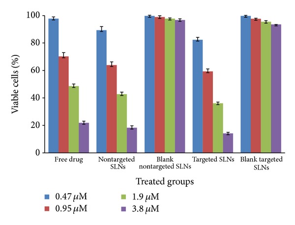
The obtained results of MTT cytotoxicity assay have been illustrated in Table 2 and Figure 3.
Table 2.
IC50 of etoposide loaded in non-targeted and hyaluronate targeted SLNs in SK-OV-3 cells.
| Free drug | Non-targeted SLNs | AH-targeted SLNs | |
|---|---|---|---|
| IC50 (µM) | 1.49 ± 0.08 | 1.24 ± 0.13 | 0.78 ± 0.12 |
Figure 3.

Percentage of viable cells of SK-OV-3 determined by the MTT assay after treatment with etoposide loaded nontargeted and hyaluronate targeted SLNs in comparison to blank nontargeted and targeted SLNs and free drug (n = 3).
All drug-loaded nanoparticles caused higher cytotoxicity compared to the free etoposide at the same concentration and their respective blank SLNs. The mechanism of enhanced cytotoxicity of drug-loaded lipid nanoparticles has been previously reported [30, 31]. It is well understood that improvement in the cytotoxicity is because of the elevated drug concentrations within the cells. As we can see in Figure 3, nontargeted drug-loaded SLNs have lower cell survival compared to the free etoposide solution. For example, the observed cell survival after treatment with targeted nanoparticles was 36.08 ± 0.88%, while it was 42.73 ± 1.49% and 48.57 ± 1.61% for nontargeted SLNs and free drug solution, respectively, at the concentration of 1.9 μM (P < 0.05).
The results verified that targeted and nontargeted SLNs of etoposide have reduced IC50 to 52% and 83% of free drug, respectively (Table 2). In a study, Saliou et al. [32] reported that lipid nanocapsules of etoposide reduced the IC50 of the drug from 100 to 2.5 μM in H209 cells. These lipid nanocapsules also could reduce the IC50 of etoposide to about 4–30 times in glioma cell lines [33]. In an experiment conducted by Nasti et al. [34] chitosan/triphosphate nanoparticles coated with HA showed the IC50 of about half of the noncoated nanoparticles on murine fibroblasts of L929 and macrophage cells of J774.2. Han et al. [35] successfully overcame on drug resistance of MCF-7/ADR cells with 4.3-fold reduction in IC50 of doxorubicin by SiRNA polyamidoamine-hyaluronic acid complex.
It could be concluded that the internalization of the drug into cells was enhanced when the drug was encapsulated in SLNs. This phenomenon might be the result of the high affinity of lipid materials of SLNs for the cell membrane and the nanoscaled size of SLNs.
The correlation between nanoparticles size and intracellular concentration has been observed in the study performed by Zhang et al. [36] and their results indicated that the less the particle size is, the more the intracellular drug concentration and cytotoxicity is.
In addition, comparing the targeted and nontargeted nanoparticles determines that the cytotoxicity in the targeted nanoparticles has been increased, probably due to the presence of HA on targeted nanoparticles which could interact with CD44 receptors and make them internalized into cells more easily. Cho et al. [37] have surveyed NPLs containing docetaxel targeted by HA upon cancer cell line MCF-7 and showed that they were endocytosed by CD44 receptors.
3.3. Cellular Uptake Studies
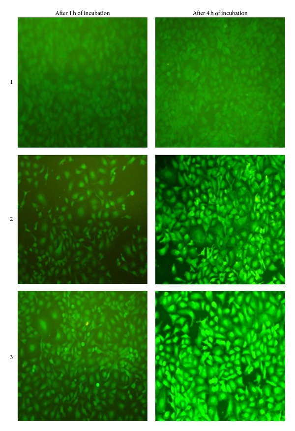
After incubating for 1 hour, only targeted nanoparticles made a slight fluorescence in the cells (Figure 4). After 4 hours of incubation, the fluorescence was remarkably higher in the cells which were treated by targeted SLNs rather than those which were treated by nontargeted nanoparticles, and there was no observable fluorescence in cells incubated with pure sodium fluorescein (Figure 4). Therefore, it could be concluded that increased cytotoxicity in results obtained from MTT assay has resulted from special uptake of targeted nanoparticles due to presence of HA as targeting agent.
Figure 4.

Fluorescence images of SK-OV-3 cells after 1 and 4 hours of incubation with (1) free sodium fluorescein, (2) sodium fluorescein containing nontargeted SLNs, and (3) sodium fluorescein containing targeted SLNs.
4. Conclusion
Hyaluronate targeted SLNs containing etoposide increase the cytotoxicity of etoposide in SK-OV-3 cells and could be a valuable method for reducing the prescribed dose and also systemic side effects.
Acknowledgment
The authors would like to thank Vice Chancellor for Research of Isfahan University of Medical Sciences who provided project finances.
Conflict of Interests
The authors declare that there is no conflict of interests.
References
- 1.Guppy AE, Nathan PD, Rustin GJS. Epithelial ovarian cancer: a review of current management. Clinical Oncology. 2005;17(6):399–411. doi: 10.1016/j.clon.2005.05.009. [DOI] [PubMed] [Google Scholar]
- 2.O’Dwyer PJ, Leyland-Jones B, Alonso MT. Etoposide (VP-16-213). Current status of an active anticancer drug. New England Journal of Medicine. 1985;312(11):692–700. doi: 10.1056/NEJM198503143121106. [DOI] [PubMed] [Google Scholar]
- 3.Taylor RE, McElwain TJ, Barrett A, Peckham MJ. Etoposide as a single agent in relapsed advanced lymphomas. A phase II study. Cancer Chemotherapy and Pharmacology. 1982;7(2-3):175–177. doi: 10.1007/BF00254543. [DOI] [PubMed] [Google Scholar]
- 4.Mukherjee S, Ray S, Thakur RS. Solid lipid nanoparticles: a modern formulation approach in drug delivery system. Indian Journal of Pharmaceutical Sciences. 2009;71(4):349–358. doi: 10.4103/0250-474X.57282. [DOI] [PMC free article] [PubMed] [Google Scholar]
- 5.Li H, Zhao X, Ma Y, Zhai G, Li L, Lou H. Enhancement of gastrointestinal absorption of quercetin by solid lipid nanoparticles. Journal of Controlled Release. 2009;133(3):238–244. doi: 10.1016/j.jconrel.2008.10.002. [DOI] [PubMed] [Google Scholar]
- 6.Uner M, Yener G. Importance of solid lipid nanoparticles (SLN) in various administration routes and future perspectives. International Journal of Nanomedicine. 2007;2(3):289–300. [PMC free article] [PubMed] [Google Scholar]
- 7.Mehnert W, Mäder K. Solid lipid nanoparticles: production, characterization and applications. Advanced Drug Delivery Reviews. 2001;47(2-3):165–196. doi: 10.1016/s0169-409x(01)00105-3. [DOI] [PubMed] [Google Scholar]
- 8.Müller RH, Mäder K, Gohla S. Solid lipid nanoparticles (SLN) for controlled drug delivery-a review of the state of the art. European Journal of Pharmaceutics and Biopharmaceutics. 2000;50(1):161–177. doi: 10.1016/s0939-6411(00)00087-4. [DOI] [PubMed] [Google Scholar]
- 9.Yadav KS, Chuttani K, Mishra AK, Sawant KK. Long circulating nanoparticles of etoposide using PLGA-MPEG and PLGA-pluronic block copolymers: characterization, drug-release, blood-clearance, and biodistribution studies. Drug Development Research. 2010;71(4):228–239. [Google Scholar]
- 10.Reddy LH, Sharma RK, Chuttani K, Mishra AK, Murthy RR. Etoposide-incorporated tripalmitin nanoparticles with different surface charge: formulation, characterization, radiolabeling, and biodistribution studies. The AAPS Journal. 2004;6(3):p. e23. doi: 10.1208/aapsj060323. [DOI] [PMC free article] [PubMed] [Google Scholar]
- 11.Nevius BA, Chen YP, Ferry JL, Decho AW. Surface-functionalization effects on uptake of fluorescent polystyrene nanoparticles by model biofilms. Ecotoxicology. 2012;21(8):2205–2213. doi: 10.1007/s10646-012-0975-3. [DOI] [PubMed] [Google Scholar]
- 12.Kremser G, Rath T, Kunert B, et al. Structural characterisation of alkyl amine-capped zinc sulphide nanoparticles. Journal of Colloid and Interface Science. 2012;369(1):154–159. doi: 10.1016/j.jcis.2011.12.032. [DOI] [PMC free article] [PubMed] [Google Scholar]
- 13.Sun Y. Synthesis of gold-amine nanoparticles of various sizes using two different methods [M.S. thesis] Manhattan, Kan, USA: Department of Chemistry, Kansas State University; 2010. [Google Scholar]
- 14.Luo Y, Bernshaw NJ, Lu Z-R, Kopecek J, Prestwich GD. Targeted delivery of doxorubicin by HPMA copolymer-hyaluronan bioconjugates. Pharmaceutical Research. 2002;19(4):396–402. doi: 10.1023/a:1015170907274. [DOI] [PubMed] [Google Scholar]
- 15.Akima K, Ito H, Iwata Y, et al. Evaluation of antitumor activities of hyaluronate binding antitumor drugs: synthesis, characterization and antitumor activity. Journal of Drug Targeting. 1996;4(1):1–8. doi: 10.3109/10611869609046255. [DOI] [PubMed] [Google Scholar]
- 16.Rosato A, Banzato A, De Luca G, et al. HYTAD1-p20: a new paclitaxel-hyaluronic acid hydrosoluble bioconjugate for treatment of superficial bladder cancer. Urologic Oncology: Seminars and Original Investigations. 2006;24(3):207–215. doi: 10.1016/j.urolonc.2005.08.020. [DOI] [PubMed] [Google Scholar]
- 17.Peer D, Margalit R. Loading mitomycin C inside long circulating hyaluronan targeted nano-liposomes increases its antitumor activity in three mice tumor models. International Journal of Cancer. 2004;108(5):780–789. doi: 10.1002/ijc.11615. [DOI] [PubMed] [Google Scholar]
- 18.Lee H, Mok H, Lee S, Oh Y, Park TG. Target-specific intracellular delivery of siRNA using degradable hyaluronic acid nanogels. Journal of Controlled Release. 2007;119(2):245–252. doi: 10.1016/j.jconrel.2007.02.011. [DOI] [PubMed] [Google Scholar]
- 19.Yun YH, Goetz DJ, Yellen P, Chen W. Hyaluronan microspheres for sustained gene delivery and site-specific targeting. Biomaterials. 2004;25(1):147–157. doi: 10.1016/s0142-9612(03)00467-8. [DOI] [PubMed] [Google Scholar]
- 20.Varshosaz J, Mohammadi Ghalaei P, Hassanzadeh F. Hyaluronate targeted solid lipid nanoparticles of etoposide: optimization and in vitro characterization. Journal of Nanomaterials. 2014;2014:12 pages.345845 [Google Scholar]
- 21.Varshosaz J, Eskandari S, Tabakhian M. Production and optimization of valproic acid nanostructured lipid carriers by the Taguchi design. Pharmaceutical Development and Technology. 2010;15(1):89–96. doi: 10.3109/10837450903013568. [DOI] [PubMed] [Google Scholar]
- 22.Ataman-Önal Y, Munier S, Ganée A, et al. Surfactant-free anionic PLA nanoparticles coated with HIV-1 p24 protein induced enhanced cellular and humoral immune responses in various animal models. Journal of Controlled Release. 2006;112(2):175–185. doi: 10.1016/j.jconrel.2006.02.006. [DOI] [PubMed] [Google Scholar]
- 23.Teeranachaideekul V, Souto EB, Junyaprasert VB, Müller RH. Cetyl palmitate-based NLC for topical delivery of Coenzyme Q10-development, physicochemical characterization and in vitro release studies. European Journal of Pharmaceutics and Biopharmaceutics. 2007;67(1):141–148. doi: 10.1016/j.ejpb.2007.01.015. [DOI] [PubMed] [Google Scholar]
- 24.Itamochi H, Oishi T, Shimada M, et al. Inhibiting the mTOR pathway synergistically enhances cytotoxicity in ovarian cancer cells induced by etoposide through upregulation of c-Jun. Clinical Cancer Research. 2011;17(14):4742–4750. doi: 10.1158/1078-0432.CCR-11-0190. [DOI] [PubMed] [Google Scholar]
- 25.Strober W. Monitoring Cell Growth. Current Protocols InImmunology. Washington, DC, USA: John Wiley & Sons; 2001. [Google Scholar]
- 26.Bae YH, Park K. Targeted drug delivery to tumors: myths, reality and possibility. Journal of Controlled Release. 2011;153(3):198–205. doi: 10.1016/j.jconrel.2011.06.001. [DOI] [PMC free article] [PubMed] [Google Scholar]
- 27.Hobbs SK, Monsky WL, Yuan F, et al. Regulation of transport pathways in tumor vessels: role of tumor type and microenvironment. Proceedings of the National Academy of Sciences of the United States of America. 1998;95(8):4607–4612. doi: 10.1073/pnas.95.8.4607. [DOI] [PMC free article] [PubMed] [Google Scholar]
- 28.Yuan F, Dellian M, Fukumura D, et al. Vascular permeability in a human tumor xenograft: molecular size dependence and cutoff size. Cancer Research. 1995;55(17):3752–3756. [PubMed] [Google Scholar]
- 29.Choi KY, Min KH, Na JH, et al. Self-assembled hyaluronic acid nanoparticles as a potential drug carrier for cancer therapy: synthesis, characterization, and in vivo biodistribution. Journal of Materials Chemistry. 2009;19(24):4102–4107. [Google Scholar]
- 30.Miglietta A, Cavalli R, Bocca C, Gabriel L, Rosa Gasco M. Cellular uptake and cytotoxicity of solid lipid nanospheres (SLN) incorporating doxorubicin or paclitaxel. International Journal of Pharmaceutics. 2000;210(1-2):61–67. doi: 10.1016/s0378-5173(00)00562-7. [DOI] [PubMed] [Google Scholar]
- 31.Serpe L, Catalano MG, Cavalli R, et al. Cytotoxicity of anticancer drugs incorporated in solid lipid nanoparticles on HT-29 colorectal cancer cell line. European Journal of Pharmaceutics and Biopharmaceutics. 2004;58(3):673–680. doi: 10.1016/j.ejpb.2004.03.026. [DOI] [PubMed] [Google Scholar]
- 32.Saliou B, Thomas O, Lautram N, et al. Development and in vitro evaluation of a novel lipid nanocapsule formulation of etoposide. European Journal of Pharmaceutical Sciences. 2013;50(2):172–180. doi: 10.1016/j.ejps.2013.06.013. [DOI] [PubMed] [Google Scholar]
- 33.Lamprecht A, Benoit J. Etoposide nanocarriers suppress glioma cell growth by intracellular drug delivery and simultaneous P-glycoprotein inhibition. Journal of Controlled Release. 2006;112(2):208–213. doi: 10.1016/j.jconrel.2006.02.014. [DOI] [PubMed] [Google Scholar]
- 34.Nasti A, Zaki NM, De Leonardis P, et al. Chitosan/TPP and chitosan/TPP-hyaluronic acid nanoparticles: systematic optimisation of the preparative process and preliminary biological evaluation. Pharmaceutical Research. 2009;26(8):1918–1930. doi: 10.1007/s11095-009-9908-0. [DOI] [PubMed] [Google Scholar]
- 35.Han M, Lv Q, Tang XJ, et al. Overcoming drug resistance of MCF-7/ADR cells by altering intracellular distribution of doxorubicin via MVP knockdown with a novel siRNA polyamidoamine-hyaluronic acid complex. Journal of Controlled Release. 2012;163(2):136–144. doi: 10.1016/j.jconrel.2012.08.020. [DOI] [PubMed] [Google Scholar]
- 36.Zhang XG, Miao J, Dai YQ, Du YZ, Yuan H, Hu FQ. Reversal activity of nanostructured lipid carriers loading cytotoxic drug in multi-drug resistant cancer cells. International Journal of Pharmaceutics. 2008;361(1-2):239–244. doi: 10.1016/j.ijpharm.2008.06.002. [DOI] [PubMed] [Google Scholar]
- 37.Cho HJ, Yoon HY, Koo H, et al. Self-assembled nanoparticles based on hyaluronic acid-ceramide (HA-CE) and Pluronic® for tumor-targeted delivery of docetaxel. Biomaterials. 2011;32(29):7181–7190. doi: 10.1016/j.biomaterials.2011.06.028. [DOI] [PubMed] [Google Scholar]


