Abstract
Ocean acidification (OA) due to atmospheric CO2 rise is expected to influence marine primary productivity. In order to investigate the interactive effects of OA and light changes on diatoms, we grew Phaeodactylum tricornutum, under ambient (390 ppmv; LC) and elevated CO2 (1000 ppmv; HC) conditions for 80 generations, and measured its physiological performance under different light levels (60 µmol m−2 s−1, LL; 200 µmol m−2 s−1, ML; 460 µmol m−2 s−1, HL) for another 25 generations. The specific growth rate of the HC-grown cells was higher (about 12–18%) than that of the LC-grown ones, with the highest under the ML level. With increasing light levels, the effective photochemical yield of PSII (Fv′/Fm′) decreased, but was enhanced by the elevated CO2, especially under the HL level. The cells acclimated to the HC condition showed a higher recovery rate of their photochemical yield of PSII compared to the LC-grown cells. For the HC-grown cells, dissolved inorganic carbon or CO2 levels for half saturation of photosynthesis (K1/2 DIC or K1/2 CO2) increased by 11, 55 and 32%, under the LL, ML and HL levels, reflecting a light dependent down-regulation of carbon concentrating mechanisms (CCMs). The linkage between higher level of the CCMs down-regulation and higher growth rate at ML under OA supports the theory that the saved energy from CCMs down-regulation adds on to enhance the growth of the diatom.
Introduction
Atmospheric CO2 concentration is expected to reach 800–1000 ppmv by the end of this century due to relentless consumptions of fossil fuels and exacerbated deforestation [1]; at the same time the oceans are taking up CO2 from the atmosphere at a rate of about 1 million tons per hour, leading to ocean acidification (OA) [2]. While calcifying algae are known to be sensitive to OA that decreases their calcification [3]–[7], diatoms, which account for approximately 40% of the total primary production in the oceans, show diversified or controversial responses [8]–[22]. Elevated CO2 concentrations are shown to enhance [8]–[14], have no effect [8], [12], [20], [21] or even inhibit [10], [12], [14], [20], [22] growth rates of diatom species. Although elevated CO2 in the ocean increases its availability to photosynthetic organisms, the reduced pH can influence the acid-base balance of cells [23]. In addition, the elevated CO2 and reduced pH levels can interact with solar radiation and temperature, showing synergistic, antagonistic or balanced effects [24]. Consequently, the mechanisms involved in the responses to OA of diatoms need to be further explored.
Diatoms in different waters experience fluctuations of light, temperature as well as changes in seawater carbonate chemistry. With the ongoing of OA, diatoms are exposed to declining pH and increased sunlight exposures in the upper mixing layer, which is shoaled due to enhanced stratification along with ocean warming [24]. It is known that phytoplankton cells exhibit different growth rates or photosynthetic performances under fluctuating light than constant light regimes [25], [26]. Therefore, it is important to see responses of diatoms to OA under different light levels or with fluctuating sunlight. Previously, we showed that OA under low or moderate levels of sunlight that fluctuates during diurnal cycles enhanced the growth of the diatoms Phaeodactylum tricornutum, Skeletonema costatum and Thalassiosira pseudonana, but led to growth inhibition under high (>40%) incident sunlight levels [12]. In this study, we grew the model diatom, P. tricornutum, in laboratory cultures under different but constant light levels (without diurnal change) to further explore the interactive effects of OA and light on its growth and photosynthetic performances.
Materials and Methods
Statement of Ethics
Phaeodactylum tricornutum Bohlin (strain CCMA 106, isolated from the South China Sea, SCS, in 2004) was obtained from the Center for Collections of Marine Bacteria and Phytoplankton of the State Key Laboratory of Marine Environmental Science, Xiamen University. No specific permits were required for using this species.
Algal Culture Conditions
Cells of P. tricornutum were grown in 0.22 µm filtered natural seawater collected from the SCS (SEATS station: 116°E, 18°N) and enriched with Aquil medium [27]. The cultures (triplicate per CO2 level) were continuously aerated (350 ml min−1) with ambient CO2 level (LC, 390 ppmv) or CO2-enriched (HC, 1000 ppmv) air, maintained in a CO2 plant incubator (HP1000G-D, Ruihua Instrument and Equipment Co., Ltd., Wuhan, China), and illuminated with cool white fluorescent tubes that provided 70 µmol photons m−2 s−1 of PAR (12L: 12D) at 20°C. Semi-continuous cultures were operated by dilution with the CO2-equilibrated media every 24 h, and the concentration of cells was maintained lower than 25×104 cells ml−1, so that the seawater carbonate chemistry parameters were stable (Table 1) with pH variations <0.05 units. The pH was measured with a pH meter (Mettler Toledo DL15 Titrator, Sweden) calibrated daily with a standard National Bureau of Standards (NBS) buffer (Hanna). Other parameters of the seawater carbonate system (Table 1) were calculated using the CO2SYS software [28] taking into account the salinity, pCO2, pH, nutrient concentrations and temperature; the equilibrium constants K1 and K2 for carbonic acid dissociation [29] and KB for boric acid [30] were referred.
Table 1. Chemical parameters of seawater carbonate system.
| pCO2 | pHNBS | DIC (µmol kg−1) | HCO3 − (µmol kg−1) | CO3 2− (µmol kg−1) | CO2 (µmol kg−1) | Total alkalinity (µmol kg−1) |
| LC | 8.18±0.02a | 1994.7±75.5a | 1802.1±63.0a | 180.0±12.5a | 12.6a | 2263.6±90.8a |
| HC | 7.82±0.02b | 2188.4±78.6b | 2064.0±72.2b | 92.1±6.4b | 32.3b | 2303.6±86.4a |
Carbonate chemistry parameters of the medium for LC (ambient, 390 ppmv CO2) and HC (enriched, 1000 ppmv CO2) cultures. These parameters were averaged for 21 replicate measurements (n = 3, seven measurements for each culture). Different superscript letters represent significant difference between LC and HC.
The cultures were maintained in the above conditions for approximately 80 generations before being used in the following experiments.
Experimental Design
The light levels were set as: photosynthesis-limited (60 µmol m−2 s−1; LL), half-saturated (200 µmol m−2 s−1; ML) and saturated light (460 µmol m−2 s−1; HL) [11], with the photoperiod of 12 h d−1. Before and after dilution, cell concentrations were determined every 24 h with a particle count and size analyzer (Z2 Coulter, Beckman, USA). The specific growth rate was calculated as:  , where N1 and N2 represent the cell concentrations at times T1 (after the dilution) and T2 (before the next dilution, T2−T1 = 24 h).
Carotenoids and Chlorophyll Determination
The concentration of chlorophyll was measured by filtering the cultures onto a GF/F filter (Φ25 mm, Whatman), which was then extracted in 5 ml absolute methanol and maintained in darkness at least overnight at 4°C, before centrifugation (10 min at 5000 g; Universal 320R, Hettich, Germany). The absorption spectrum of the supernatant was obtained by scanning the sample from 250 to 750 nm with a scanning spectrophotometer (DU 800, Beckman, Fullerton, California, USA). Chlorophyll concentration was calculated according to [31] and that of carotenoids according to [32], as follows:
 |
 |
Chlorophyll Fluorescence Measurements
Chl fluorescence was determined using a Xenon-Pulse Amplitude Modulated fluorometer (XE- PAM, Walz, Germany). To assess the photochemical responses of LC- and HC-grown cells to the changes of light, the rapid light curve (RLC), effective photochemical quantum yield (Fv′/Fm′) and non-photochemical quenching (NPQ) were determined. The cells harvested during the middle photoperiod were illuminated for 3 min with actinic light similar to the growth light level before measurement of RLC and Fv′/Fm′ to avoid effects on the photosystems caused by quasi-dark adaptation during manipulation [33]. Fv′/Fm′ was determined under the growth light level. The RLCs were determined at eight PAR levels (0, 29, 395, 592, 832, 1228, 1606 and 2180 µmol photons m−2 s−1), each of which lasted for 10 s and were separated by a 0.8 s saturating white light pulse (5000 µmol photons m−2 s−1). Parameters characteristic of the RLCs were estimated according to [34] as follows:, where a, b and c are the parameters and E is the photon flux density (µmol m−2 s−1). The maximal rate of relative electron transport (rETRmax), the light harvesting efficiency (α) and the initial light saturation point (Ek) were calculated according to [34] from the fitted RLC:  ;
;  ;
;  . After 15 min dark adaptation, fluorescence induction curves were measured with the actinic light of 1228 µmol m−2 s−1. The NPQ was calculated as:
  , where Fm represents the maximum fluorescence yield after dark adaptation (15 min) and Fm′, the maximum fluorescence yield determined at the actinic light levels.
In order to determine the effects of the dim light on the recovery of the PSII, the changes of Fv/Fm were determined when the cells were transferred to the dark or dim light (10 µmol m−2 s−1) conditions.
Assessment of Photosynthetic Affinity for Dissolved Inorganic Carbon (DIC)
We applied Chl fluorescence technique to obtain the relationship of photosynthesis with DIC levels according to Wu et al. [11]. Briefly, the cells were harvested, washed with, and re-suspended in DIC-free seawater medium buffered with 20 mM Tris (pH 8.18) [35] at a final density of ∼3×10⁴ cell ml−1, and then were incubated at 400 µmol m−2 s−1 for 15 min to exhaust intracellular DIC before sodium bicarbonate solution was injected to obtain different DIC concentrations between 0–2200 µmol L−1. The relative electron transport (rETR) was determined as mentioned above, and the K1/2 (reciprocal of photosynthetic affinity) values for DIC or CO2 were calculated using the Michaelis-Menten equation.
Data Analysis
The two way ANOVA (Tukey-test) was used to establish differences among the treatments, and the significance level was set at p<0.05.
Results
Carbonate Chemistry System
The pH levels in the LC and HC cultures were 8.18 (±0.02) and 7.82 (±0.02). In the HC cultures, DIC, HCO3 − and CO2 levels were significantly higher (by 9.7%, 14.5% and 156.3%, respectively) and the CO3 2− level was lowered by 48.8%. There was no significant difference in total alkalinity between the LC and HC cultures (Table 1).
Growth Rate and Photosynthetic Pigments
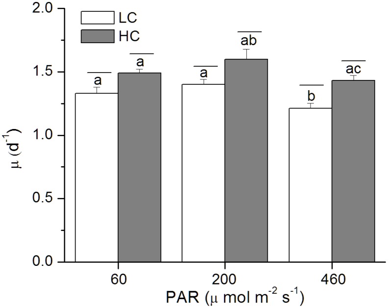
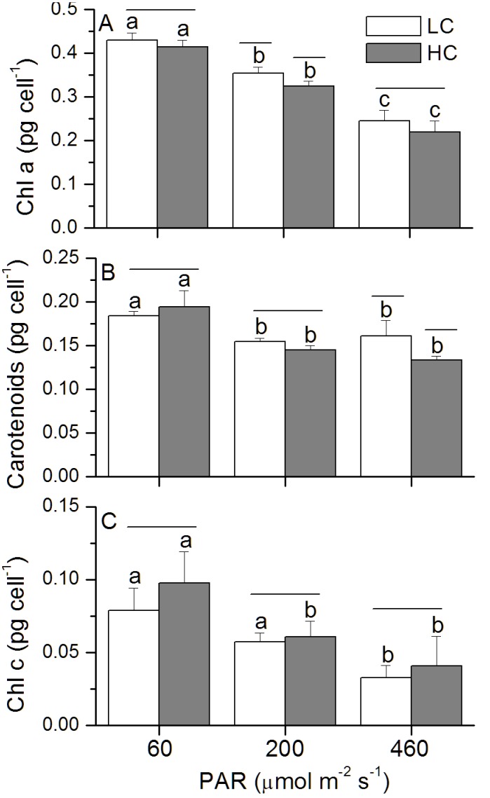
Compared to the LC-grown cells, elevated CO2 enhanced the specific growth rate by 12, 14 and 18% under the low, medium and high light levels (Fig. 1). The growth rate was highest at the medium but lower at the low and high light levels (Fig. 1). Chl a concentration per cell sharply declined (p<0.01) with increased light levels, with a significant difference between LC- and HC-grown cells (p = 0.02; Fig. 2A) under the medium light level and insignificant effects under the low (p = 0.18) and high light levels (p = 0.22) (Fig. 2A). For the content of carotenoids, significant difference between the two CO2 levels was observed under the HL level (p = 0.01; Fig. 2B). Chl c content increased in the HC-grown cells, although the difference was only marginally significant (p = 0.08–0.71) (Fig. 2C). HL-grown cells showed the highest ratio between the carotenoids and Chl a, but the lowest ratio of Chl c to Chl a, and the HC-grown cells had lager values for the ratio of Chl c to Chl a, although the difference between the CO2 treatment was not significant (Table 2).
Figure 1. Specific growth rates of P. tricornutum.
Growth rates of P. tricornutum cells grown under the LC (390 ppmv, pH 8.18) and HC (1000 ppmv, pH 7.82) and then both acclimated to different light levels (60, 200 and 460 µmol m−2 s−1) for 25–36 generations. Values are means ± SD, n = 3. The short-lines above the histogram bars indicate significant difference between LC and HC, and the different letters indicate significant differences among the light treatments within the HC- or LC-grown cells at p<0.05.
Figure 2. Pigmentation of P. tricornutum.
Chl α (A), Carotenoids (B) and Chl c (C) of P. tricornutum cells grown under the LC (390 ppmv, pH 8.18) and HC (1000 ppmv, pH 7.82) and then both acclimated to different light levels (60, 200 and 460 µmol m−2 s−1) for 25–36 generations. Values are means ± SD, n = 3. The short-lines above the histogram bars indicate significant difference between LC and HC, and the different letters indicate significant differences among the light treatments within the HC- or LC-grown cells at p<0.05.
Table 2. Ratios between carotenoids, Chl c and Chl a concentrations of P. tricornutum.
| PAR (µmol m−2 s−1) | Carotenoids/Chl a | Chl c/Chl a | ||
| LC | HC | LC | HC | |
| 60 | 0.43±0.027a | 0.47±0.048a | 0.18±0.030a | 0.23±0.046ab |
| 200 | 0.44±0.006a | 0.45±0.029a | 0.16±0.008ac | 0.19±0.027a |
| 460 | 0.65±0.039b | 0.61±0.083b | 0.13±0.015ac | 0.18±0.044a |
The ratio between carotenoids, Chl c and Chl a concentrations of P. tricornutum cells grown under the LC (390 ppmv, pH 8.18) and HC (1000 ppmv, pH 7.82) and then both acclimated to different light levels (60, 200 and 460 µmol m−2 s−1) for 25–36 generations. Values are means ± SD, n = 3. Different superscript letters indicate significant differences among different treatments within the ratio between carotenoids or Chl c and Chl a at p<0.05.
Photochemical and Non-photochemical Responses
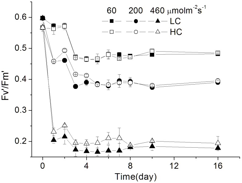
The effective quantum yield of the PSII, Fv′/Fm′, showed constant values, at 0.47, 0.40, 0.18 under the low, medium and high light levels, and was significantly enhanced by elevated CO2 under the HL condition (p<0.01; Fig. 3).
Figure 3. The effective photochemical yield (Fv .
′/Fm′) of P. tricornutum . The effective photochemical yield (Fv′/Fm′) of P. tricornutum cells grown under the LC (390 ppmv, pH 8.18) and HC (1000 ppmv, pH 7.82) and then both acclimated to different light levels (60, 200 and 460 µmol m−2 s−1) for different generations (from 0 to 36). Values are means ± SD, n = 3.
In views of rapid light curves (RLC), the saturation PAR level (Ek) significantly increased with the increase of growth light levels (p<0.01), but decreased with the acclimation time from day 1 to day 16, while the apparent photosynthetic efficiency (α) showed the opposite trends (p<0.01) (Table 3). After acclimation to different light levels for 25–36 generations, the apparent light use efficiency (α) was enhanced in the HC-grown cells under the low (p = 0.004), medium (p = 0.001) and high (p = 0.02) light levels, but the maximal electron transport (rETRmax) showed no significant variations between the LC and HC cultures (Table 3).
Table 3. The fitted parameters derived from the rapid light curves of P. tricornutum.
| Generations and parameters | LC | HC | |||||
| 60 | 200 | 460 | 60 | 200 | 460 | ||
| Ek | 370±15.7a | 672±9.9a | 1313±146.7a* | 426±72.1a | 680±38.8a | 1157±76.4a* | |
| 1–2 | rETRmax | 105±1.1a | 133±0.3a | 127±7.9a* | 113±7.5a | 133±7.4a | 138±3.8a* |
| α | 0.28±0.005a | 0.20±0.003a | 0.10±0.017a | 0.27±0.027a | 0.20±0.001a | 0.12±0.011a | |
| Ek | 314±18.7a | 629±12.7a* | 1167±28.1b* | 342±9.1a | 654±16.1a* | 1076±82.1ab* | |
| 12–18 | rETRmax | 77±5.1b | 102±1.6b | 107±8.7b | 81±2.6b | 117±0.4b | 118±5.4b |
| α | 0.24±0.001b | 0.16±0.001b | 0.10±0.005a | 0.24±0.014b | 0.18±0.005a | 0.11±0.007a | |
| Ek | 261±6.4b | 546±17.2b | 1060±41.0c | 248±20.0b | 481±1.3b | 982±79.8b | |
| 25–36 | rETRmax | 81±0.6b | 115±2.9c | 118±4.8a | 82±0.9b | 113±0.9b | 127±4.5b |
| α | 0.30±0.010a* | 0.20±0.001a* | 0.10±0.001a* | 0.33±0.031c* | 0.24±0.002b* | 0.13±0.019a* | |
The fitted parameters derived from the rapid light curves of P. tricornutum cells grown under the LC (390 ppmv, pH 8.18) and HC (1000 ppmv, pH 7.82) and then both acclimated to different light levels (60, 200 and 460 µmol m−2 s−1) for different generations (from 1 to 36 generations): Ek, the initial light saturation point (µmol m−2 s−1); rETRmax, the maximal rate of relative electron transport; α, the apparent light use efficiency. Values are means ± SD, n = 3 (triplicate cultures). Different superscript letters represent significant among the different generations within the LC or HC-grown cells and the asterisks indicate significant difference between LC and HC at p<0.05.
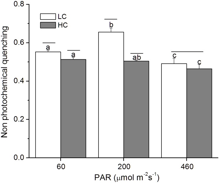
The LL-grown cells showed higher non photochemical quenching (NPQ) when exposed to a light stress, which decreased with the increase of growth light levels (Fig. 4). In addition, the NPQ was significantly reduced by the elevated CO2 under the low (p = 0.006) and medium (p<0.01) light levels (Fig. 4).
Figure 4. Non-photochemical quenching of P. tricornutum.
The changes in non-photochemical quenching of P. tricornutum cells grown under the LC (390 ppmv, pH 8.18) and HC (1000 ppmv, pH 7.82) and then both acclimated to different light levels (60, 200 and 460 µmol m−2 s−1) for 25–36 generations under high actinic light (1228 µmol m−2 s−1). Values are means ± SD, n = 3. The short-lines above the histogram bars indicate significant difference between LC and HC, and the different letters indicate significant differences among the light treatments within the HC- or LC-grown cells at p<0.05.
Photosynthetic Affinity for Inorganic Carbon
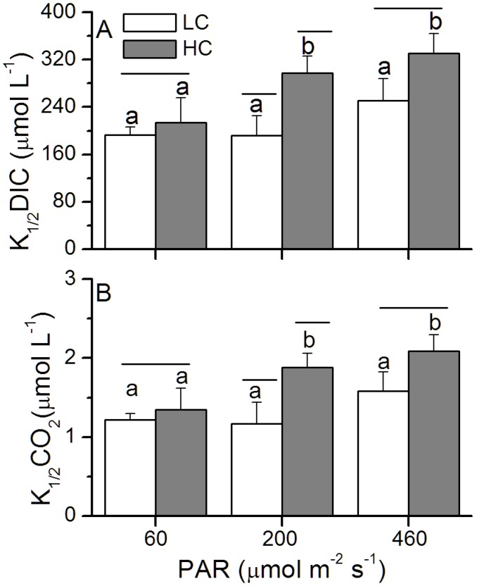
For the HC-grown cells, dissolved inorganic carbon (DIC) or CO2 levels for half saturation of photosynthesis (K1/2 DIC or K1/2 CO2) increased by 11%, 55% and 32% under the low, medium and high light levels, respectively, and the differences between the LC and HC was only significant at ML level (p = 0.02, Fig. 5), marginally significant at HL (p = 0.05), insignificant at LL level (p = 0.45). In addition, values of rETRmax increased with the increase of growth light levels.
Figure 5. Half-saturation constants (K1/2) for DIC (A) and CO2 (B) of P. tricornutum.
Half-saturation constants (K1/2) for DIC (A) and CO2 (B) of P. tricornutum cells grown under the LC (390 ppmv, pH 8.18) and HC (1000 ppmv, pH 7.82) and then both acclimated to different light levels (60, 200 and 460 µmol m−2 s−1) for 25–36 generations under middle actinic light (830 µmol m−2 s−1). Values are means ± SD, n = 3. The short-lines above the histogram bars indicate significant difference between LC and HC, and the different letters indicate significant differences among the light treatments within the HC- or LC-grown cells at p<0.05.
The Recovery of the Photochemical Yield under Dim Light
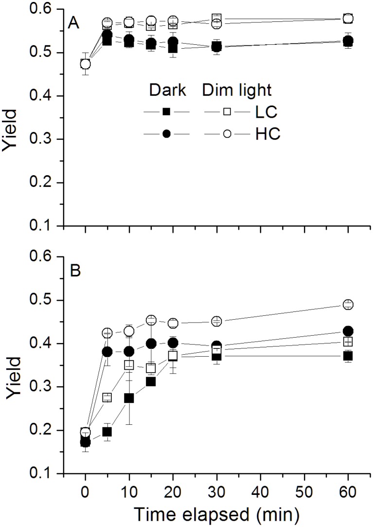
Under the dark or dim light level, the maximal photochemical yield of PSII, Fv/Fm, increased rapidly, reaching the largest value after 10 min, and then remained constant (Fig. 6A, B). Compared with the dark condition, faster recovery of Fv/Fm was observed in the presence of dim light and the cells acclimated to the HC condition showed a higher recovery rate compared to that of the LC-grown ones, especially for the cells grown under HL level (Fig. 6B).
Figure 6. The changes in quantum yield (Fv/Fm) of P. tricornutum.
The changes in quantum yield of P. tricornutum cells grown under the LC (390 ppmv, pH 8.18) and HC (1000 ppmv, pH 7.82) and then both acclimated to different light levels (A: 60 µmol m−2 s−1; B: 460 µmol m−2 s−1) for 25–36 generations under dark or dim light (10 µmol m−2 s−1). Values are means ± SD, n = 3.
Discussion
In this study, growth rate of P. tricornutum was enhanced by the elevated CO2 concentration under photosynthesis-limited, half-saturated and saturated light levels, being consistent with previous studies [11], [12], [36], [37]. Nevertheless, the present study provided the first evidence that the enhanced extent of growth under OA increased with acclimated light levels up to 460 µmol m−2 s−1. This result is consistent with our previous results obtained under low sunlight levels, but contradictory to that under high sunlight levels with daytime average PAR over 200 µmol m−2 s−1 (Table 4), that led to inhibited growth rate under the OA condition [12]. In the present study, when the cells had been grown at PAR of 460 µmol m−2 s−1, their growth rate was still enhanced by the same OA condition by 18% (Fig. 1; Table 4), However, the growth rates were comparatively lower as shown in the precious study under fluctuation sunlight of the same level (daytime average, 460 µmol m−2 s−1). Such a discrepancy must be attributed to the difference between the indoor constant and outdoor fluctuating light sources. Under the outdoor fluctuating solar radiation, during a day when the mean PAR level of 200 or 460 µmol m−2 s−1, the maximal PAR during noon time could exceed 1000 or 2000 µmol m−2 s−1, which caused higher levels of NPQ in the HC- than in the LC- grown cells, indicating that outdoor growth, even with the same level of light dose and with UV radiation screened off, could have brought more light stress to the cells grown under OA conditions [12]. Nevertheless, in the present study, compared to the LC-grown cells, HC-grown cells showed lower NPQ (Fig. 4) and higher Fv′/Fm′ even under the HL level (Fig. 3). This hints that compared to those grown fluctuating sunlight, the HC-grown cells under HL spent less energy to cope with light stress and photo-acclimation with daily solar radiation changes, therefore, their growth rates were enhanced by elevated CO2. Previously, mitochondrial respiration and carbon fixation were shown to be enhanced in P. tricornutum [11] and Thalassiosira pesudonana [15] under the same OA condition. Enhanced respiration can supplies more adenosine triphosphate (ATP), its consumption can reduce transmembrane thylakoidal pH gradient (ΔpH), therefore, NPQ could be reduced (Fig. 4). Consequently, the OA might have led to more ATP consumption with decreased NPQ [38].
Table 4. Mean specific growth rates of P. tricornutum.
| PAR (µmol m−2 s−1) | Mean specific growth rate (µ; day−1) | |||
| Indoor (constant light level) | Outdoor (solar radiation) | |||
| LC | HC | LC | HC | |
| 60 | 1.33 | 1.49 (12%) | 0.81 | 0.95 (17%) |
| 200 | 1.40 | 1.59 (14%) | 1.01 | 1.00 (−0.1%) |
| 460 | 1.21 | 1.43 (18%) | 1.07 | 0.88 (−18%) |
Mean specific growth rates of P. tricornutum cells grown under LC and HC conditions and both acclimated (for 25–36 generations) to the constant growth light levels in the present study compared to that observed in the previous study under fluctuating sunlight (acclimated for 21–25 generations) [12]. The data in parentheses represent the percentage change between the LC and HC conditions. Note, the µ values are much smaller under fluctuating than under the constant light regimes.
In addition, a common structural adaptation to light increase involves the reduction in antenna size or the light harvesting ability in order to reduce the absorption of light energy [39], or to increase the carotenoid content to dissipate excessive energy [40]. In our study, compared to LL- or HL-grown cells, the cells grown at medium light level showed higher growth rates, and with increased light level, Chl a per cell and the ratio of Chl c to Chl a sharply declined (Fig. 2A), being consistent with that previously reported in diatoms [41], [42], that altered the proportions of chlorophyll a/c and chlorophyll a/fucoxanthin protein-pigment complexes. The highest ratio between the carotenoids and Chl a concentration at the high light level reflects higher capacity of photoprotection (Table 2). Additionally, cells grown at the LL with the lowest initial Ek and higher apparent α (Table 3) imply higher light use efficiency. The fact that HC-grown cells showed higher α and lower Ek values compared to that of LC-grown ones implies that OA enhanced light use efficiency (Table 3), thus, growth rates were enhanced under the LL and ML levels. The apparent α of HL-grown cells was also enhanced, supporting the enhanced growth rate of the cells under OA, though the absolute specific growth rate was lower than that under LL and ML.
Most diatoms operate CO2 concentrating mechanisms (CCMs) [43] to accumulate intracellular CO2 and increase the CO2/O2 ratio around Rubisco [44]. The activity of the enzyme carbonic anhydrase (CA), which accelerates the inter-conversion between HCO3 − and CO2, is known to be down-regulated by elevated CO2, leading to a reduction of the active transport or use of HCO3 − [45]. The HC-grown cells had higher K1/2 DIC and K1/2 CO2 which were increased by 11–55% under the different light levels (Fig. 5), indicating a light dependent down-regulation of CCMs. Given that the operating of CCMs is energetically costly, phytoplankton cells (such as Chlorella vulgaris, Anabaena variabilis, Dunaliella tertiolecta) grown under low light levels usually show a decreased capacity and/or affinity for DIC transport [43], [46]–[51]. Such a down-regulation has been thought to be due to energy limitation under low light [43], [50], [51]. However, in the present study, the CCM of P. tricornutum was down-regulated under 1000 ppmv to a less extent in the low light compared to that under medium and high light levels (Fig. 5). Under the photosynthesis-oversaturating light level (HL), the CCM down-regulation was about 3 times that under the limited light level (LL) (Fig. 5). The cells grown at the low light level showed increased Chl a content (Fig. 2A) and higher apparent α (Table 3), suggesting that the increased light capture efficiency to meet the energy allocation to CCM operation, especially for the LC-grown cells, which need more energy for operation of CCMs.
Diatoms are subject to photoinactivation of their PSII reaction centers [52], [53], therefore, augmented capacity for their PSII repair is required to maintain photosynthesis [13]. If repair of photodamaged PSII fails to catch up with photoinactivation, PSII would suffer photoinhibition [54]–[56]. OA treatment is known to increase the susceptibility to photoinactivation of PSII [13] and then increased the capacity of PSII repair [19]. Such an increased capacity provides an explanation for the faster recovery of the Fv/Fm under dim light (Fig. 6). Considering dynamic light environment that phytoplankton cells are exposed to, the cells grown under OA conditions with higher recovery rate will suffer less photodamage [19].
With progressive ocean changes, enhanced stratification due to global warming [24], [57] will expose phytoplankton cells in the upper mixing layer (UML) to increased integrated sunlight levels and doses. Such an ocean change may enhance light stress to phytoplankton cells within UML. On the other hand, ocean acidification can result in further light stress for surface phytoplankton assemblages [12]. Therefore, ocean changes due to increased CO2 concentration in the atmosphere are likely to trigger higher photoinhibition, though repairing processes in diatoms could be stimulated.
In conclusion, the OA condition under 1000 µatm CO2 stimulated the growth of P. tricornutum rates under either light limiting or photosynthesis over-saturating light levels, with higher extent of the growth enhancement under the high light level, though the higher specific growth rate was found under the low and medium light levels. The discrepancy between the present and previous study [12] in growth response to OA under fluctuating sunlight could attribute to additional energy requirement for the diatom to cope with light stress and photo-acclimation with diurnal solar changes. Such a hypothetical theory, though requires further experimental testing, is supported by a recent finding in the diatom Chaetoceros debilis that showed OA increased cellular quota of particulate organic carbon under constant light levels but decreased it under changing light regimes [58]. This hints that the diatom cells grown under sunlight or fluctuating light regimes may be stressed to allocate more energy for photoprotection and acclimation and resulted less carboxylation and cellular carbon storage.
Funding Statement
This study was supported by National Basic Research Program of China (2011CB200902), National Natural Science Foundation (41120164007), Program for Changjiang Scholars and Innovative Research Team (IRT0941), SOA (GASI-03-01-02-04), and China-Japan collaboration project from MOST (S2012GR0290). The funders had no role in study design, data collection and analysis, decision to publish, or preparation of the manuscript.
References
- 1.Intergovemmental panel on climate change (IPCC) (2001) Climate change 2001: Contribution of working groups and to the third assessment report of the intergovemental panel on climate change, the scientific basis. Cambridge: Cambridge University Press: 398.
- 2. Sabine CL, Feely RA, Gruber N, Key RM, Lee K, et al. (2004) The oceanic sink for anthropogenic CO2 . Science 305 (5682): 367–371. [DOI] [PubMed] [Google Scholar]
- 3. Gao K, Ruan Z, Villafañe VE, Helbling EW (2009) Ocean acidification exacerbates the effect of UV radiation on the calcifying phytoplankter Emiliania huxleyi . Limnol Oceanogr 54 (6): 1855–1862. [Google Scholar]
- 4. Gao K, Zheng Y (2010) Combined effects of ocean acidification and solar UV radiation on photosynthesis, growth, pigmentation and calcification of the coralline alga Corallina sessilis (Rhodophyta). Glob Change Biol 16 (8): 2388–2398. [Google Scholar]
- 5. Beaufort L, Probert I, de Garidel-Thoron T, Bendif EM, Ruiz-Pino D, et al. (2011) Sensitivity of coccolithophores to carbonate chemistry and ocean acidification. Nature 476 (7358): 80–83. [DOI] [PubMed] [Google Scholar]
- 6.Riebesell U, Tortell PD (2011) Effects of ocean acidification on pelagic organisms and ecosystems, in: Ocean acidification, edited by: Gattuso, J.-P. and Hansson, L., Oxford University Press, UK, 99–116.
- 7. Venn AA, Tambutté E, Holcomb M, Laurent J, Allemand D, et al. (2013) Impact of seawater acidification on pH at the tissue–skeleton interface and calcification in reef corals. Proc Nati Acad of Sci 110 (5): 1634–1639. [DOI] [PMC free article] [PubMed] [Google Scholar]
- 8. Kim JM, Lee K, Shin K, Kang JH, Lee HW (2006) The effect of seawater CO2 concentration on growth of a natural phytoplankton assemblage in a controlled mesocosm experiment. Limnol Oceanogr 51 (4): 1629–1636. [Google Scholar]
- 9. King AL, Sanudo-Wilhelmy SA, Leblanc K, Hutchins DA, Fu F (2011) CO2 and vitamin B12 interactions determine bioactive trace metal requirements of a subarctic pacific diatom. ISME J 5: 1388–1396. [DOI] [PMC free article] [PubMed] [Google Scholar]
- 10. Low-Décarie E, Fussmann GF, Bell G (2011) The effect of elevated CO2 on growth and competition in experimental phytoplankton communities. Glob Change Biol 17: 2525–2535. [Google Scholar]
- 11. Wu Y, Gao K, Riebesell U (2010) CO2-induced seawater acidification affects physiological performance of the marine diatom Phaeodactylum tricornutum . Biogeosciences 7 (9): 2915–2923. [Google Scholar]
- 12. Gao K, Xu JT, Gao G, Li YH, Hutchins DA, et al. (2012) Rising CO2 and increased light exposure synergistically reduce marine primary productivity. Nature Climate Change 2: 519–523. [Google Scholar]
- 13. McCarthy A, Rogers SP, Duffy SJ, Campbell DA (2012) Elevated carbon dioxide differentially alters the photophysiology of Thalassiosira pseudonana (Bacillariophyceae) and Emiliania huxleyi (Haptophyta). J Phycol 48 (3): 635–646. [DOI] [PubMed] [Google Scholar]
- 14. Li G, Campbell DA (2013) Rising CO2 interacts with growth light and growth rate to alter photosystem II photoinactivation of the coastal diatom Thalassiosira pseudonana . PLoS ONE 8: e55562. [DOI] [PMC free article] [PubMed] [Google Scholar]
- 15. Yang G, Gao K (2012) Physiological responses of the marine diatom Thalassiosira pseudonana to increased pCO2 and seawater acidity. Mar Environ Res 79: 142–151. [DOI] [PubMed] [Google Scholar]
- 16. Riebesell U, Schulz KG, Bellerby RG, Botros M, Fritsche P, et al. (2007) Enhanced biological carbon consumption in a high CO2 ocean. Nature 450 (7169): 545–548. [DOI] [PubMed] [Google Scholar]
- 17. Sugie K, Yoshimura T (2013) Effects of pCO2 and iron on the elemental composition and cell geometry of the marine diatom Pseudo-nitzschia pseudodelicatissima (Bacillariophyceae). J Phycol 49: 475–488. [DOI] [PubMed] [Google Scholar]
- 18.Gao K, Campbell DA (2013) Photophysiological responses of marine diatoms to elevated CO2 and decreased pH: a review. Funct Plant Biol. [DOI] [PubMed]
- 19. Li Y, Gao K, Villafañe VE, Helbling EW (2012) Ocean acidification mediates photosynthetic response to UV radiation and temperature increase in the diatom Phaeodactylum tricornutum . Biogeosciences 9: 3931–3942. [Google Scholar]
- 20. Ihnken S, Roberts S, Beardall J (2001) Differential responses of growth and photosynthesis in the marine diatom Chaetoceros muelleri to CO2 and light availability. Phycologia 50 (2): 182–193. [Google Scholar]
- 21. Boelen P, van de Poll WH, van der Strate HJ, Neven IA, Beardall J, et al. (2011) Neither elevated nor reduced CO2 affects the photophysiological performance of the marine antarctic diatom Chaetoceros brevis . J Exp Mar Biol Ecol 406: 38–45. [Google Scholar]
- 22. Torstensson A, Chierici M, Wulff A (2012) The influence of increased temperature and carbon dioxide levels on the benthic/sea ice diatom Navicula directa . Polar Biol 35: 205–214. [Google Scholar]
- 23. Flynn KJ, Blankford JC, Baird ME, Raven JA, Clark DR, et al. (2012) Changes in pH at the exterior surface of plankton with ocean acidification. Nature Climate Change 2: 510–513. [Google Scholar]
- 24. Gao K, Helbling EW, Häder DP, Hutchins DA (2012) Responses of marine primary producers to interactions between ocean acidification, solar radiation, and warming. Marine Ecol Prog Ser 470: 167–189. [Google Scholar]
- 25. Lavaud J, Strzepek RF, Kroth PG (2007) Photoprotection capacity differs among diatoms: Possible consequences on the spatial distribution of diatoms related to fluctuations in the underwater light climate. Limnol Oceanogr 52: 1188–1194. [Google Scholar]
- 26. Jin P, Gao K, Villafane VE, Campbell DA, Helbling W (2013) Ocean acidification alters the photosynthetic responses of a coccolithophorid to fluctuating UV and visible radiation. Plant Physiol 162: 2084–2094. [DOI] [PMC free article] [PubMed] [Google Scholar]
- 27. Morel FMM, Rueter JG, Anderson DM, Guillard RRL (1979) Aquil: A chemically defined phytoplankton culture medium for trace metal studies. J Phycol 15 (2): 135–141. [Google Scholar]
- 28.Lewis E, Wallance DWR (1998) Program developed for CO2 system calculations. ORNL/CDIAC-105. Carbon Dioxide Information Analysis Center, Oak Ridge National Laboratory, US Department of Energy, Oak Ridge, Tennessee.
- 29. Roy RN, Roy LN, Vogel KM, Porter-Moore C, Pearson T, et al. (1993) The dissociation constants of carbonic acid in seawater at salinities 5 to 45 and temperature 0 to 45°C. Mar Chem 44: 249–267. [Google Scholar]
- 30. Dickson AG (1990) Standard potential of the reaction: AgCl (s) +1/2 H2 (g) = Ag (s) + HCl (aq), and the standard acidity constant of the ion HSO4 − in synthetic seawater from 273.15 to 318.15 K. J Chem Thermodyn. 22: 113–127. [Google Scholar]
- 31. Ritchie RJ (2006) Consistent sets of spectrophotometric chlorophyll equations for acetone, methanol and ethanol solvents. Photosynth Res 89: 27–41. [DOI] [PubMed] [Google Scholar]
- 32. Strickland JDH, Parsons TR (1968) A practical handbook of seawater analysis. B Fish Res Board Can 167: 49–80. [Google Scholar]
- 33. Ralph PJ, Gademann R (2005) Rapid light curves: A powerful tool to assess photosynthetic activity. Aquatic Botany 82: 222–237. [Google Scholar]
- 34. Eilers PHC, Peeters JCH (1988) A model for the relationship between light intensity and the rate of photosynthesis in phytoplankton. Ecol Model 42 (3): 199–215. [Google Scholar]
- 35. Gao K, Aruga Y, Asada K, Ishihara T, Akano T, et al. (1993) Calcification in the articulated coralline alga corallina pilulifera with special reference to the effect of elevated CO2 concentration. Mar Biol 117: 129–132. [Google Scholar]
- 36. Schippers P, Lürling M, Scheffer M (2004) Increase of atmospheric CO2 promotes. phytoplankton productivity. Ecology Letters 7: 446–451. [Google Scholar]
- 37. Bartual A, Gálvez JA (2003) Short- and long-term effects of irradiance and CO2 availability on carbon fixation by two marine diatoms. Can J Bot 81: 191–200. [Google Scholar]
- 38. Kanazawa A, Kramer DM (2002) In vivo modulation of nonphotochemical exciton quenching (NPQ) by regulation of the chloroplast ATP synthase. Proc Nati Acad of Sci 99: 12789–12794. [DOI] [PMC free article] [PubMed] [Google Scholar]
- 39. Gordillo FJL, Figueroa FL, Niell FX (2003) Photon- and carbon-use efficiency in Ulva rigida at different CO2 and N levels. Planta 218: 315–322. [DOI] [PubMed] [Google Scholar]
- 40. Horton P, Ruban AV, Walters RG (1994) Regulation of light harvesting in green plants (Indication by nonphotochemical quenching of chlorophyll fluorescence). Plant Physiol 106 (2): 415–420. [DOI] [PMC free article] [PubMed] [Google Scholar]
- 41. Perry MJ, Talbot MC, Alberte RS (1981) Photoadaption in marine phytoplankton: response of the photosynthetic unit. Mar Biol 62: 91–101. [Google Scholar]
- 42. Janssen M, Bathke L, Marquardt J, Krumbein WE, Rhiel E (2001) Changes in the photosynthetic apparatus of diatoms in response to low and high light intensities. International Microbiology 4: 27–33. [DOI] [PubMed] [Google Scholar]
- 43. Giordano M, Beardall J, Raven JA (2005) CO2 concentrating mechanisms in algae: mechanisms, environmental modulation, and evolution. Annu Rev Plant Biol 56: 99–131. [DOI] [PubMed] [Google Scholar]
- 44. Burkhardt S, Amoroso G, Riebesell U, Sültemeyer D (2001) CO2 and HCO3 − uptake in marine diatom acclimated to different CO2 concentrations. Limnol Oceanogr 46: 1378–1391. [Google Scholar]
- 45. Trimborn S, Wolf-Gladrow DA, Richter K-U, Rost B (2009) The effect of pCO2 on carbon acquisition and intracellular assimilation in four marine diatoms. J Exp Mar Biol Ecol 376: 26–36. [Google Scholar]
- 46. Shiraiwa Y, Miyachi S (1983) Factors controlling induction of carbonic anhydrase and efficiency of photosynthesis in Chlorella vulgaris llh cells. Plant Cell Physiol 24 (5): 919–923. [Google Scholar]
- 47. Beardall J, Roberts S, Millhouse J (1991) Effects of nitrogen limitation on uptake of inorganic carbon and specific activity of ribulose-1, 5-bisphosphate carboxylase/oxygenase in green microalgae. Can J Bot 69: 1146–1150. [Google Scholar]
- 48. Beardall J, Giordano M (2002) Ecological implications of microalgal and cyanobacterial CO2 concentrating mechanisms, and their regulation. Funct Plant Biol 29: 335–347. [DOI] [PubMed] [Google Scholar]
- 49.Beardall J, Raven JA (2013) Limits to phototrophic growth in dense culture: CO2 supply and light, in: Algae for Biofuels and Energy, edited by: Borowitzka, M. A. and Moheimani, N. R., 2013.
- 50. Raven JA, Giordano M, Beardall J, Maberly SC (2011) Algal and aquatic plant carbon concentrating mechanisms in relation to environmental change. Photosynth Res 109: 281–296. [DOI] [PubMed] [Google Scholar]
- 51. Young EB, Beardall J (2005) Modulation of photosynthesis and inorganic carbon acquisition in a marine microalga by nitrogen, iron, and light availability. Can J Bot 83: 917–928. [Google Scholar]
- 52. Six C, Finkel ZV, Irwin AJ, Campbell DA (2007) Light variability illuminates niche-partitioning among marine picocyanobacteria. PLoS ONE 2: e1341. [DOI] [PMC free article] [PubMed] [Google Scholar]
- 53. Edelman M, Mattoo AK (2008) D1-protein dynamics in photosystem II: the lingering enigma. Photosynth Res 98: 609–620. [DOI] [PubMed] [Google Scholar]
- 54. Aro EM, Suorsa M, Rokka A, Allahverdiyeva Y, Paakkarinen V, et al. (2005) Dynamics of photosystem II: a proteomic approach to thylakoid protein complexes. J Exp Bot 56: 347–356. [DOI] [PubMed] [Google Scholar]
- 55. Nishiyama Y, Allakhverdiev SI, Murata N (2006) A new paradigm for the action of reactive oxygen species in the photoinhibition of photosystem II. Biochim Biophys Acta 1757: 742–749. [DOI] [PubMed] [Google Scholar]
- 56. Murata N, Takahashi S, Nishiyama Y, Allakhverdiev SI (2007) Photoinhibition of photosystem II under environmental stress. Biochim Biophys Acta 1767: 414–421. [DOI] [PubMed] [Google Scholar]
- 57. Doney SC, Ruckelshaus M, Emmett Duffy J, Barry JP, Chan F, et al. (2012) Climate change impacts on marine ecosystems. Annu Rev Mar Sci 4: 11–37. [DOI] [PubMed] [Google Scholar]
- 58.Hoppe CJM, Holtz LM, Trimborn S, Rost B (2014) Contrasting responses of Chaetoceros debilis (Bacillariophyceae) to ocean acidification under constant and dynamic light. New Phytologist.


