Abstract
Background
Autotransporters form a large family of outer membrane proteins specifying diverse biological traits of Gram-negative bacteria. In this study, we report the identification and characterization of a novel autotransporter gene product of Burkholderia mallei (locus tag BMA1027 in strain ATCC 23344).
Results
Database searches identified the gene in at least seven B. mallei isolates and the encoded proteins were found to be 84% identical. Inactivation of the gene encoding the autotransporter in the genome of strain ATCC 23344 substantially reduces adherence to monolayers of HEp-2 laryngeal cells and A549 type II pneumocytes, as well as to cultures of normal human bronchial epithelium (NHBE). Consistent with these findings, expression of the autotransporter on the surface of recombinant E. coli bacteria increases adherence to these cell types by 5–7 fold. The gene specifying the autotransporter was identified in the genome of 29 B. pseudomallei isolates and disruption of the gene in strain DD503 reduced adherence to NHBE cultures by 61%. Unlike B. mallei, the mutation did not impair binding of B. pseudomallei to A549 or HEp-2 cells. Analysis of sera from mice infected via the aerosol route with B. mallei and B. pseudomallei revealed that animals inoculated with as few as 10 organisms produce antibodies against the autotransporter, therefore indicating expression in vivo.
Conclusions
Our data demonstrate that we have identified an autotransporter protein common to the pathogenic species B. mallei and B. pseudomallei which mediates adherence to respiratory epithelial cells and is expressed in vivo during the course of aerosol infection.
Background
Autotransporter proteins are the largest known family of virulence factors expressed by Gram-negative bacteria and play prominent roles in processes such as invasion [1], serum resistance [2,3], phospholipolysis [4-6], cytotoxicity [7], adherence [8,9], survival within eukaryotic cells [10], intracellular motility [11], cell-to-cell aggregation [12,13], and biofilm formation [14,15]. These molecules display conserved structural features including an N-terminal surface-exposed domain responsible for the biological function and a hydrophobic C-terminus that tethers the autotransporter to the outer membrane (OM). Based on the structure of the C-terminus, autotransporters can be classified as conventional or oligomeric [16-21]. The C-terminus of conventional autotransporters consists of ~300 amino acids (aa) forming 10–12 antiparallel β-strands, while that of oligomeric autotransporters is substantially shorter (~70 aa) and specifies only 4 β-strands. Because of their structure and role in virulence, autotransporters are attractive targets for developing countermeasures against pathogenic organisms. Large portions of autotransporters are located on the bacterial surface and therefore readily accessible for recognition by the immune system. Additionally, autotransporters play important roles in pathogenesis, thus targeting them may hinder the ability to cause disease. This hypothesis is supported by several studies demonstrating the effectiveness of autotransporter-based countermeasures. For example, immunization with Neisseria meningitidis NadA elicits antibodies (Abs) binding to the bacterial surface and promoting complement-mediated killing [22,23], which is key to protection against this organism. Antibodies against Haemophilus influenzae Hap block adherence to epithelial cells and immunization with Hap protects mice in nasopharyngeal colonization studies [24,25]. Vaccination with the Proteus mirabilis autotransporter cytotoxin Pta yields Abs that not only reduce bacterial burden in a murine urinary tract infection model, but also neutralize the cytotoxic activity of Pta for bladder cells [26]. Moreover, Bordetella pertussis Pertactin, an autotransporter adhesin, is a component of licensed vaccines for whooping cough (http://www.cdc.gov/vaccines/pubs/pinkbook/downloads/pert.pdf).
Burkholderia mallei and Burkholderia pseudomallei are closely related Gram-negative organisms for which developing efficacious countermeasures is highly desirable. Both species are classified as Tier 1 agents by the U.S. Federal Select Agent Program because of concerns regarding their use as bioweapons, especially since B. mallei has been utilized in this manner on more than one occasion [27-31]. Burkholderia mallei is a host-adapted pleomorphic coccobacillus that does not persist in the environment outside of its natural equine reservoir. The bacterium causes the highly contagious zoonotic disease glanders, which primarily affects horses, and is endemic to parts of Asia, Africa, South America and the Middle East [27,32-38]. In humans, infection typically occurs via the cutaneous or aerosol route upon contact with infected animals. Clinical manifestations include fever, pneumonia, necrosis of the trachea and bronchi, bacteremia, and dissemination of B. mallei to organs where it causes necrotizing abscesses. Burkholderia pseudomallei is a saprophyte of wet soils and is endemic to countries bordering the equator. The organism can infect most mammals and causes the disease melioidosis in humans, a febrile illness that varies greatly in its clinical presentation. Disease states range from flu-like malaise to septicemia, chronic abscess formation in deep tissues, or bacteremic pneumonia [33,39-45]. Infection is generally acquired by percutaneous inoculation, ingestion and inhalation of aerosols, and the risk of contracting disease is proportionate to the concentration of B. pseudomallei in soil. Burkholderia pseudomallei is a leading cause of sepsis and bacteremic pneumonia in Southeast Asia and Australia, and melioidosis is increasingly recognized as an emerging infectious diseases in many tropical regions of the world [40,46,47].
Glanders and melioidosis have high mortality rates (up to 50%) despite aggressive antimicrobial therapy. The recommended treatment involves the use of ceftazidime and meropenem (intensive phase) and TMP-SMX and co-amoxiclav (eradication phase) for several months [48]. Response to treatment is slow and eradication of the agents is difficult, often resulting in protracted alternating bouts of remission and exacerbation. There are no vaccines available to protect against either Burkholderia species. Clearly, there is a need to identify and characterize targets for developing countermeasures for these organisms. The genomes of B. mallei and B. pseudomallei have been reported to encode multiple autotransporters [49-51]. In this study, we examined one of these gene products and evaluated it role in adherence in vitro and virulence in a mouse aerosol model of infection.
Results
Identification of a gene encoding a potential autotransporter adhesin shared by B. mallei and B. pseudomallei
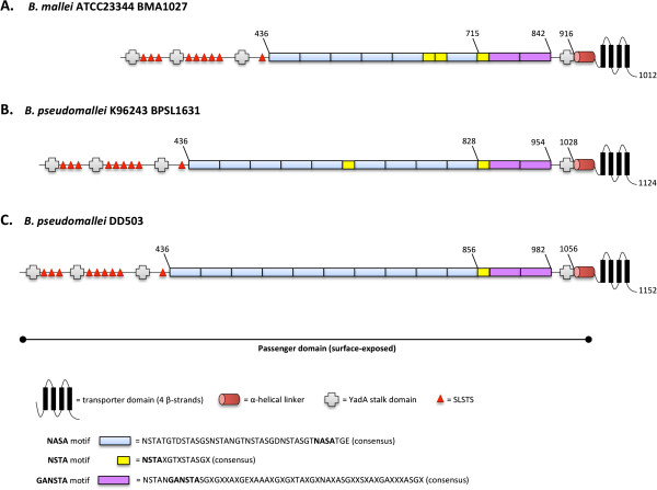
Comparative sequence analyses identified a gene product in the published genome of B. mallei strain ATCC 23344 (locus tag # BMA1027) that resembles the adhesins Yersinia enterocolitica YadA [2,21,52], Moraxella catarrhalis Hag [8,53,54], B. pseudomallei BoaA and BoaB [55], and B. mallei BoaA [55]. These molecules belong to the oligomeric coiled-coil adhesin (Oca) sub-family of oligomeric autotransporter proteins and have a characteristic modular organization consisting of: (i) a surface-exposed region specifying adhesive properties termed passenger domain, (ii) a short linker region predicted to form an α helix, and (iii) a hydrophobic C-terminus composed of four β-strands anchoring the autotransporter in the OM designated transporter domain [16,19-21]. As shown in Figure 1A, BMA1027 is predicted to possess these structural features.
Figure 1.
Structural features of BMA1027 and orthologous gene products. Different regions of the protein encoded by B. mallei ATCC 23344 BMA1027 (A), B. pseudomallei K96243 BPSL1631 (B) and the B. pseudomallei DD503 BMA1027 ortholog (C) are depicted with the positions of residues defining selected domains. Transporter domains (OM anchors) and helical linkers were identified using the PSIPRED secondary structure prediction algorithm. The colored boxes, red triangles, and grey crosses show the relative position and number of repeated aa motifs.
Searches using the Pfam database revealed that the region encompassing aa 936–1012 of BMA1027 shows similarity to a YadA anchor domain (PF3895.10; expect value of 6.3e−22), which is present in most Oca and described as important for oligomerization and targeting autotransporters to the OM. Pfam searches also indicated that BMA1027 contains four YadA stalk domains (PF05662, formerly designated HIM; expect values ranging from 2.2e−4 to 1.5e−9; grey crosses in Figure 1A). This motif is associated with invasins and haemagglutinins and is present in YadA as well as Hag [2,8,52,53]. YadA contains one stalk domain, which has been shown to be necessary for protein stability and adhesive properties. Further sequence analysis revealed that the passenger domain of BMA1027 specifies repeated aa motifs, a trait noted in several oligomeric autotransporters including YadA [2,52], Hag [8,53], BoaA and BoaB [55], the B. pseudomallei biofilm factor BbfA [56], and the M. catarrhalis UspA1, UspA2, and UspA2H proteins [57-60]. As illustrated in Figure 1A, the passenger domain of BMA1027 contains nine copies of the 5-mer SLSTS (red triangles) and several repeated elements beginning with residues NSTA (colored boxes). Additional characteristics of the predicted protein are listed in Table 1.
Table 1.
Characteristics a of BMA1027 orthologous genes and their encoded products
| Strain b | Locus tag | Predicted protein (aa) | MW (kDa) | Potential signal sequence cleavage site c |
|---|---|---|---|---|
|
B. pseudomallei
|
|
|
|
|
| 1026b/DD503* |
BP1026B_I1575 |
1,152 |
107.4 |
ASA37▼G, AMA69▼A |
| K96243 |
BPSL1631 |
1,124 |
104.8 |
ASA37▼G, AMA69▼A |
|
B. mallei
|
|
|
|
|
| ATCC 23344 | BMA1027 | 1,012 | 94.7 | ASA37▼G, AMA69▼A |
aSequence analyses were performed using Vector NTI (Life Technologies™) and online tools available through the ExPASy Bioinformatics Resources Portal.
bGenomic sequences are available through the NCBI genomic BLAST service.
cThe putative signal sequence cleavage sites were determined using the SignalP 4.1 server.
*The B. pseudomallei strain DD503 is a derivative of isolate 1026b in which the AmrAB-OprA antibiotic efflux pump has been deleted to facilitate mutant construction [61]. The BMA1027 orthologs of strains DD503 and 1026b are identical (confirmed by nucleotide sequence analysis, data not shown).
The published genomic sequence of the B. pseudomallei strain K96243 was found to specify a BMA1027 ortholog (locus tag # BPSL1631, Figure 1B) that is 89% identical to that of B. mallei ATCC 23344. The BMA1027 ortholog was sequenced from the B. pseudomallei strain used in our laboratory, DD503, and was predicted to encode a protein that is 97% and 87% identical to that of B. pseudomallei K96243 and B. mallei ATCC 23344, respectively (Figure 1C). Database searches with the NCBI genomic BLAST service also identified orthologs in several B. pseudomallei and B. mallei isolates. Seven B. mallei and twenty-nine B. pseudomallei strains for which sequences are available through the service were found to have the gene. Characteristics of these ORFs are listed in the Additional files 1 and 2. Overall, the proteins are 87-100% identical and differ primarily in the number and/or arrangement of SLST repeats, YadA stalk domains, and/or NSTA elements in their passenger domains. Based on these results, we conclude that BMA1027 orthologs are well-conserved gene products shared by B. mallei and B. pseudomallei. While preparing this article, Campos and colleagues published a report in which they functionally characterized autotransporter genes specified by the B. pseudomallei strain 1026b [51]. One of these molecules corresponds to the BMA1027 ortholog (locus tag # BP1026B_1575), which the authors designated bpaC. Henceforth, BMA1027 and orthologs will be referred to as BpaC.
Expression and functional properties of the BpaC protein in E. coli
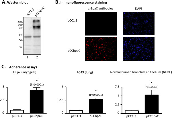
Because of sequence and structural similarities to known bacterial adhesins, we speculated that BpaC mediates adherence to epithelial cells. To test this hypothesis, the bpaC gene of B. pseudomallei DD503 was cloned and expressed in the E. coli strain EPI300. This organism does not adhere well to epithelial cells [8,53,55,62] and therefore provides a suitable heterologous genetic background to study the adherence properties of BpaC. To verify protein expression, whole cell lysates were prepared from E. coli EPI300 harboring the plasmid pCC1.3 (control) or pCCbpaC (specifies B. pseudomallei DD503 bpaC) and analyzed by western blot. Figure 2A shows that α-BpaC Abs (directed against aa 392–1098, part of surface-exposed passenger domain) react specifically with a band of 100-kDa in E. coli expressing the bpaC gene (lane 2), which is consistent with the predicted molecular mass of the gene product (Table 1). The Abs also specifically reacted with an antigen of high molecular weight (≥250 kDa), which likely corresponds to an oligomeric form of BpaC. Immunofluorescence-labeling of non-permeabilized E. coli cells was used to demonstrate that BpaC is displayed on the surface of recombinant bacteria. As shown in Figure 2B, E. coli carrying pCCbpaC is labeled by α-BpaC Abs while recombinant bacteria harboring the control plasmid pCC1.3 are not. Staining of nucleic acids with DAPI verified that equivalent numbers of bacteria were examined.
Figure 2.
Analysis of E. coli recombinant strains. Panel A: Whole cell lysates were resolved by SDS-PAGE, transferred to PVDF membranes and analyzed by western blot with Abs against BpaC. Lane 1, E. coli (pCC1.3); lane 2, E. coli (pCCbpaC). MW markers are shown to the left in kilodaltons. Panel B: Non-permeabilized E. coli strains were fixed onto glass slides and fluorescently-labeled with DAPI (blue) and with α-BpaC Abs (red). Bacteria were visualized by microscopy using a Zeiss LSM 510 Meta confocal system. Representative microscopic fields are shown. Panel C: E. coli strains were incubated with epithelial cells for 3-hr. Cells were then washed to remove unbound bacteria, lysed, diluted and spread onto agar plates to enumerate bound bacteria. The results are expressed as the mean percentage (±standard error) of inoculated bacteria attached to epithelial cells. Asterisks indicate that the increased adherence of E. coli (pCCbpaC), compared to that of E. coli carrying the control plasmid pCC1.3, is statistically significant (P value shown in parentheses). Adherence assays were performed in duplicate on at least 4 independent occasions.
Quantitative adherence assays revealed that E. coli expressing BpaC binds to HEp-2 (laryngeal) and A549 (lung) human epithelial cells at levels 7- and 5-fold greater than bacteria carrying pCC1.3, respectively (Figure 2C). BpaC expression was also found to increase adherence by 7-fold to normal human bronchial epithelium (NHBE) cultured in an air-liquid interface system, which has been shown to represent an environment similar to the airway lumen in vivo [54,63,64]. These results demonstrate that BpaC mediates adherence to respiratory epithelial cells. Burkholderia pseudomallei and B. mallei are facultative intracellular bacteria that replicate within several eukaryotic cell types. Moreover, autotransporter adhesins frequently perform additional functions including invasion [1], intracellular motility [11], and survival inside host cells [10]. For these reasons, we examined the ability of E. coli expressing BpaC to invade epithelial cells and survive within murine macrophages. The results of these experiments indicated that BpaC does not substantially increase invasion of epithelial cells, phagocytosis of recombinant bacteria by J774A.1 murine macrophages, or survival inside these immune cells (data not shown).
In vitro characterization of B. mallei and B. pseudomallei mutant strains
To study the functional properties of the bpaC gene product in Burkholderia, we constructed isogenic bpaC mutants of B. pseudomallei DD503 and B. mallei ATCC 23344. Whole cell lysates and sarkosyl-insoluble OM proteins were prepared from these strains and analyzed by western blot to verify lack of BpaC expression in the mutants. However, α-BpaC Abs did not react with protein preparations of parent or mutant strains (data not shown). Other methods such as immunoprecipitation and immunofluorescence-labeling also failed to detect BpaC expression. These results indicate that the bpaC gene is expressed at very low levels under the laboratory growth conditions we used to propagate the organisms.
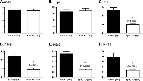
Because adherence assays with recombinant bacteria revealed that BpaC expression increases the binding of E. coli to NHBE cultures and monolayers of A549 and HEp-2 cells (Figure 2C), we compared the ability of Burkholderia parent and bpaC mutant strains to attach to these respiratory cells. Figure 3C shows that inactivation of the bpaC gene in B. pseudomallei DD503 affects adherence to NHBE cultures, reducing levels by 61%. The B. pseudomallei mutant bound to A549 and HEp-2 cells at wild-type levels. The bpaC mutation significantly impaired the ability of B. mallei ATCC 23344 to attach to A549 cells (66% reduction, Figure 3D), HEp-2 monolayers (72% reduction, Figure 3E), and NHBE cultures (66% reduction, Figure 3F). These results demonstrate that the bpaC gene product contributes to the adherence of B. mallei and B. pseudomallei to epithelial cells derived from the human respiratory tract.
Figure 3.

Adherence of B. mallei and B. pseudomallei strains to human respiratory epithelial cells. The effect of a bpaC mutation on the adherence of B. pseudomallei (Bp) DD503 and B. mallei (Bm) ATCC 23344 to monolayers of A549 (panels A and D) and HEp-2 (panels B and E) cells and cultures of NHBE (panels C and F) was measured in duplicate on at least 3 separate occasions. Strains were incubated with epithelial cells for 3-hr. Cells were then washed to remove unbound bacteria, lysed, diluted and spread onto agar plates to enumerate bound bacteria. The results are expressed as the mean percentage (±standard error) of inoculated bacteria adhering to epithelial cells. Asterisks indicate that the difference between the adherence of the bpaC KO mutant and that of the parent strain is statistically significant (P value shown in parentheses).
As stated earlier, autotransporter adhesins often perform multiple biological functions including invasion [1] and survival within host cells [10]. In addition, B. pseudomallei and B. mallei are facultative intracellular pathogens that effectively replicate inside professional phagocytic cells. Therefore, we measured the ability of Burholderia mutant and parent strains to invade epithelial cells (A549 and HEp-2) and replicate within J774A.1 murine macrophages. The results of these experiments indicated that the bpaC mutation does not cause a defect in invasion of epithelial cells, phagocytosis of recombinant bacteria by J774A.1 murine macrophages, or growth inside these cells (data not shown). The bpaC mutants did not show defects in resistance to the bactericidal activity of normal human serum (data not shown), which is another biological function commonly associated with Oca autotransporters [2,3,19,65,66].
Virulence of B. mallei and B. pseudomallei mutant strains and BpaC expression in vivo
To determine whether BpaC contributes to virulence, we calculated the median lethal dose (LD50) of B. pseudomallei and B. mallei mutant strains in a mouse model of aerosol infection. The model entails the use of a Microsprayer® to deliver bacteria directly into the murine lungs [67]. The device generates aerosol particles from the tip of a bent, 23-gauge nebulizing tube attached to a high-pressure stainless steel syringe that contains bacteria. BALB/c mice were anesthetized and placed in a custom-designed acrylic holder inside a Class II Biosafety cabinet. A modified pediatric otoscope equipped with a light source was then used to introduce the nebulizing tube portion of the Microsprayer® into the trachea of animals, and 50-μL of bacterial suspension was aerosolized into the lungs by pushing the plunger of the high-pressure syringe. Following infection, mice were observed daily for clinical signs of illness and morbidity. As shown in Table 2, the bpaC mutation did not have an impact on the LD50 values of B. mallei ATCC 23344 or B. pseudomallei DD503. Tissues (i.e. lungs, spleen, liver) from mice that survived the acute phase of infection did not show differences in bacterial loads (data not shown). Based on these results, we conclude that the bpaC mutation does not affect the virulence of B. mallei ATCC 23344 or B. pseudomallei DD503 via the aerosol route of infection.
Table 2.
Median lethal dose determination of B. mallei and B. pseudomallei WT and mutant strains
| Organism | Strain | Inoculating dose (CFU) | Group size | % death | LD 50 (CFU) |
|---|---|---|---|---|---|
|
B. mallei
a
|
ATCC 23344 (WT) |
9,100 |
5 |
100 |
|
| |
|
5,550 |
5 |
100 |
|
| |
|
910 |
9 |
78 |
346 |
| |
|
455 |
5 |
40 |
|
| |
|
91 |
9 |
11 |
|
|
B. mallei
a
|
bpaC KO (mutant) |
10,400 |
5 |
100 |
|
| |
|
5,200 |
6 |
83 |
|
| |
|
1,040 |
9 |
100 |
238 |
| |
|
520 |
5 |
40 |
|
| |
|
104 |
9 |
22 |
|
| PBS (control)a |
|
0 |
5 |
0 |
|
|
B. pseudomallei
b
|
DD503 (WT) |
380,000 |
5 |
100 |
|
| |
|
38,000 |
5 |
100 |
1,202 |
| |
|
3,800 |
5 |
100 |
|
| |
|
380 |
5 |
0 |
|
|
B. pseudomallei
b
|
bpaC KO (mutant) |
350,000 |
5 |
100 |
|
| |
|
35,000 |
5 |
100 |
1,107 |
| |
|
3,500 |
5 |
100 |
|
| |
|
350 |
5 |
0 |
|
| PBS (control)b | 0 | 5 | 0 |
amice were monitored daily for clinical signs of illness/morbidity for 10 days post-inoculation.
bmice were monitored daily for clinical signs of illness/morbidity for 6 days post-inoculation.
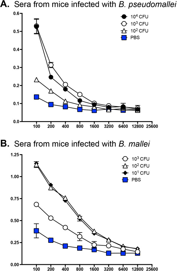
To gain insight into the immune response to BpaC during infection, we tested sera from mice that survived aerosol challenge with B. mallei ATCC 23344 and B. pseudomallei 1026b [67] by ELISA for the presence of Abs against the adhesin. Panels A and B in Figure 4 show that mice inoculated with doses as low as 10 CFU produced Abs against BpaC, which in turn demonstrates that this autotransporter is expressed by both B. mallei and B. pseudomallei during infection.
Figure 4.
ELISA with sera from mice that survived aerosol challenge with various doses of B. pseudomallei 1026b and B. mallei ATCC 23344. Serum samples were serially diluted and placed in duplicate wells of plates coated with purified His-tagged protein encompassing aa 392–1068 of B. pseudomallei 1026b BpaC. Goat α-mouse Abs conjugated to HRP were used as secondary Abs. The y-axis shows absorbance at a wavelength of 650 nm, which is indicative of antibody binding to antigens coating the plates. The x-axis represents two-fold dilutions of sera starting at 1:100 to 1:12,800. The results are expressed as the mean absorbance (±standard deviation). Closed circles show sera from mice inoculated with 104B. pseudomallei bacteria (panel A). Open circles show sera from mice infected with 103 organisms (panels A and B). Open triangles show sera from mice inoculated with 102 bacteria (panels A and B). Closed diamonds show sera from mice infected with 101 CFU of B. mallei ATCC 23344 (panel B). Blue squares represent sera from control mice that were inoculated with 50 μL of PBS using the Microsprayer (panels A and B). Of note, sera from mice that survived acute infection by B. pseudomallei and B. mallei are described elsewhere [67].
Discussion
The genome of B. mallei ATCC 23344 has been reported to specify eight autotransporter gene products [49] and of these, only BoaA (adhesin, [55]) and BimA (intracellular motility protein, [11,68]) have been functionally characterized. Both are classified as oligomeric autotransporters because they possess a short C-terminal transporter module predicted to form 4 β-strands, which anchor the molecules on the bacterial surface. In the present study, we characterized a third B. mallei ATCC 2334 oligomeric autotransporter, BpaC (BMA1027). Comparative sequence analyses indicate that the gene product is conserved among B. mallei isolates (see Additional files 1 and 2) and resembles members of the Oca (oligomeric coiled-coil adhesin) sub-family of oligomeric autotransporter proteins [16,19-21]. Consistent with this, inactivation of bpaC in the genome of B. mallei ATCC 23344 reduces adherence to monolayers of A549 (lung) and HEp-2 (larynx) cells grown in submerged cultures (Figure 3D and E, respectively). Though these cells are relevant to aerosol infection by B. mallei, they lack key features of the airway mucosa such as cilia and mucociliary activity. Ciliated cells contribute to preventing colonization of the respiratory tract by pathogenic agents by moving secretions (and trapped organisms) toward the laryngopharynx for expectoration or swallowing to the stomach (where the acidic pH neutralizes organisms). For these reasons, we measured the adherence of the B. mallei bpaC mutant to cultures of NHBE grown in an air-liquid interface system. These cultures mimic the structure and function of the airway mucosa as they form a pseudostratified epithelium with tight junctions, contain ciliated and mucus-producing goblet cells, and display mucociliary activity [63,64]. Quantitative assays using this system revealed that adherence of the bpaC mutant was reduced by 66% (Figure 3F).
Orthologs of BpaC were identified in 29 B. pseudomallei isolates (see Additional files 1 and 2). The genome of some of these strains has not been completed, resulting in the passenger domain and transporter module of BpaC seemingly specified by two different ORFs (e. g. B7210, 112, BPC006, 354e). Inactivation of bpaC in the genome of the B. pseudomallei strain DD503 caused a 2.6-fold reduction in adherence to NHBE cultures (Figure 3C), which is consistent with the phenotype of the B. mallei bpaC mutant (Figure 3F). However, the bpaC mutation did not affect adherence of B. pseudomallei to A549 or HEp-2 cells (Figure 3A and B, respectively). One possible explanation for this lack of effect is that other adhesins expressed by the B. pseudomallei DD503 bpaC mutant provide a high background of adherence to A549 and HEp-2 monolayers. For instance, BoaA and BoaB have been shown to mediate binding of B. pseudomallei DD503 to HEp-2 and A549 cells [55]. Moreover, it was recently demonstrated that the B. pseudomallei gene products BpaA, BpaB, BpaD, BpaE and BpaF all play a role in adherence to A549 cells [51]. The genes encoding these molecules are present in the genome of strain DD503.
While preparing this article, Campos and colleagues published a study in which they demonstrate that BpaC is an adhesin for A549 cells [51]. The authors reported that a mutation in the bpaC gene of B. pseudomallei strain 340 causes an ~ 10-fold reduction in adherence. These results are in contrast with our data showing that a B. pseudomallei DD503 bpaC mutant binds to A549 cells at wild-type levels (Figure 3A). One possible explanation for this phenotypic difference is that we performed adherence assays using plate-grown bacteria, and infected A549 cells for 3 hours before washing off unbound B. pseudomallei and measuring cell-binding. Campos et al. used overnight broth cultures to inoculate A549 cells and infected monolayers for only 2 hours. The method used to construct mutants might have impacted the experimental outcome of adherence assays as well. In the present study, an internal portion of the bpaC ORF was replaced with a zeocin resistance marker and this mutation was introduced in the genome of B. pseudomallei DD503 via allelic exchange. In contrast, the bpaC gene of B. pseudomallei strain 340 was disrupted via co-integration of a large plasmid (~9-kb) in the genome [51]. It is unlikely that the strain background in which the mutations were constructed has any effect on the phenotype of bpaC mutants since strains DD503 [61] and 340 [69] are both antibiotic-sensitive derivatives of the same clinical isolate, B. pseudomallei 1026b. Despite these differences, our data constitute independent proof of the role of BpaC as an adhesin. These results are substantiated by showing that expression of BpaC on the surface of recombinant E. coli bacteria increases adherence to NHBE, A549 and HEp-2 cells (Figure 2).
Given the phenotype of mutants in assays with NHBE cultures and that adherence is a key step in pathogenesis by most infectious agents, we expected the bpaC mutation to reduce the virulence of B. mallei and/or B. pseudomallei in a mouse model of aerosol infection. However, the results of our animal experiments indicate that the mutants are as virulent as wild-type strains (Table 2). Presumably, other adhesins expressed by the bpaC mutants provide sufficient adherence to the murine airway mucosa to allow colonization at wild-type levels and for the normal course of disease to ensue. It is unlikely that the lack of phenotype we observed in vivo is due to non-expression of BpaC. Though we discovered that B. pseudomallei DD503 and B. mallei ATCC 23344 do not produce detectable amounts of BpaC under routine laboratory growth conditions, ELISA with sera from mice that survived acute aerosol infection with the agents show that animals produce Abs against the protein (Figure 4A and B). Moreover, sera from horses with experimental glanders have been shown to contain high antibody titers against BpaC [70]. These results are particularly significant as horses are the natural host and reservoir for B. mallei and arguably the most relevant surrogate to study glanders. Together, these data demonstrate that BpaC is expressed in vivo and elicits the production of Abs during infection.
The infection model we used to examine the effect of the bpaC mutation might have impacted the outcome of virulence experiments. This hypothesis is supported by the Campos et al. study in which they show that the bpaC mutation reduces the ability of B. pseudomallei strain 340 to disseminate and/or survive in the liver [51]. In these experiments, BALB/c mice were infected intranasally with 500 CFU of the agent and bacterial loads in tissues were determined 48 hours post-infection. In contrast, we inoculated BALB/c mice intratracheally using a Microsprayer®, which nebulizes bacteria directly into the lungs, infected animals with doses ranging from 102 to 105 CFU, and determined bacterial burden in survivors 6–10 days post-infection (Table 2). It is also known that the choice of bacterial strains [71], inoculation route [72], and animal background [73] can significantly affect the course of disease by B. pseudomallei and B. mallei. For example, the LD50 value of the same B. pseudomallei isolate has been shown to differ by several orders of magnitude in C57BL/6 mice and BALB/c mice [74]. Therefore, a complete understanding of the role of BpaC in pathogenesis may require the use of multiple infection models.
Conclusion
This study demonstrates that BpaC is an autotransporter protein, which mediates adherence of B. mallei and B. pseudomallei to host cells that are relevant to pathogenesis by the organisms. We show that BpaC is conserved among isolates of both Burkholderia species, is expressed in vivo, and elicits production of Abs during infection. Hence, BpaC displays many properties of an important virulence factor and potential target for developing countermeasures. Though our animal experiments indicate that a mutation in bpaC does not impact the virulence of B. mallei or B. pseudomallei, adherence to host surfaces is a key early step in pathogenesis by most infectious agents. To accomplish this, pathogenic organisms typically express multiple adhesins to ascertain host colonization. It is likely that disruption of multiple genes specifying adherence factors, including bpaC, will result in decreased virulence and clarify the role of the autotransporter in the pathogenesis of B. mallei and B. pseudomallei. Continued investigation of BpaC will yield important information regarding the complex biology and virulence of these organisms, and may contribute to development of comprehensive countermeasures targeting autotransporters and their roles in pathogenesis.
Methods
Strains, plasmids, tissue culture cell lines and growth conditions
The strains and plasmids used in this study are listed in Table 3. For construction of the B. pseudomallei bpaC mutant, Low Salt Luria Bertani (LSLB) agar (Teknova) supplemented with antibiotics was utilized as selective medium. For all other experiments, B. pseudomallei was cultured on Trypticase Soy Agar (BD) at 37°C. Brucella Agar (BD) supplemented with 5% glycerol was used to grow Burkholderia mallei at 37°C. Where indicated, antibiotics were added to the culture media at the following concentrations: 7.5 μg/mL (for B. mallei) and 100 μg/mL (for B. pseudomallei) Polymixin B (MP Biomedicals), 7.5 μg/mL (for B. mallei) and 50 μg/mL (for B. pseudomallei) kanamycin (MP Biomedicals), 7.5 μg/mL (for B. mallei) and 100 μg/mL (for B. pseudomallei) zeocin™ (Life Technologies™). Plate-grown bacteria (40-hr for B. mallei, 20-hr for B. pseudomallei) were used for all experiments. For conjugative transfer of plasmids from E. coli to Burkholderia, MgSO4 was added to culture media at a final concentration of 10 mM.
Table 3.
Strains and plasmids
| Strain/plasmid | Description | Reference |
|---|---|---|
|
B. pseudomallei |
|
|
| DD503 |
Parental strain; polymixin B resistant, zeocin sensitive, kanamycin sensitive (derived from clinical isolate 1026b) |
[61] |
|
bpaC KO |
Isogenic bpaC mutant strain of DD503; polymixin B resistant, zeocin resistant, kanamycin sensitive |
This study |
|
B. mallei |
|
|
| ATCC 23344 |
Wild-type strain; polymixin B resistant, zeocin sensitive, kanamycin sensitive |
[75] |
|
bpaC KO |
Isogenic bpaC mutant strain of ATCC 23344; polymixin B resistant, zeocin resistant, kanamycin sensitive |
This study |
|
E. coli |
|
|
| EPI300 |
Cloning strain |
epicentre® Illumina® |
| TUNER™ |
Expression strain for purification of His-tagged BpaC |
EMD Millipore |
| S17 |
Strain used for conjugational transfer of pKASbpaC.zeo to B. pseudomallei and B. mallei |
[76] |
| pCC1™ |
Cloning vector, chloramphenicol resistant |
epicentre® Illumina® |
| pCCbpaC |
pCC1 containing the B. pseudomallei DD503 bpaC gene, chloramphenicol resistant |
This study |
| pCCbpaC.zeo |
pCCbpaC in which a zeocin resistance cassette was introduced near the middle of the bpaC ORF; chloramphenicol and zeocin resistant |
This study |
| pCC1.3 |
pCC1-based plasmid control, does not confer adherence to human epithelial cells; chloramphenicol resistant |
[77] |
| pKAS46 |
Mobilizable suicide plasmid; kanamycin resistant |
[78] |
| pKASbpaC.zeo |
pKAS46 containing the insert from pCCbpaC.zeo |
This study |
| pEM7ZEO |
Source of the zeocin resistance marker |
Life Technologies™ |
| pELHisBPSL1631-BMA1027 | Plasmid expressing aa 392–1068 of B. pseudomallei 1026b BpaC fused to six N-terminal histidine residues, introduced in E. coli TUNER and used to purify His-tagged BpaC protein for antibody production and ELISA experiments; chloramphenicol resistant. | [67] |
Escherichia coli was cultured at 37°C using LSLB supplemented with 15 μg/mL chloramphenicol, 50 μg/mL kanamycin, or 50 μg/mL zeocin, where indicated. For conjugation experiments, LSLB was supplemented with 10 mM MgSO4. For assays with E. coli clones carrying pCC1-based plasmids, the CopyControl™ Induction Solution (epicentre® Illumina®) was added to LSLB as previously reported [9].
The cell lines HEp-2 (human laryngeal epithelium; ATCC CCL-23), A549 (type II alveolar epithelium; ATCC CCL85) and J774A.1 (murine macrophages; ATCC TIB-67) were cultured as outlined by others [5,55]. Normal human bronchial epithelium (NHBE; LONZA) was expanded, cryopreserved and cultured in an air-liquid interface system as previously described [54,63,64]. The apical surface of the NHBE was exposed to air for a minimum of 3 weeks prior to use in adherence assays to ascertain proper cellular differentiation and the development of functional cilia.
Recombinant DNA methodology
Standard molecular biology techniques were performed as described elsewhere [79]. Genomic DNA was purified from Burkholderia using the Easy-DNA™ Kit (Life Technologies™). Plasmid DNA was isolated with the QIAprep Spin Miniprep kit (QIAGEN). The Platinum® Pfx DNA Polymerase (Life Technologies™) was used to amplify the 3.8-kb bpaC gene of B. pseudomallei DD503 with primers P1 (5’-ATA CCC AAA TCG GCG TTC TCT GGT-3′) and P2 (5′-TGC GCG AAT CAA TCG AGA TAC CCA-3′) and the PCR product was used as a template in sequencing reactions. The amplicon was also cloned in the vector pCC1™ using the CopyControl™ PCR cloning kit (epicentre® Illumina®), producing the plasmid pCCbpaC (Table 3). The latter was sequenced to determine that PCR did not introduce mutations resulting in aa substitutions in the bpaC gene product.
Construction of isogenic mutant strains of B. mallei and B. pseudomallei
The plasmid pCCbpaC was digested with the enzyme NsiI (New England BioLabs®, Inc.) to remove a 0.6-kb fragment internal to the bpaC ORF, treated with the End-It™ DNA End Repair Kit (epicentre® Illumina®), and ligated with a 0.4-kb zeocin resistance cassette to yield the construct pCCbpaC.zeo. This plasmid was restricted with BamHI (New England BioLabs®, Inc.) and a 3.4-kb fragment corresponding to the bpaC ORF disrupted by the insertion of the zeocin resistance cassette was excised from an agarose gel, purified with the High Pure PCR Product Purification kit (Roche Applied Science), and treated with the End-It™ DNA End Repair Kit. This blunt DNA fragment was then cloned in the suicide vector pKAS46. The resulting plasmid, designated pKASbpaC.zeo, was introduced in the E. coli strain S17 by electroporation, and subsequently transferred into B. mallei ATCC 23344 or B. pseudomallei DD503 by conjugation, as previously reported [55,80]. Upon conjugation, Burkholderia colonies were selected for resistance to zeocin. These putative mutants were then screened by PCR using Platinum® Pfx DNA Polymerase with primers P1 and P2. The primers yielded a PCR product of 3.8-kb in the parent strains and a smaller amplicon of 3.6-kb in bpaC mutants. The PCR products from mutant strains were sequenced to verify proper allelic exchange and successful disruption of bpaC.
Nucleotide sequence and bioinformatic analyses
PCR products and plasmids were sequenced at the University of Michigan Sequencing Core (http://seqcore.brcf.med.umich.edu). Chromatograms were assembled using the Sequencher® 5 software (Gene Codes Corporation). Sequence analyses were performed using Vector NTI (Life Technologies™) and the various online tools available through the EsPASy Bioinformatics Resource Portal (http://www.expasy.org). Signal sequence cleavage sites were determined using the SignalP 4.1 server (http://www.cbs.dtu.dk/services/SignalP). The B. mallei ATCC 23344 bpaC gene product (locus tag # BMA1027) was identified by searching the genome of the organism for the presence of a YadA anchor domain (Pfam database number PF3895.10) through the NCBI genomic BLAST service using the blastp program (http://www.ncbi.nlm.nih.gov/sutils/genom_table.cgi). The other bpaC gene products described in this study were identified using the predicted aa sequence of the B. mallei ATCC 23344 BpaC protein to search the genomes of the B. mallei and B. pseudomallei strains available through the NCBI genomic BLAST service utilizing the tblastn and blastp programs. Structural features of the BpaC proteins (helical regions, hydrophobic β-strands) were identified with the PSIPRED Protein Sequence Analysis Workbench service (http://bioinf.cs.ucl.ac.uk/psipred/).
Experiments with epithelial cells and J774 murine macrophages
Adherence, invasion, and intracellular survival assays were performed as previously reported by our laboratory [53-55]. Cells were inoculated with bacteria at a multiplicity of infection (MOI) of 100. Duplicate assays were performed on at least 3 occasions. Statistical analyses were performed using the Mann–Whitney test (GraphPad Prism® 6 software) and P values < 0.05 are reported as statistically significant.
Bactericidal assays
The method used to examine the effect of bpaC mutations on the ability of Burkholderia to resist the bactericidal activity of complement is outlined elsewhere [9,77,81]. We used final concentrations of 50% and 25% serum in assays with B. pseudomallei and B. mallei, respectively.
Protein preparations, western blot, purification of recombinant BpaC protein, and antibody production
Sarkosyl-insoluble OM protein preparations were obtained as described by Carlone et al. [82]. The methods used to prepare whole cell lysates and perform western blot experiments are described elsewhere [8,53,54,57,83,84]. His-tagged recombinant BpaC was obtained from cultures of E. coli TUNER carrying the plasmid pELHisBPSL1631-BMA1027, as previously outlined by our laboratory [67]. To obtain polyclonal Abs directed against BpaC, the purified His-tagged protein was emulsified in Freund’s adjuvants (SIGMA-ALDRICH®) and used to immunize female BALB/c mice as reported by Lafontaine and colleagues [85].
Immunofluorescence labeling of E. coli and microscopy
Expression of BpaC on the surface of E. coli recombinant bacteria was visualized by immunofluorescence microscopy as outlined by Balder et al. [55]. Briefly, paraformaldehyde-fixed E. coli cells were spotted onto glass slides. These bacteria were probed with α-BpaC polyclonal Abs, followed by incubation with a goat α-mouse antibody labeled with Alexa Fluor 546® (Life Technologies™) and the nucleic acid dye DAPI (Life Technologies™). Slides were examined by microscopy using a Zeiss LSM 510 Meta confocal system.
ELISA
Duplicate wells of Immulon™ 2HB plates (Thermo Scientific Nunc) were coated overnight at 4C° with 1 μg of His-tagged BpaC. Excess unbound antigen was removed by washing the wells with PBS + 0.05% Tween 20 (PBST), and the wells were then blocked with PBS + 0.05% containing 3% dry milk (blocking buffer) for 1 hour at room temperature. After washing with PBST, the wells were probed overnight at 4°C with sera from mice that survived acute aerosol infection with B. mallei ATCC 23344 and B. pseudomallei 1026b [67] diluted in blocking buffer. After this incubation, the wells were washed with PBST and incubated overnight with a goat α-mouse antibody conjugated to Horse Radish Peroxidase (SouthernBiotech) diluted in blocking buffer. After washing off the excess secondary antibody with PBST, 100 μL of the SureBlue™ TMB Microwell Peroxidase Substrate (KPL) was added to the wells. Color development, which is indicative of Abs binding to BpaC, was measured spectrophotometrically by determining the absorbance of well contents at a wavelength of 650 nm.
Animal experiments
Female BALB/c mice (6–8 weeks of age) were purchased from Frederick National Laboratory for Cancer Research. The animals were anesthetized by injecting a dose of 250 mg/kg of 2,2,2 tribromoethanol (SIGMA-ALDRICH®) intraperitoneally. Once anesthetized, mice were inoculated intratracheally with 50 μL of bacterial suspensions using a Microsprayer® model I-1C (PennCentury™) as previously reported by our laboratory [67]. Infected animals were monitored twice daily. Humane end-points were strictly observed. Mice exhibiting signs of moderate to severe discomfort were euthanized. This was accomplished by anesthetizing the animals with 2,2,2 tribromoethanol followed by cervical dislocation, in accordance with the AVMA Guidelines on euthanasia. Food and water were provided ad libitum. Analgesics were not used as they may have affected the experimental outcomes of the studies. Survival data were analyzed using the Kaplan-Meier method and the LD50 values were calculated according to Reed and Muench [86].
Compliance and animal research ethic statements
All experiments with live B. pseudomallei and B. mallei were performed inside a Class II Biosafety Cabinet in a BSL3 laboratory and in compliance with the rules and regulations of the U.S. Federal Select Agent Program. The experiments were approved by the University of Georgia’s Institutional Biosafety Committee (IBC).
Animal experiments were carried out in strict accordance with the recommendations in the Guide for the Care and Use of Laboratory Animals of the National Institutes of Health. The experiments were approved by the University of Georgia’s Institutional Animal Care and Use Committee (IACUC). All efforts were made to minimize animal suffering.
Abbreviations
aa: Amino acid; OM: Outer membrane; Oca: Oligomeric coiled-coil adhesin; Bp: Burkholderia pseudomallei; Bm: Burkholderia mallei; KO: Knock-out; Abs: Antibodies; LD50: Median lethal dose; LSLB: Low salt luria bertani; CFU: Colony forming units; ORF: Open reading frame; NHBE: Normal human bronchial epithelium.
Competing interests
ERL, RB, FM and RJH do not have financial or non-financial competing interests. In the past five years, the authors have not received reimbursements, fees, funding, or salary from an organization that may in any way gain or lose financially from the publication of this manuscript, either now or in the future. Such an organization is not financing this manuscript. The authors do not hold stocks or shares in an organization that may in any way gain or lose financially from the publication of this manuscript, either now or in the future. The authors do not hold and are not currently applying for any patents relating to the content of the manuscript. The authors have not received reimbursements, fees, funding, or salary from an organization that holds or has applied for patents relating to the content of the manuscript. The authors do not have non-financial competing interests (political, personal, religious, ideological, academic, intellectual, commercial or any other) to declare in relation to this manuscript.
Authors’ contributions
Conceived and designed the experiments: ERL and RJH. Performed the experiments: ERL, FM, RB. Analyzed the data: ERL, RB, RJH. Wrote the manuscript: ERL and RJH. All authors read and approve the final manuscript.
Supplementary Material
Comparison of the structural features specified by B. pseudomallei and B. mallei bpaC gene products.
Characteristics a of BMA1027 orthologous genes and their encoded products.
Contributor Information
Eric R Lafontaine, Email: elafon10@uga.edu.
Rachel Balder, Email: Rachel.Balder@gmail.com.
Frank Michel, Email: fmichel@uga.edu.
Robert J Hogan, Email: jhogan@uga.edu.
Acknowledgements
The study was supported by NIAID award AI062775 to ERL and by institutional funds from the College of Veterinary Medicine at the University of Georgia (UGA) to ERL and RJH. We thank Donald Woods (University of Calgary) for providing strains. We thank Laura Wiese (UGA), Sean Buskirk (UGA), Lauren Snipes (UGA), Xiudan Gao (UGA) and Serena Lipski (University of Toledo) for technical assistance. We thank Shawn Zimmerman (UGA) and Tomislav Jelesijevic (UGA) for their assistance redacting the manuscript.
References
- Capecchi B, Adu-Bobie J, Di Marcello F, Ciucchi L, Masignani V, Taddei A, Rappuoli R, Pizza M, Arico B. Neisseria meningitidis NadA is a new invasin which promotes bacterial adhesion to and penetration into human epithelial cells. Mol Microbiol. 2005;55(3):687–698. doi: 10.1111/j.1365-2958.2004.04423.x. [DOI] [PubMed] [Google Scholar]
- Roggenkamp A, Ackermann N, Jacobi CA, Truelzsch K, Hoffmann H, Heesemann J. Molecular analysis of transport and oligomerization of the Yersinia enterocolitica adhesin YadA. J Bacteriol. 2003;185(13):3735–3744. doi: 10.1128/JB.185.13.3735-3744.2003. [DOI] [PMC free article] [PubMed] [Google Scholar]
- Attia AS, Lafontaine ER, Latimer JL, Aebi C, Syrogiannopoulos GA, Hansen EJ. The UspA2 protein of Moraxella catarrhalis is directly involved in the expression of serum resistance. Infect Immun. 2005;73(4):2400–2410. doi: 10.1128/IAI.73.4.2400-2410.2005. [DOI] [PMC free article] [PubMed] [Google Scholar]
- Farn JL, Strugnell RA, Hoyne PA, Michalski WP, Tennent JM. Molecular characterization of a secreted enzyme with phospholipase B activity from Moraxella bovis. J Bacteriol. 2001;183(22):6717–6720. doi: 10.1128/JB.183.22.6717-6720.2001. [DOI] [PMC free article] [PubMed] [Google Scholar]
- Lipski SL, Akimana C, Timpe JM, Wooten RM, Lafontaine ER. The Moraxella catarrhalis autotransporter McaP is a conserved surface protein that mediates adherence to human epithelial cells through its N-terminal passenger domain. Infect Immun. 2007;75(1):314–324. doi: 10.1128/IAI.01330-06. [DOI] [PMC free article] [PubMed] [Google Scholar]
- Timpe JM, Holm MM, Vanlerberg SL, Basrur V, Lafontaine ER. Identification of a Moraxella catarrhalis outer membrane protein exhibiting both adhesin and lipolytic activities. Infect Immun. 2003;71(8):4341–4350. doi: 10.1128/IAI.71.8.4341-4350.2003. [DOI] [PMC free article] [PubMed] [Google Scholar]
- Maroncle NM, Sivick KE, Brady R, Stokes FE, Mobley HL. Protease activity, secretion, cell entry, cytotoxicity, and cellular targets of secreted autotransporter toxin of uropathogenic Escherichia coli. Infect Immun. 2006;74(11):6124–6134. doi: 10.1128/IAI.01086-06. [DOI] [PMC free article] [PubMed] [Google Scholar]
- Bullard B, Lipski S, Lafontaine ER. Regions important for the adhesin activity of Moraxella catarrhalis Hag. BMC Microbiol. 2007;7:65. doi: 10.1186/1471-2180-7-65. [DOI] [PMC free article] [PubMed] [Google Scholar]
- Lipski SL, Holm MM, Lafontaine ER. Identification of a Moraxella catarrhalis gene that confers adherence to various human epithelial cell lines in vitro. FEMS Microbiol Lett. 2007;267(2):207–213. doi: 10.1111/j.1574-6968.2006.00549.x. [DOI] [PubMed] [Google Scholar]
- Fexby S, Bjarnsholt T, Jensen PO, Roos V, Hoiby N, Givskov M, Klemm P. Biological Trojan horse: antigen 43 provides specific bacterial uptake and survival in human neutrophils. Infect Immun. 2007;75(1):30–34. doi: 10.1128/IAI.01117-06. [DOI] [PMC free article] [PubMed] [Google Scholar]
- Stevens JM, Ulrich RL, Taylor LA, Wood MW, Deshazer D, Stevens MP, Galyov EE. Actin-binding proteins from Burkholderia mallei and Burkholderia thailandensis can functionally compensate for the actin-based motility defect of a Burkholderia pseudomallei bimA mutant. J Bacteriol. 2005;187(22):7857–7862. doi: 10.1128/JB.187.22.7857-7862.2005. [DOI] [PMC free article] [PubMed] [Google Scholar]
- Klemm P, Hjerrild L, Gjermansen M, Schembri MA. Structure-function analysis of the self-recognizing Antigen 43 autotransporter protein from Escherichia coli. Mol Microbiol. 2004;51(1):283–296. doi: 10.1046/j.1365-2958.2003.03833.x. [DOI] [PubMed] [Google Scholar]
- Heras B, Totsika M, Peters KM, Paxman JJ, Gee CL, Jarrott RJ, Perugini MA, Whitten AE, Schembri MA. The antigen 43 structure reveals a molecular Velcro-like mechanism of autotransporter-mediated bacterial clumping. Proc Natl Acad Sci U S A. 2014;111(1):457–462. doi: 10.1073/pnas.1311592111. [DOI] [PMC free article] [PubMed] [Google Scholar]
- Valle J, Mabbett AN, Ulett GC, Toledo-Arana A, Wecker K, Totsika M, Schembri MA, Ghigo JM, Beloin C. UpaG, a new member of the trimeric autotransporter family of adhesins in uropathogenic Escherichia coli. J Bacteriol. 2008;190(12):4147–4167. doi: 10.1128/JB.00122-08. [DOI] [PMC free article] [PubMed] [Google Scholar]
- Sherlock O, Schembri MA, Reisner A, Klemm P. Novel roles for the AIDA adhesin from diarrheagenic Escherichia coli: cell aggregation and biofilm formation. J Bacteriol. 2004;186(23):8058–8065. doi: 10.1128/JB.186.23.8058-8065.2004. [DOI] [PMC free article] [PubMed] [Google Scholar]
- Cotter SE, Surana NK, 3rd St Geme JW. Trimeric autotransporters: a distinct subfamily of autotransporter proteins. Trends Microbiol. 2005;13(5):199–205. doi: 10.1016/j.tim.2005.03.004. [DOI] [PubMed] [Google Scholar]
- Henderson IR, Navarro-Garcia F, Nataro JP. The great escape: structure and function of the autotransporter proteins. Trends Microbiol. 1998;6(9):370–378. doi: 10.1016/S0966-842X(98)01318-3. [DOI] [PubMed] [Google Scholar]
- Stathopoulos C, Hendrixson DR, Thanassi DG, Hultgren SJ, 3rd St Geme JW, 3rd Curtiss R. Secretion of virulence determinants by the general secretory pathway in gram-negative pathogens: an evolving story. Microbes Infect. 2000;2(9):1061–1072. doi: 10.1016/S1286-4579(00)01260-0. [DOI] [PubMed] [Google Scholar]
- Henderson IR, Navarro-Garcia F, Desvaux M, Fernandez RC, Ala’Aldeen D. Type V protein secretion pathway: the autotransporter story. Microbiol Mol Biol Rev. 2004;68(4):692–744. doi: 10.1128/MMBR.68.4.692-744.2004. [DOI] [PMC free article] [PubMed] [Google Scholar]
- Linke D, Riess T, Autenrieth IB, Lupas A, Kempf VA. Trimeric autotransporter adhesins: variable structure, common function. Trends Microbiol. 2006;14(6):264–270. doi: 10.1016/j.tim.2006.04.005. [DOI] [PubMed] [Google Scholar]
- Hoiczyk E, Roggenkamp A, Reichenbecher M, Lupas A, Heesemann J. Structure and sequence analysis of Yersinia YadA and Moraxella UspAs reveal a novel class of adhesins. Embo J. 2000;19(22):5989–5999. doi: 10.1093/emboj/19.22.5989. [DOI] [PMC free article] [PubMed] [Google Scholar]
- Ciabattini A, Giomarelli B, Parigi R, Chiavolini D, Pettini E, Arico B, Giuliani MM, Santini L, Medaglini D, Pozzi G. Intranasal immunization of mice with recombinant Streptococcus gordonii expressing NadA of Neisseria meningitidis induces systemic bactericidal antibodies and local IgA. Vaccine. 2008;26(33):4244–4250. doi: 10.1016/j.vaccine.2008.05.049. [DOI] [PubMed] [Google Scholar]
- Bowe F, Lavelle EC, McNeela EA, Hale C, Clare S, Arico B, Giuliani MM, Rae A, Huett A, Rappuoli R, Dougan G, Mills KH. Mucosal vaccination against serogroup B meningococci: induction of bactericidal antibodies and cellular immunity following intranasal immunization with NadA of Neisseria meningitidis and mutants of Escherichia coli heat-labile enterotoxin. Infect Immun. 2004;72(7):4052–4060. doi: 10.1128/IAI.72.7.4052-4060.2004. [DOI] [PMC free article] [PubMed] [Google Scholar]
- Liu DF, Mason KW, Mastri M, Pazirandeh M, Cutter D, Fink DL, 3rd St Geme JW, Zhu D, Green BA. The C-terminal fragment of the internal 110-kilodalton passenger domain of the Hap protein of nontypeable Haemophilus influenzae is a potential vaccine candidate. Infect Immun. 2004;72(12):6961–6968. doi: 10.1128/IAI.72.12.6961-6968.2004. [DOI] [PMC free article] [PubMed] [Google Scholar]
- Cutter D, Mason KW, Howell AP, Fink DL, Green BA, 3rd St Geme JW. Immunization with Haemophilus influenzae Hap adhesin protects against nasopharyngeal colonization in experimental mice. J Infect Dis. 2002;186(8):1115–1121. doi: 10.1086/344233. [DOI] [PubMed] [Google Scholar]
- Alamuri P, Eaton KA, Himpsl SD, Smith SN, Mobley HL. Vaccination with proteus toxic agglutinin, a hemolysin-independent cytotoxin in vivo, protects against Proteus mirabilis urinary tract infection. Infect Immun. 2009;77(2):632–641. doi: 10.1128/IAI.01050-08. [DOI] [PMC free article] [PubMed] [Google Scholar]
- Waag DM, Deshazer D. In: Biological Weapons Defense: Infectious Diseases and Counterbioterrorism. Lindler LE LF, Korch GW, editor. Totowa, New Jersey: Humana Press Inc; 2004. Glanders: New Insights into an old Disease. [Google Scholar]
- Alibek K, Handelman S. Biohazard: The Chilling True Story of the Largest Covert Biological Weapons Programin the World. New York: Random House; 1999. [Google Scholar]
- Lehavi O, Aizenstien O, Katz LH, Hourvitz A. [Glanders--a potential disease for biological warfare in humans and animals] Harefuah. 2002;141:119. Spec No:88–91. [PubMed] [Google Scholar]
- Wheelis M. First shots fired in biological warfare. Nature. 1998;395(6699):213. doi: 10.1038/26089. [DOI] [PubMed] [Google Scholar]
- Wheelis M. In: Biological and Toxin Weapons: Research, Development, and use from the Middle Ages to 1945. Geissler EMoon JEC, editor. Oxford: Oxford University Press; 1999. Biological Sabotage in World War I; pp. 35–72. [Google Scholar]
- Bondi SK, Goldberg JB. Strategies toward vaccines against Burkholderia mallei and Burkholderia pseudomallei. Expert Rev Vaccines. 2008;7(9):1357–1365. doi: 10.1586/14760584.7.9.1357. [DOI] [PMC free article] [PubMed] [Google Scholar]
- Galyov EE, Brett PJ, Deshazer D. Molecular insights into Burkholderia pseudomallei and Burkholderia mallei pathogenesis. Annu Rev Microbiol. 2010;64:495–517. doi: 10.1146/annurev.micro.112408.134030. [DOI] [PubMed] [Google Scholar]
- Arun S, Neubauer H, Gurel A, Ayyildiz G, Kuscu B, Yesildere T, Meyer H, Hermanns W. Equine glanders in Turkey. Vet Rec. 1999;144(10):255–258. doi: 10.1136/vr.144.10.255. [DOI] [PubMed] [Google Scholar]
- Neubauer H, Meyer H, Finke EJ. Human glanders. Revue Internationale des Services de Sante des Forces Armees. 1997;70:258–265. [Google Scholar]
- Whitlock GC, Estes DM, Torres AG. Glanders: off to the races with Burkholderia mallei. FEMS Microbiol Lett. 2007;277(2):115–122. doi: 10.1111/j.1574-6968.2007.00949.x. [DOI] [PubMed] [Google Scholar]
- Srinivasan A, Kraus CN, DeShazer D, Becker PM, Dick JD, Spacek L, Bartlett JG, Byrne WR, Thomas DL. Glanders in a military research microbiologist. N Engl J Med. 2001;345(4):256–258. doi: 10.1056/NEJM200107263450404. [DOI] [PubMed] [Google Scholar]
- Gregory BC, Waag DM. Medical Aspects of Biological Warfare. Washington DC: Borden Institute Walter Reed Army Medical Center; 2007. Glanders; pp. 121–146. (U.S Army Medical Department Borden Insitute Textbooks of Biological Warfare). [Google Scholar]
- Cheng AC, Currie BJ. Melioidosis: epidemiology, pathophysiology, and management. Clin Microbiol Rev. 2005;18(2):383–416. doi: 10.1128/CMR.18.2.383-416.2005. [DOI] [PMC free article] [PubMed] [Google Scholar]
- Wiersinga WJ, Currie BJ, Peacock SJ. Melioidosis. N Engl J Med. 2012;367(11):1035–1044. doi: 10.1056/NEJMra1204699. [DOI] [PubMed] [Google Scholar]
- Wiersinga WJ, van der Poll T, White NJ, Day NP, Peacock SJ. Melioidosis: insights into the pathogenicity of Burkholderia pseudomallei. Nat Rev Microbiol. 2006;4(4):272–282. doi: 10.1038/nrmicro1385. [DOI] [PubMed] [Google Scholar]
- Currie BJ, Fisher DA, Howard DM, Burrow JN, Lo D, Selva-Nayagam S, Anstey NM, Huffam SE, Snelling PL, Marks PJ Stephens DP, Lum GD, Jacups SP, Krause VL. Endemic melioidosis in tropical northern Australia: a 10-year prospective study and review of the literature. Clin Infect Dis. 2000;31(4):981–986. doi: 10.1086/318116. [DOI] [PubMed] [Google Scholar]
- Currie BJ, Fisher DA, Anstey NM, Jacups SP. Melioidosis: acute and chronic disease, relapse and re-activation. Trans R Soc Trop Med Hyg. 2000;94(3):301–304. doi: 10.1016/S0035-9203(00)90333-X. [DOI] [PubMed] [Google Scholar]
- Adler NR, Govan B, Cullinane M, Harper M, Adler B, Boyce JD. The molecular and cellular basis of pathogenesis in melioidosis: how does Burkholderia pseudomallei cause disease? FEMS Microbiol Rev. 2009;33(6):1079–1099. doi: 10.1111/j.1574-6976.2009.00189.x. [DOI] [PubMed] [Google Scholar]
- Vietri NJ, Deshazer D. Medical Aspects of Biological Warfare. Washington DC: Borden Institute Walter Reed Army Medical Center; 2007. Melioidosis; pp. 147–166. (U.S Army Medical Department Borden Insitute Textbooks of Biological Warfare). [Google Scholar]
- Dance DA. Melioidosis as an emerging global problem. Acta Trop. 2000;74(2–3):115–119. doi: 10.1016/s0001-706x(99)00059-5. [DOI] [PubMed] [Google Scholar]
- Rolim DB, Vilar DC, Sousa AQ, Miralles IS, de Oliveira DC, Harnett G, O’Reilly L, Howard K, Sampson I, Inglis TJ. Melioidosis, northeastern Brazil. Emerg Infect Dis. 2005;11(9):1458–1460. doi: 10.3201/eid1109.050493. [DOI] [PMC free article] [PubMed] [Google Scholar]
- Lipsitz R, Garges S, Aurigemma R, Baccam P, Blaney DD, Cheng AC, Currie BJ, Dance DA, Gee JE, Larsen J, Limmathurotsakul D, Morrow MG, Norton R, O’Mare E, Peacock SJ, Pesik N, Rogers LP, Schweizer HP, Steinmetz I, Tan G, Tan P, Wiersinga WJ, Wuthiekanun V, Smith TL. Workshop on Treatment of and Postexposure Prophylaxis for Burkholderia pseudomallei and B. mallei infection, 2010. Emerg Infect Dis. 2012;18(12) doi: 10.3201/eid1812.120638. online report. [DOI] [PMC free article] [PubMed] [Google Scholar]
- Lazar Adler NR, Stevens JM, Stevens MP, Galyov EE. Autotransporters and their role in the virulence of Burkholderia pseudomallei and Burkholderia mallei. Front Microbiol. 2011;2:151. doi: 10.3389/fmicb.2011.00151. [DOI] [PMC free article] [PubMed] [Google Scholar]
- Campos CG, Borst L, Cotter PA. Characterization of BcaA, a putative classical autotransporter protein in Burkholderia pseudomallei. Infect Immun. 2013;81(4):1121–1128. doi: 10.1128/IAI.01453-12. [DOI] [PMC free article] [PubMed] [Google Scholar]
- Campos CG, Byrd MS, Cotter PA. Functional characterization of Burkholderia pseudomallei trimeric autotransporters. Infect Immun. 2013;81(8):2788–2799. doi: 10.1128/IAI.00526-13. [DOI] [PMC free article] [PubMed] [Google Scholar]
- Nummelin H, Merckel MC, Leo JC, Lankinen H, Skurnik M, Goldman A. The Yersinia adhesin YadA collagen-binding domain structure is a novel left-handed parallel beta-roll. Embo J. 2004;23(4):701–711. doi: 10.1038/sj.emboj.7600100. [DOI] [PMC free article] [PubMed] [Google Scholar]
- Bullard B, Lipski SL, Lafontaine ER. Hag directly mediates the adherence of Moraxella catarrhalis to human middle ear cells. Infect Immun. 2005;73(8):5127–5136. doi: 10.1128/IAI.73.8.5127-5136.2005. [DOI] [PMC free article] [PubMed] [Google Scholar]
- Balder R, Krunkosky TM, Nguyen CQ, Feezel L, Lafontaine ER. Hag mediates adherence of Moraxella catarrhalis to ciliated human airway cells. Infect Immun. 2009;77(10):4597–4608. doi: 10.1128/IAI.00212-09. [DOI] [PMC free article] [PubMed] [Google Scholar]
- Balder R, Lipski S, Lazarus JJ, Grose W, Wooten RM, Hogan RJ, Woods DE, Lafontaine ER. Identification of Burkholderia mallei and Burkholderia pseudomallei adhesins for human respiratory epithelial cells. BMC Microbiol. 2010;10:250. doi: 10.1186/1471-2180-10-250. [DOI] [PMC free article] [PubMed] [Google Scholar]
- Lazar Adler NR, Dean RE, Saint RJ, Stevens MP, Prior JL, Atkins TP, Galyov EE. Identification of a Predicted Trimeric Autotransporter Adhesin Required for Biofilm Formation of Burkholderia pseudomallei. PLoS One. 2013;8(11):e79461. doi: 10.1371/journal.pone.0079461. [DOI] [PMC free article] [PubMed] [Google Scholar]
- Cope LD, Lafontaine ER, Slaughter CA, Hasemann CA Jr, Aebi C, Henderson FW, McCracken GH Jr, Hansen EJ. Characterization of the Moraxella catarrhalis uspA1 and uspA2 genes and their encoded products. J Bacteriol. 1999;181(13):4026–4034. doi: 10.1128/jb.181.13.4026-4034.1999. [DOI] [PMC free article] [PubMed] [Google Scholar]
- Brooks MJ, Sedillo JL, Wagner N, Laurence CA, Wang W, Attia AS, Hansen EJ, Gray-Owen SD. Modular arrangement of allelic variants explains the divergence in Moraxella catarrhalis UspA protein function. Infect Immun. 2008;76(11):5330–5340. doi: 10.1128/IAI.00573-08. [DOI] [PMC free article] [PubMed] [Google Scholar]
- Brooks MJ, Sedillo JL, Wagner N, Wang W, Attia AS, Wong H, Laurence CA, Hansen EJ, Gray-Owen SD. Moraxella catarrhalis binding to host cellular receptors is mediated by sequence-specific determinants not conserved among all UspA1 protein variants. Infect Immun. 2008;76(11):5322–5329. doi: 10.1128/IAI.00572-08. [DOI] [PMC free article] [PubMed] [Google Scholar]
- Lafontaine ER, Cope LD, Aebi C, Latimer JL, McCracken GH Jr, Hansen EJ. The UspA1 protein and a second type of UspA2 protein mediate adherence of Moraxella catarrhalis to human epithelial cells in vitro. J Bacteriol. 2000;182(5):1364–1373. doi: 10.1128/JB.182.5.1364-1373.2000. [DOI] [PMC free article] [PubMed] [Google Scholar]
- Moore RA, DeShazer D, Reckseidler S, Weissman A, Woods DE. Efflux-mediated aminoglycoside and macrolide resistance in Burkholderia pseudomallei. Antimicrob Agents Chemother. 1999;43(3):465–470. doi: 10.1128/aac.43.3.465. [DOI] [PMC free article] [PubMed] [Google Scholar]
- Balder R, Hassel J, Lipski S, Lafontaine ER. Moraxella catarrhalis strain O35E expresses two filamentous hemagglutinin-like proteins that mediate adherence to human epithelial cells. Infect Immun. 2007;75(6):2765–2775. doi: 10.1128/IAI.00079-07. [DOI] [PMC free article] [PubMed] [Google Scholar]
- Krunkosky TM, Fischer BM, Martin LD, Jones N, Akley NJ, Adler KB. Effects of TNF-alpha on expression of ICAM-1 in human airway epithelial cells in vitro. Signaling pathways controlling surface and gene expression. Am J Respir Cell Mol Biol. 2000;22(6):685–692. doi: 10.1165/ajrcmb.22.6.3925. [DOI] [PubMed] [Google Scholar]
- Krunkosky TM, Jordan JL, Chambers E, Krause DC. Mycoplasma pneumoniae host-pathogen studies in an air-liquid culture of differentiated human airway epithelial cells. Microb Pathog. 2007;42(2–3):98–103. doi: 10.1016/j.micpath.2006.11.003. [DOI] [PubMed] [Google Scholar]
- Pearson MM, Hansen EJ. Identification of gene products involved in Biofilm production by Moraxella catarrhalis ETSU-9 in vitro. Infect Immun. 2007;75(9):4316–4325. doi: 10.1128/IAI.01347-06. [DOI] [PMC free article] [PubMed] [Google Scholar]
- Wang W, Pearson MM, Attia AS, Blick RJ, Hansen EJ. A UspA2H-negative variant of Moraxella catarrhalis strain O46E has a deletion in a homopolymeric nucleotide repeat common to uspA2H genes. Infect Immun. 2007;75(4):2035–2045. doi: 10.1128/IAI.00609-06. [DOI] [PMC free article] [PubMed] [Google Scholar]
- Lafontaine ER, Zimmerman S, Shaffer TL, Michel F, Gao X, Hogan RJ. Use of a safe, reproducible and rapid aerosol delivery method to study infection by Burkholderia pseudomallei and Burkholderia mallei in mice. PLoS One. 2013;8(10):e76804. doi: 10.1371/journal.pone.0076804. [DOI] [PMC free article] [PubMed] [Google Scholar]
- Stevens MP, Stevens JM, Jeng RL, Taylor LA, Wood MW, Hawes P, Monaghan P, Welch MD, Galyov EE. Identification of a bacterial factor required for actin-based motility of Burkholderia pseudomallei. Mol Microbiol. 2005;56(1):40–53. doi: 10.1111/j.1365-2958.2004.04528.x. [DOI] [PubMed] [Google Scholar]
- Mima T, Schweizer HP. The BpeAB-OprB efflux pump of Burkholderia pseudomallei 1026b does not play a role in quorum sensing, virulence factor production, or extrusion of aminoglycosides but is a broad-spectrum drug efflux system. Antimicrob Agents Chemother. 2010;54(8):3113–3120. doi: 10.1128/AAC.01803-09. [DOI] [PMC free article] [PubMed] [Google Scholar]
- Tiyawisutsri R, Holden MT, Tumapa S, Rengpipat S, Clarke SR, Foster SJ, Nierman WC, Day NP, Peacock SJ. Burkholderia Hep_Hap autotransporter (BuHA) proteins elicit a strong antibody response during experimental glanders but not human melioidosis. BMC Microbiol. 2007;7:19. doi: 10.1186/1471-2180-7-19. [DOI] [PMC free article] [PubMed] [Google Scholar]
- Goodyear A, Bielefeldt-Ohmann H, Schweizer H, Dow S. Persistent gastric colonization with Burkholderia pseudomallei and dissemination from the gastrointestinal tract following mucosal inoculation of mice. PLoS One. 2012;7(5):e37324. doi: 10.1371/journal.pone.0037324. [DOI] [PMC free article] [PubMed] [Google Scholar]
- Revelli DA, Boylan JA, Gherardini FC. A non-invasive intratracheal inoculation method for the study of pulmonary melioidosis. Front Cell Infect Microbiol. 2012;2:164. doi: 10.3389/fcimb.2012.00164. [DOI] [PMC free article] [PubMed] [Google Scholar]
- Hoppe I, Brenneke B, Rohde M, Kreft A, Haussler S, Reganzerowski A, Steinmetz I. Characterization of a murine model of melioidosis: comparison of different strains of mice. Infect Immun. 1999;67(6):2891–2900. doi: 10.1128/iai.67.6.2891-2900.1999. [DOI] [PMC free article] [PubMed] [Google Scholar]
- Leakey AK, Ulett GC, Hirst RG. BALB/c and C57Bl/6 mice infected with virulent Burkholderia pseudomallei provide contrasting animal models for the acute and chronic forms of human melioidosis. Microb Pathog. 1998;24(5):269–275. doi: 10.1006/mpat.1997.0179. [DOI] [PubMed] [Google Scholar]
- Nierman WC, DeShazer D, Kim HS, Tettelin H, Nelson KE, Feldblyum T, Ulrich RL, Ronning CM, Brinkac LM, Daugherty SC, Davidsen TD, Deboy RT, Dimitrov G, Dodson RJ, Durkin AS, Gwinn ML, Haft DH, Khouri H, Kolonay JF, Madupu R, Mohammoud Y, Nelson WC, Radune D, Romero CM, Sarria S, Selengut J, Shamblin C, Sullivan SA, White O, Yu Y. et al. Structural flexibility in the Burkholderia mallei genome. Proc Natl Acad Sci U S A. 2004;101(39):14246–14251. doi: 10.1073/pnas.0403306101. [DOI] [PMC free article] [PubMed] [Google Scholar]
- Simon R, Priefer U, Puhler A. A broad host range mobilisation system for in vivo genetic engineering: transposon mutagenesis in gram-negative bacteria. Bio/Technology. 1983;1:784–791. doi: 10.1038/nbt1183-784. [DOI] [Google Scholar]
- Holm MM, Vanlerberg SL, Foley IM, Sledjeski DD, Lafontaine ER. The Moraxella catarrhalis porin-like outer membrane protein CD is an adhesin for human lung cells. Infect Immun. 2004;72(4):1906–1913. doi: 10.1128/IAI.72.4.1906-1913.2004. [DOI] [PMC free article] [PubMed] [Google Scholar]
- Skorupski K, Taylor RK. Positive selection vectors for allelic exchange. Gene. 1996;169(1):47–52. doi: 10.1016/0378-1119(95)00793-8. [DOI] [PubMed] [Google Scholar]
- Sambrook J, Russell DW. Molecular Cloning: A Laboratory Manual (Third Edition) 3. Woodbury NY: Cold Spring Harbor Laboratory Press; 2001. [Google Scholar]
- Burtnick M, Bolton A, Brett P, Watanabe D, Woods D. Identification of the acid phosphatase (acpA) gene homologues in pathogenic and non-pathogenic Burkholderia spp. facilitates TnphoA mutagenesis. Microbiology. 2001;147(Pt 1):111–120. doi: 10.1099/00221287-147-1-111. [DOI] [PubMed] [Google Scholar]
- Aebi C, Lafontaine ER, Cope LD, Latimer JL, Lumbley SL, McCracken GH Jr, Hansen EJ. Phenotypic effect of isogenic uspA1 and uspA2 mutations on Moraxella catarrhalis 035E. Infect Immun. 1998;66(7):3113–3119. doi: 10.1128/iai.66.7.3113-3119.1998. [DOI] [PMC free article] [PubMed] [Google Scholar]
- Carlone GM, Thomas ML, Rumschlag HS, Sottnek FO. Rapid microprocedure for isolating detergent-insoluble outer membrane proteins from Haemophilus species. J Clin Microbiol. 1986;24(3):330–332. doi: 10.1128/jcm.24.3.330-332.1986. [DOI] [PMC free article] [PubMed] [Google Scholar]
- Shaffer TL, Balder R, Buskirk SW, Hogan RJ, Lafontaine ER. Use of the Chinchilla model to evaluate the vaccinogenic potential of the Moraxella catarrhalis filamentous hemagglutinin-like proteins MhaB1 and MhaB2. PLoS One. 2013;8(7):e67881. doi: 10.1371/journal.pone.0067881. [DOI] [PMC free article] [PubMed] [Google Scholar]
- Patrick CC, Kimura A, Jackson MA, Hermanstorfer L, Hood A, McCracken GH Jr, Hansen EJ. Antigenic characterization of the oligosaccharide portion of the lipooligosaccharide of nontypable Haemophilus influenzae. Infect Immun. 1987;55(12):2902–2911. doi: 10.1128/iai.55.12.2902-2911.1987. [DOI] [PMC free article] [PubMed] [Google Scholar]
- Lafontaine ER, Wagner NJ, Hansen EJ. Expression of the Moraxella catarrhalis UspA1 protein undergoes phase variation and is regulated at the transcriptional level. J Bacteriol. 2001;183(5):1540–1551. doi: 10.1128/JB.183.5.1540-1551.2001. [DOI] [PMC free article] [PubMed] [Google Scholar]
- Reed LJ, Muench H. A simple method for estimating fifty percent end points. Am J Hyg. 1938;27:793–497. [Google Scholar]
Associated Data
This section collects any data citations, data availability statements, or supplementary materials included in this article.
Supplementary Materials
Comparison of the structural features specified by B. pseudomallei and B. mallei bpaC gene products.
Characteristics a of BMA1027 orthologous genes and their encoded products.


